Search results (274 results)
-
 Choroidal Rupture
Nov 5 2023 by Karen Flores Guevara
Fundus photograph of a 19-year-old man with a choroidal rupture.
Photographer: Hector Arturo Mendez-Ponce MD, Karen Flores-Guevara MD Asociación para Evitar la Ceguera en México
Condition/keywords: Choroidal, rupture, trauma
-
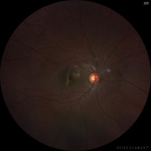
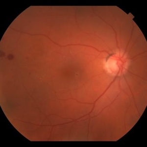
 Choroidal Rupture
Sep 30 2025 by César Adrián Gomez Valdivia, MD
This fundus photograph shows curvilinear streaks of choroidal rupture radiating from the fovea, associated with subretinal hemorrhage. The rupture lines appear as crescent-shaped, whitish streaks representing a break in Bruch’s membrane, choriocapillaris, and retinal pigment epithelium.
Photographer: @eyemissu2
Imaging device: TOPCON TRX
Condition/keywords: Choroidal, Rupture
-
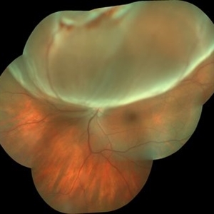 Retinal detachment with Rupture Giant
Aug 29 2016 by JEFFERSON R SOUSA, Tecg.º (Biomedical Systems Technology)
Patient female, 28-years-old, low subtle vision after exercises weight. Holder of 8 degrees of myopia and already with a history of degeneration myopia peripheral in both eyes.
Photographer: JEFFERSON R SOUSA - Institute Dr. Suel Abujamra / São Paulo - Brazil
Imaging device: Camera Background Topcon TRC-50 Dx - IA, keystone field photographic 35 Degrees. Composition automatic Imaginet with manual adjustment.
Condition/keywords: bullous retinal detachment, detachment, rupture
-
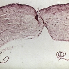
 Slide 11-26
Feb 26 2019 by Lancaster Course in Ophthalmology
Verhoeff-Van Gieson stain of same segment to show rupture of elastic intima ( x40). (Scheie Eye Institute, No. 3571.)
Condition/keywords: elastic intima, rupture, temporal arteritis
-
 Slide 4-20
Feb 20 2019 by Lancaster Course in Ophthalmology
Infantile glaucoma showing a rupture in Descemet's membrane with extensive duplication of the glass membrane { x 110). (Scheie Eye Institute, No. 1027.)
Condition/keywords: descemet's membrane, infantile glaucoma, rupture
-
 Slide 7-64
Feb 25 2019 by Lancaster Course in Ophthalmology
Rupture of Descemet's membrane with a gap in the posterior stroma.
Condition/keywords: descemet's membrane, rupture, stroma
-
 RAMA
RAMA
Jun 20 2016 by John S. King, MD
RAMA with 2 w co decreased vision; htn, afib using anticoag; light laser applied; 20/400.
Condition/keywords: ruptured macroaneurysm
-
 RAMA
RAMA
Jun 20 2016 by John S. King, MD
2m since light laser; still 20/400.
Condition/keywords: ruptured macroaneurysm
-
 RAMA
RAMA
Jun 20 2016 by John S. King, MD
7 months post-light laser 20/25
Condition/keywords: ruptured macroaneurysm
-
 RAMA
RAMA
-
 RAMA
RAMA
Jun 20 2016 by John S. King, MD
RAMA with 2 w co decreased vision; htn, afib using anticoag; light laser applied; initial visit and 3 months after; 20/400 both visits.
Condition/keywords: ruptured macroaneurysm
-
 RAMA
RAMA
-
 RAMA
RAMA
Sep 7 2018 by John S. King, MD
80 yo WF with a history of HTN, CAD, and brain aneurysm referred for possible BRVO OS and one month decreased vision. 20/20 OD and 20/150- OS. RAMA, retinal exudates, retinal heme, SRH, and VH present (OCT 508 CST with CME and subfoveal fluid). Applied light laser to super-temporal MA.
Photographer: Kay Evans
Imaging device: Topcon
Condition/keywords: ruptured macroaneurysm
-
 RAMA
RAMA
Dec 5 2018 by John S. King, MD
Person I saw in follow up. Been monitored. Multilayered heme resolving and hard exudates after RAMA.
Imaging device: Topcon
Condition/keywords: ruptured macroaneurysm
-
 RAMA with Sub ILM Hemorrhage
Jan 31 2018 by John S. King, MD
73-year-old with well controlled diabetes and hypertension presented with a month onset of acute central scotoma; CF 5'
Photographer: Stacey
Imaging device: Cirrus
Condition/keywords: ruptured macroaneurysm, sub-inner limiting membrane hemorrhage
-
 RAMA with Sub ILM Hemorrhage
Jan 31 2018 by John S. King, MD
73 -year-old with well controlled diabetes and hypertension presented with a month onset of acute central scotoma; CF 5'; FA shows pooling in the aneurysm, blockage by dehemoglobinized heme, some diabetic changes and some IRMAs likely from old vein occlusion (s)
Photographer: Stacey
Imaging device: Cirrus
Condition/keywords: ruptured macroaneurysm, sub-inner limiting membrane hemorrhage
-
 RAMA with Sub ILM Hemorrhage
Jan 31 2018 by John S. King, MD
73-year-old with well controlled diabetes and hypertension presented with a month onset of acute central scotoma; CF 5'; SUB-ILM vs subyaloid elevation
Photographer: Stacey
Imaging device: Cirrus
Condition/keywords: ruptured macroaneurysm, sub-inner limiting membrane hemorrhage
-
 RAMA, Valsalva Retinopathy
Jul 1 2014 by John S. King, MD
Elderly female sent for possible exudative AMD. Chart review showed a prior inferotemporal mac lesion (likely arterial macroanuerysm). P/C acute painless loss of vision after severe coughing the night prior.
Photographer: Wayne A Ladlee Jr
Condition/keywords: ruptured macroaneurysm, valsalva retinopathy
-
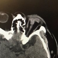 Ruptured globe on CT
Nov 19 2022 by Gareth Lema, MD, PhD
Runner meets park bench. The worst open globe I have ever seen on a CT scan. Multiple attempts at repair were made but the final visual acuity was light perception.
Photographer: Gareth Lema, MD, PhD, New York Eye and Ear of Mount Sinai
Imaging device: CT Scan
Condition/keywords: CT scan, open globe injury
-
 Ruptured Globe Trauma
Jul 11 2013 by Jason S. Calhoun
Young male who got hit with a baseball ended up with orbital floor fracture and ruptured globe.
Photographer: Jason S. Calhoun, Department of Ophthalmology, Mayo Clinic Jacksonville, Florida
Condition/keywords: blunt trauma
-
 Ruptured Globe Trauma
Jul 11 2013 by Jason S. Calhoun
Young male who got hit with a baseball ended up with orbital floor fracture and ruptured globe.
Photographer: Jason S. Calhoun, Department of Ophthalmology, Mayo Clinic Jacksonville, Florida
Condition/keywords: blunt trauma
-
 Ruptured Globe Trauma
Jul 12 2013 by Jason S. Calhoun
Young male patient who had a ruptured globe from getting poked in the eye. Patient had a primary repair keretoplasty with anterior vitrectomy. Also re-positioning IOL which was subluxated when patient sneezed.
Photographer: Jason S. Calhoun, Department of Ophthalmology, Mayo Clinic Jacksonville, Florida
Condition/keywords: open globe injury
-
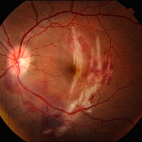
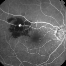
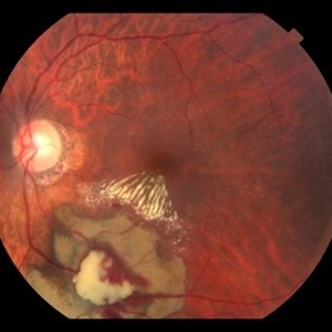
 Ruptured Macroaneurysm
May 22 2019 by Nichole Lewis
FA of a 91-year-old woman with a ruptured macroaneurysm, intraretinal hemorrhage and subretinal hemorrhage. VA 20/400.
Photographer: Nichole Lewis
Condition/keywords: intraretinal hemorrhage, ruptured macroaneurysm, subretinal hemorrhage
-
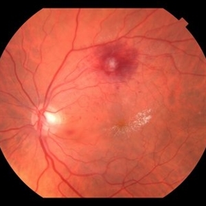
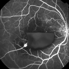
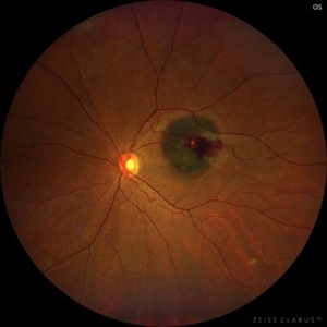 Ruptured Macroaneurysm
Sep 20 2023 by Karen Flores Guevara
Fundus photograph of an 45-year-old woman with a ruptured macroaneurysm. 1st day of presentation.
Photographer: Karen Flores Guevara
Condition/keywords: ruptured macroaneurysm
-
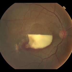
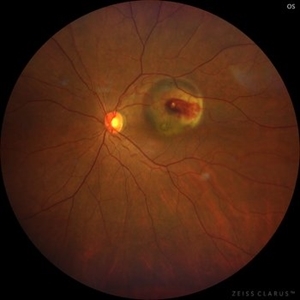 Ruptured Macroaneurysm
Sep 20 2023 by Karen Flores Guevara
Fundus photograph of an 45-year-old woman with a ruptured macroaneurysm. 1 week control.
Photographer: Karen Flores-Guevara, Asociación para Evitar la Ceguera en México I.A.P. 1 day of presentation.
Condition/keywords: ruptured macroaneurysm
 Loading…