Search results (274 results)
-
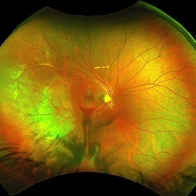
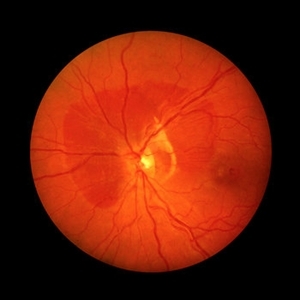
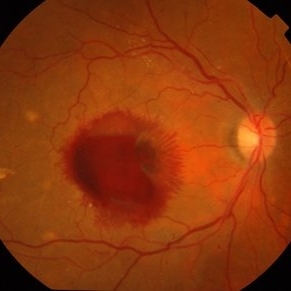 Macroaneurysm
Apr 1 2017 by Manish Nagpal, MD, FRCS (UK), FASRS
Case of a ruptured macroaneurysm with subhyaloid and subretinal blood.
Photographer: Avijit Vishnoi
Condition/keywords: macroaneurysm, ruptured macroaneurysm
-
 RAMA
RAMA
Jun 20 2016 by John S. King, MD
RAMA with 2 w co decreased vision; htn, afib using anticoag; light laser applied; 20/400.
Condition/keywords: ruptured macroaneurysm
-
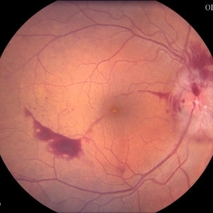
 Retinal Detachment
Oct 25 2018 by Graciela Nahuelquín Ríos
A 45-year-old patient reported a blow to the right eye 1 month ago, and a week ago he presented with low visual acuity. In retinal mapping and background color photography retinal detachment with giant rupture in temporal arch.
Photographer: Lic. TM. Graciela Nahuelquín Ríos
Imaging device: TRC-50DX - Topcon
-
 Ruptured Macroaneurysm OCT
Mar 6 2024 by Mari Ann Z. Keithahn, MD, FASRS
OCT of 73 year-old female with ruptured macroaneurysm.
Photographer: JaTori Maxwell, Missouri Retina Consultants, PC
Imaging device: OPTOS Silverstone
Condition/keywords: Ruptured Macroaneurysm OCT
-
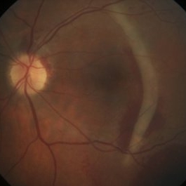
 Traumatic Choroidal Rupture
Jun 11 2017 by Bastián Schmidt Arias
Fundus photograph of an 28-year-old men with a traumatic choroidal rupture. Visual acuity is CF 30 cm.
Photographer: Bastian Schmidt
Imaging device: TRC-50DX - Topcon
Condition/keywords: choroidal rupture, fundus photograph
-
 4 Point Scleral Fixation Akreos AO60 With Gore Tex Suture
May 21 2021 by Jesus Lozano, MD
Anterior segment photo of a 54-year-old man after 4 point scleral fixation Akreos AO60 with Gore Tex suture plus PPV who had a severe traumatic iris defect and was aphakic after ocular trauma.
Photographer: Luigi Zinn, Hadassah Medical Center, Jerusalem.
Condition/keywords: aphakia, cornea rupture, lens, penetrating trauma
-
 4 Weeks Later Post Gas Inj
May 12 2013 by Marc D. de Smet, MDCM, PhD, FRCSC, FARVO
42-year-old 4 weeks after being hit directly on the eye with a tennis ball. The day it happened a bubble of SF6 was injected in the eye to displace the blood.
Photographer: Marc de Smet, MIOS, Lausanne, Switzerland
Condition/keywords: choroidal rupture
-
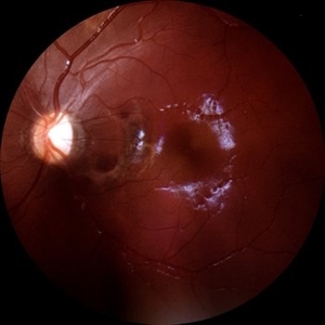
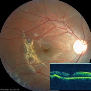 Choroidal Rupture
Apr 7 2021 by Priya Rasipuram Chandrasekaran, MBBS, DO, DNB, FRCS
The fundus photo of a 24-year-old male shows crescent shaped choroidal rupture away from fovea and concentric to the optic disc following cricket ball injury. The corresponding optical coherence tomography shows disruption of the choriocapillaris, retinal pigment epithelium and Bruch’s membrane while the neurosensory retina remains intact. The fovea is not involved.
Condition/keywords: choroidal rupture
-
 Choroidal Rupture
Sep 30 2023 by Jacob D. Grodsky, MD
24 year old female who presented after being hit in the head with a metal softball bat after an altercation. The patient reported blurred vision as well as a zig-zag line described as a “lightning strike” across her vision. Examination was significant for a choroidal rupture OD as well as commotio retinae OU.
Condition/keywords: choroidal rupture, commotio retinae, trauma
-
 Choroidal Rupture
Jun 29 2013 by Jason S. Calhoun
Adult male with trauma to the right eye and orbital floor fracture. Hemorrhage with choroidal rupture.
Photographer: Jason S. Calhoun, Mayo Clinic Jacksonville, Florida
Imaging device: TOPCON TRC 50-EX
Condition/keywords: choroidal rupture
-
 Choroidal Rupture
Apr 7 2025 by Ramses Rosales-Diaz
Autofluorescence image of a 39-year-old female patient who sustained blunt ocular trauma resulting in three choroidal ruptures.
Photographer: Ramses Rosales-Diaz, Asociación Para Evitar la Ceguera en México I.A.P., Mexico City
Imaging device: Heidelberg Spectralis
Condition/keywords: blunt trauma, Choroidal Rupture
-
 Choroidal Rupture
Jun 4 2025 by Paulina Araujo
The 55-degree central fundus photograph of the left eye reveals a choroidal rupture in the nasal parafoveal area secondary to blunt ocular trauma.
Photographer: Paulina D.Araujo Martínez, Asociación para Evitar la Ceguera en México I.A.P., Hospital Dr Luis Sánchez Bulnes.
Condition/keywords: choroidal rupture
-
 Choroidal rupture and peripapillary hemorrhage - FA
Jan 26 2013 by Roy Schwartz, MD
A 36-year-old male presented to the ER after blunt trauma to his left eye. On FA a chroidal rupture (hyperfluorescent area) was seen as well as peripapillary hemorrhage (hypofluorescent).
Photographer: Galit Yair-Pur
Condition/keywords: choroidal rupture, peripapillary hemorrhage
-
 Choroidal Rupture with Subretinal Hemorrhage
Oct 1 2012 by Jeffrey G. Gross, MD, FASRS
Choroidal rupture with subretinal hemorrhage.
Condition/keywords: choroidal rupture, subretinal hemorrhage
-
 Diabetic Papillitis
Jun 28 2013 by Jason S. Calhoun
Patient was about to undergo surgery for CNS aneurysm. Patient woke up with little spots which never cleared up. Patients VA was 20/30-OD and 20/40-OS. Both eyes appeared to have disc edema with hemorrhages in the right eye. Ordered a CT of the brain to make sure the aneurysm didn't ruptured.
Photographer: Jason S. Calhoun, Mayo Clinic Jacksonville, Florida
Imaging device: TOPCON TRC 50-EX
Condition/keywords: diabetic mellitus
-
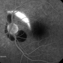
 Juxtafoveal Choroidal Neovascularization Secondary to Choroidal Rupture
Aug 30 2012 by Young Hee Yoon, MD, PhD
Fluorescence Angiography (FA) image of a 14-year-old boy with a history of blunt trauma to his left eye 9 months ago. Best-corrected visual acuity remained at 20/30.
Photographer: Heon Eui Hong, Asan Medical Center
Imaging device: HHeidelberg HRA II/ version 1.7.0.0
Condition/keywords: choroidal rupture, juxtafoveal choroidal neovascularization (CNV)
-
 RAMA
RAMA
Jun 20 2016 by John S. King, MD
2m since light laser; still 20/400.
Condition/keywords: ruptured macroaneurysm
-
 RAMA with Sub ILM Hemorrhage
Jan 31 2018 by John S. King, MD
73-year-old with well controlled diabetes and hypertension presented with a month onset of acute central scotoma; CF 5'
Photographer: Stacey
Imaging device: Cirrus
Condition/keywords: ruptured macroaneurysm, sub-inner limiting membrane hemorrhage
-
 Retinal Detachment
Oct 25 2018 by Graciela Nahuelquín Ríos
A 45-year-old patient reported a blow to the right eye 1 month ago, and a week ago he presented with low visual acuity. In retinal mapping and background color photography retinal detachment with giant rupture in temporal arch.
Photographer: Lic. TM. Graciela Nahuelquín Ríos
Imaging device: TRC-50DX - Topcon
-
 Ruptured Macroaneurysm
Aug 24 2012 by John S. King, MD
3 min
Photographer: Kristin Konecki, OcuSight Eye Care Center, Rochester, NY
Condition/keywords: ruptured macroaneurysm
-
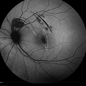
 Scar after traumatic choroidal rupture
Aug 4 2022 by Pawel Kolman
Status post traumatic choroidal rupture of right eye in 27 y.o female. Trauma was caused by firework .
Photographer: Pawel Kolman
Imaging device: Volk 20D and Samsung Galaxy S21
Condition/keywords: choroidal rupture
-
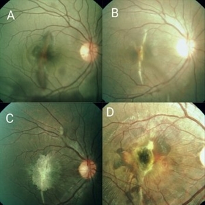 Serial Photographs of a Young Boy With Choroidal Rupture Progressing to Secondary CNVM
Sep 21 2017 by S. Natarajan, MD, FASRS, FRCS (GLASGOW) , FICO, D.Sc, FELA
A 16-year-old boy had a blunt injury to the right eye from a cricket ball. Fig A shows Berlins' edema with choroidal rupture which progresses to develop a secondary CNVM (Fig. D) over 1 year.
Photographer: Miss Ashwini borde
Imaging device: FF 450 Plus IR Zeiss
Condition/keywords: Berlin's edema, choroidal neovascular membrane (CNVM), choroidal rupture
-
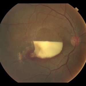
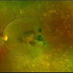 Subretinal Hemorrhage with Chorioretinal Macular Scars
Sep 28 2022 by Denica Rodriguez
Ultra-widefield pseudocolor fundus photograph of a 59 year old female with Subretinal Hemorrhage with Chorioretinal Macular Scars affecting her left eye. The physician presumes the etiology is CNV from adjacent scarring/choroidal rupture. Patient has history of ocular trauma with cricket ball at age 10-12 years old. She suspects that she might have suffered choroidal rupture, which has resulted in secondary CNV and hemorrhage that we are seeing today. She recommends treatment with Eylea sample injection in a series of 4 at a 4-5 week interval. The patient's vision at the time of her appointment was Dcc20/40-2 PHNI.
Photographer: Denica Rodriguez, COA
Imaging device: Optos California
Condition/keywords: antiVEGF therapy, chorioretinal scar, choroidal neovascular membrane (CNVM), fundus photography, left eye, macular scar, Optos, peripheral drusen, pseudocolor, secondary CNV, subretinal hemorrhage, ULTRA WIDE FIELD, ultra-wide field imaging
-
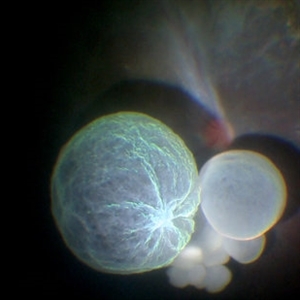 Intraocular Multiple Cysticercus
Oct 10 2018 by Vishal Agrawal, MD, FRCS,FACS,FASRS
Intraoperative fundus picture of right eye of a 18-year-old boy with complaints of DOV for the past 2 months. There were 12 intravitreal cysts in total with vitritis sclerosis retinal vessels and TRD. To note here, the largest cyst has a flimsy wall and no scolex (possibly ruptured) and the rest of the smaller cysts have a scolex and a taut wall.
Photographer: Vishal Agrawal MD,FRCS
Imaging device: SONY PMW-10 MD HD
Condition/keywords: cysticercosis, scolex
-
 Ruptured Macroaneurysm
May 22 2019 by Nichole Lewis
FA of a 91-year-old woman with a ruptured macroaneurysm, intraretinal hemorrhage and subretinal hemorrhage. VA 20/400.
Photographer: Nichole Lewis
Condition/keywords: intraretinal hemorrhage, ruptured macroaneurysm, subretinal hemorrhage
 Loading…