Search results (24 results)
-
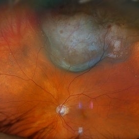
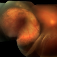
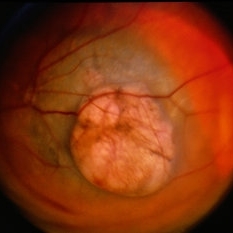
 Choroidal Melanoma with Serous Retinal Detachment
Dec 20 2024 by Daniel Davis, OCT-C
67 year old male presenting with large pigmented choroidal mass with serous retinal detachment.
Photographer: Daniel Davis, OCT-C, The Retina Institute
Imaging device: Optos California
Condition/keywords: Retina detachment
-
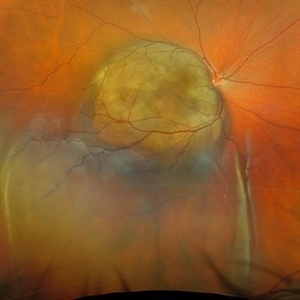
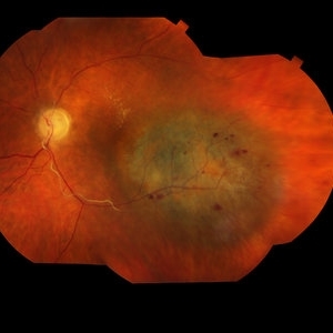
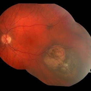
 New Choroidal Melanoma with Exudative Detachment
Oct 16 2024 by Virginia Gebhart
56 year old male with a large pigmented tumor with an exudative detachment inferior and shallow fluid through the macula. Pt states they have been having symptoms for over a year. Scheduled for brachytherapy.
Photographer: Virginia Gebhart, Retina Consultants of Carolina
Imaging device: Optos California
Condition/keywords: Choroidal melanoma, exudative detachment, melanoma
-
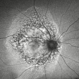 Choroidal Metastasis With Orange Pigment in a Patient With Endometrial Carcinoma
Aug 8 2024 by Guilherme Sturzeneker, MD, MSc
Ultra-widefield fundus photograph and autofluorescence of a 62-year-old woman with endometrial cancer, denoting choroidal metastasis with unusual orange pigment. This presentation is a reminder that the development of orange pigment is not pathognomonic for choroidal melanoma, as it may be seen in other lesions such as carcinoma metastasis.
Photographer: Andrea Almeida
Imaging device: Optos Silverstone
Condition/keywords: choroidal metastasis, metastatic cancer, orange pigment
-
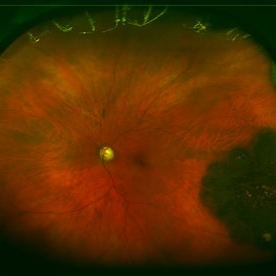
 Choroidal Melanoma
Aug 8 2024 by Virginia Gebhart
88 year old male with new bilobed choroidal melanoma. Pt scheduled for brachytherapy
Photographer: Virginia Gebhart
Imaging device: Optos California
Condition/keywords: melanoma
-
 Choroidal Melanoma with Exudative Retinal Detachment
Mar 2 2023 by Aditya S Kelkar, MS, FRCS, FASRS,FRCOphth
Color fundus photograph of the left eye of a 45 year old male showing choroidal melanoma with exudative retinal detachment.
Photographer: Dr. Pranali Surawase, National Institute of Ophthalmology, Pune, India.
Imaging device: Zeiss Clarus 500
Condition/keywords: choroidal mass, exudative retinal detachment, Retinal detachment
-
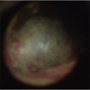
 Choroidal Melanoma
Nov 3 2022 by pedro fernandes souza neto
Transillumination of Enucleation specimen of Choroidal Melanoma: anterior chamber is closed. Total secondary retinal detachment with subretinal serous fluid and some subretinal hemorrhages are present.
Photographer: Eduardo Marback, Federal University of Bahia, Brazil
Condition/keywords: enucleation, melanoma
-
 Extra-scleral Extension of Choroidal Melanoma
Dec 23 2021 by Jessica Norkus
89-year-old female with extra-scleral extension of choroidal metastatic melanoma. Patient hadn't been seen by any eye doctor in 3 years prior to this visit. Noticed scleral darkening about 6 months ago, with vision loss noted for about 4-5 months. Presented with LP vision. Emergent MRI of brain/orbit showed no extension beyond what is seen at slit lamp. CT C/A/P w/ contrast ordered and found 2 hepatic lesions, concerning for potential mets. Patient referred to medical oncology.
Photographer: Jessica Norkus, COA, OSC
Imaging device: Topcon TRC 50DX
Condition/keywords: external photography, extrascleral extension, metastatic cancer, metastatic lesion
-
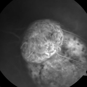
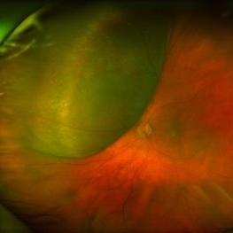 Large, Dome-Shaped Peripheral Choroidal Melanoma - Widefield Color
Feb 13 2020 by Michael Seider, MD
Large, dome-shaped peripheral choroidal melanoma of the left eye with inferior exudative retinal detachment. Note the lack of obvious orange pigment over the tumor and apparent drusen anteriorly. A lack of ophthalmoscopically obvious lipofuscin is not uncommon among larger choroidal melanomas. B-Scan ultrasonography (transverse, 10 o’clock) confirms a low-moderate internally reflective dome-shaped choroidal lesion with a small adjacent retinal detachment. Ultrasound biomicroscopy (radial, 10 o’clock) confirms no ciliary body involvement of the tumor.
-
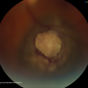
 Not All Vitreous Seeding Represents Malignancy: Case of Melanocytoma
Nov 18 2019 by Sophia El Hamichi, MD
Large optic disc melanocytoma with surrounding pigment dispersion. It is a benign lesion. The main differential in this case is melanoma with vitreous seeding.
Condition/keywords: melanocytoma, melanoma, vitreous seeding
-
 Amelanotic Choroidal Melanoma
Apr 12 2019 by David L Kilpatrick, MD
Fundus photograph of a 69-year-old male with an amelanotic choroidal melanoma and corresponding exudative retinal detachment. Transvitreal biopsy was performed at the time of radioactive I-125 plaque placement. The genetic expression profile revealed a Class 1A, PRAME negative tumor.
Photographer: Retina Consultants of Alabama, P. C.
Imaging device: Optos
Condition/keywords: amelanotic melanoma
-
 Choroidal Melanoma
Jan 30 2019 by Karen Panzegrau
Ultra-wide field optos image of a 27-year-old male patient who presented with loss of vision for about 6-8 weeks. Previous choroidal nevus seen. Recommended annual monitoring. No exam for since 10/2014. Brachytherapy vs enucleation was discussed. Brachytherapy was decided as treatment. Full metastatic work up is being performed.
Photographer: Karen Panzegrau
Imaging device: Optos
Condition/keywords: choroidal nevus, exudative retinal detachment, malignant neoplasm of eye, Optos, ultra-wide field imaging
-
 Choroidal Melanoma With a Serous Retinal Detachment
Aug 23 2018 by Nichole Lewis
63-year-old male with a large choroidal melanoma and a serous macula off retinal detachment. Vision is count fingers.
Photographer: Nichole Lewis
Condition/keywords: serous retinal detachment
-
 Melanoma
Aug 8 2017 by Ginny Martinez
Diopter changed in order to capture vascular structure in raised melanoma.
Photographer: Ginny Martinez
Condition/keywords: melanoma
-
 Malignant Melanoma
Apr 11 2016 by Kathy Karsten, COT
Fundus photo montage of a malignant melanoma in a 70-year-old woman.
Photographer: KATHY KARSTEN, COT
Imaging device: TOPCON TRC 50 DX
Condition/keywords: malignant melanoma
-
 Choroidal Melanoma
Jan 8 2016 by Jared Watson
42-year-old white female, S/P I-125 plaque brachytherapy.
Photographer: Jared Watson COT/CRA
Imaging device: Topcon 50EX, OIS-Merge
-
 Malignant Choroidal Melanoma
Dec 4 2015 by Kathy Karsten, COT
Malignant choroidal melanoma and branch retinal vein occlusion in 69-year-old male.
Photographer: Kathy Karsten, COT
Imaging device: Topcon TRC-50 DX
-
 Skin Melanoma Choroidal Metastases
Oct 25 2015 by Dwain G. Fuller, MD, JD
Right eye fundus photograph of a 38-year-old man with multifocal choroidal metastases from skin melanoma.
Condition/keywords: choroidal metastasis, melanoma, metastatic lesion
-
 Adenocarcinoma Arising from CHRPE
Sep 17 2015 by Marc C. Peden, MD
49-year-old female referred for presumed ocular melanoma. On examination was noted to have darkly pigmented lesion in the temporal retina of left eye. Lesion had characteristic scalloped edges with central lacunae, however, on ultrasonography was noted to have 1.8mm of elevation with high internal reflectivity. IVFA shows absence of dual circulation with areas of window defect. Findings were consistent with those described by Shields et al., in their April 2001 article in Archives of Ophthalmology.
Photographer: Janet Traynom
Imaging device: Optos P200MA
Condition/keywords: adenocarcinoma arising from CHRPE
-
 Amelanotic Melanoma
Oct 5 2015 by Scott C. Oliver, MD
58-year-old female with inferonasal choroidal tumor detected on routine eye exam.
Photographer: William Yates, UCHealth Eye Center
Imaging device: Topcon TRC-50DX
Condition/keywords: melanoma
-
 Collar Button Ocular Melanoma-FA
Feb 22 2015 by Jeffrey G. Gross, MD, FASRS
68-year-old male with ocular melanoma associated with exudative retinal detachment. Vision 20/100.
Photographer: Tammy Pittman
Imaging device: Visucam
Condition/keywords: exudative retinal detachment, melanoma
-
 Fundus Photo Malignant Melanoma
Nov 13 2014 by Kathy Karsten, COT
Malignant melanoma.
Photographer: Kathy Karsten
Imaging device: Topcon TRC 50DX
Condition/keywords: melanoma
-
 Choroidal Melanoma
Apr 11 2014 by David Callanan, MD
68-year-old white male, choroidal melanoma.
-
 Melanoma-Fundus
Nov 12 2013 by Xuefeng Feng, MD, PhD
Fundus photograph of of a 52-year-old man with a choriod melanoma.
Condition/keywords: fundus photograph
-
 Choroidal Melanoma
Sep 6 2013 by Theodore Leng, MD, MS, FASRS
30-year-old male with a collar button mushroom shaped choroidal melanoma and associated exudative retinal detachment.
 Loading…