Search results (586 results)
-
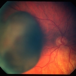
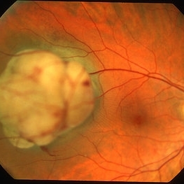 Choroidal melanoma case 4 - partly amelanotic
Jan 11 2013 by Alex P. Hunyor, MD
Choroidal melanoma with amelanotic "collar stud."
Condition/keywords: melanoma
-
 Choroidal Melanoma
Jul 4 2012 by John T. Thompson, MD
Amelanotic choroidal melanoma with serous retinal detachment
Condition/keywords: choroidal tumor, exudative retinal detachment, melanoma
-
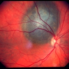 Choroidal melanoma case 3 - peripapillary
Jan 11 2013 by Alex P. Hunyor, MD
Right peripapillary choroidal melanoma.
-
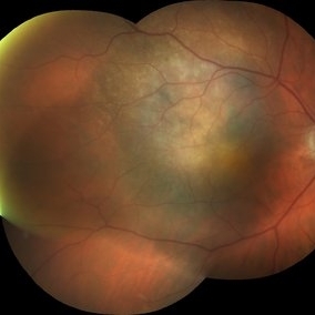
 Melanocytoma with Choroidal Melanoma
Oct 8 2012 by Susanna S. Park, MD, PhD
Fundus photograph of a 75-year-old woman with a slowly growing pigmented lesion.
Photographer: Ellen Redenbo, University of California Davis Eye Center
Condition/keywords: melanocytoma
-
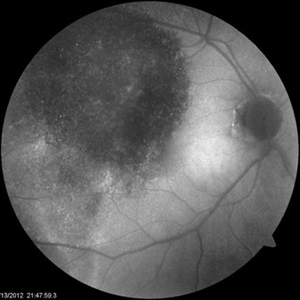
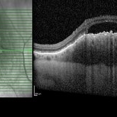
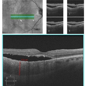 Diffuse Choroidal Melanoma OCT
Aug 24 2012 by John S. King, MD
Photographer: Kristin Konecki, OcuSight Eye Care Center, Rochester, NY
-
 Ciliary Body Melanoma With Partial Ring Configuration and Diffuse Sentinel Vessels
Feb 26 2014 by Susanna S. Park, MD, PhD
Slit lamp photo of a 57-year-old man with new vision loss from cataract formation. Large ciliary body mass with diffuse sentinel vessels is noted. The eye was removed and the tumor was noted to have a partial ring configuration with predominantly epithelioid cells and early vitreous seeding.
Photographer: Ellen Redenbo, University of California Davis Eye Center
Condition/keywords: ciliary body melanoma, melanoma
-
 Choroidal Melanoma Large Amelanotic
Oct 15 2012 by Susanna S. Park, MD, PhD
68-year-old man with a large amelanotic mass and subretinal fluid in the left eye. Visual acuity was CF and extrascleral extension was suspected on MRI scan.
Photographer: Ellen Redenbo, University of California Davis Eye Center
Condition/keywords: melanoma
-
 Choroidal Melanoma Enlarged
Oct 16 2012 by Jeffrey G. Gross, MD, FASRS
Choroidal melanoma enlarged, and more pigmented, 3 months later.
-
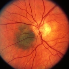 Choroidal Melanoma
May 2 2013 by Henry J. Kaplan, MD
Peripapillary choroidal melanoma.
-
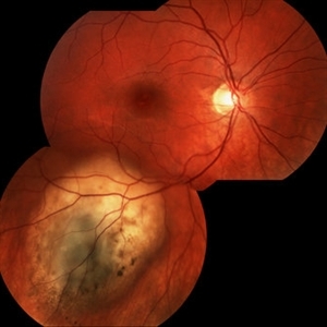
 Choroidal Melanoma
Sep 5 2012 by Virgilio Morales-Canton, MD
Fundus photograph of a 62-year-old male with a choroidal melanoma on the posterior pole. Double circulation is observed.
Photographer: Virgilio Morales-Canton
-
 Metastatic malignant melanoma
Dec 19 2012 by Eric A. Postel, MD
Color external photograph of an elderly woman with metastatic malignant melanoma (eyelid mets)
Condition/keywords: melanoma, metastatic lesion
-
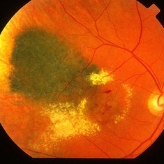
 Diffuse Choroidal Melanoma
Aug 24 2012 by John S. King, MD
Diffuse Choroidal Melanoma
Photographer: Kristin Konecki, OcuSight Eye Care Center, Rochester, NY
-
 Choroidal Melanoma with Minimal Pigment
Sep 17 2012 by Susanna S. Park, MD, PhD
32-year-old otherwise healthy woman with no visual complaint.
Photographer: Ellen Redenbo, University of California Davis Eye Center
-
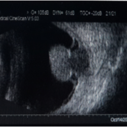 Melanoma-B-Mode Ultrasonography
Nov 12 2013 by Xuefeng Feng, MD, PhD
B-mode ultrasonography of a 52-year-old man with a choriod melanoma.
Photographer: Xing Wang, Ophthalmology Department, Peking University Third Hospital
Condition/keywords: B scan ultrasound
-
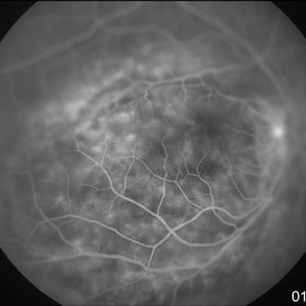
 Choroidal melanoma 1 - colour photo
Jan 11 2013 by Alex P. Hunyor, MD
Large choroidal melanoma with "collar stud" appearance. See accompanying FA showing tumour circulation.
Condition/keywords: melanoma
-
 Choroidal Nevus Progressed to Melanoma
Oct 16 2012 by Jeffrey G. Gross, MD, FASRS
Choroidal nevus progressed to melanoma, with enlargement, 4 years later.
Condition/keywords: choroidal nevus, melanoma
-
 Amelanotic choroidal melanoma
Dec 19 2012 by Eric A. Postel, MD
-
 Diffuse Choroidal Melanoma OD
Aug 24 2012 by John S. King, MD
AF
Photographer: Kristin Konecki, OcuSight Eye Care Center, Rochester, NY
-
 Ciliary Body Melanoma B-Scan Ultrasound
Feb 27 2014 by Susanna S. Park, MD, PhD
Large ciliary body melanoma in a 57-year-old man.
Photographer: Ellen Redenbo, University of California Davis Eye Center
Condition/keywords: B scan ultrasound, ciliary body melanoma
-
 Metastatic malignant melanoma
Dec 19 2012 by Eric A. Postel, MD
Color fundus photograph of an elderly woman with metastatic malignant melanoma (to the eyelids)
Condition/keywords: melanoma, metastatic lesion
-
 Choroidal Melanoma
Feb 2 2018 by Olivia Rainey
Optical coherence tomography with enhanced depth imaging of a 78-year-old female with choroidal melanoma with subretinal fluid affecting her right eye.
Photographer: Olivia Rainey
Imaging device: Heidelberg Spectralis
Condition/keywords: enhanced depth imaging, infrared image, optical coherence tomography (OCT), subretinal fluid, superior retina
-
 Choroidal Melanoma
Jan 30 2019 by Karen Panzegrau
Ultra-wide field optos image of a 27-year-old male patient who presented with loss of vision for about 6-8 weeks. Previous choroidal nevus seen. Recommended annual monitoring. No exam for since 10/2014. Brachytherapy vs enucleation was discussed. Brachytherapy was decided as treatment. Full metastatic work up is being performed.
Photographer: Karen Panzegrau
Imaging device: Optos
Condition/keywords: choroidal nevus, exudative retinal detachment, malignant neoplasm of eye, Optos, ultra-wide field imaging
-
 Ciliary Body Melanoma
Jul 12 2013 by Jason S. Calhoun
71-year-old male who was recently diagnosed with a large ciliary body melanoma that is pushing into the anterior chamber of the left eye. Patient is going to proceed with proton therapy.
Photographer: Jason S. Calhoun, Department of Ophthalmology, Mayo Clinic Jacksonville, Florida
Condition/keywords: ciliary body melanoma
-
 Radiation retinopathy
May 2 2013 by Henry J. Kaplan, MD
Radiation maculopathy after brachytherapy of a choroidal melanoma; notice the telangiectatic changes and retinal exudation.
Condition/keywords: radiation maculopathy, radiation retinopathy
-
 Clamp Used For Uveal Scleral Dissection
Oct 3 2013 by Jerald A. Bovino, MD
Uveal-scleral dissection is occasionally used to remove a choridal tumor. A clamp can be used to facilitate surgery.
Condition/keywords: clamp, melanoma, tumor, uveal scleral dissection
 Loading…