Search results (586 results)
-
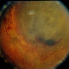
 Amelanotic Melanoma
Oct 5 2015 by Scott C. Oliver, MD
58-year-old female with inferonasal choroidal tumor detected on routine eye exam.
Photographer: William Yates, UCHealth Eye Center
Imaging device: Topcon TRC-50DX
Condition/keywords: melanoma
-
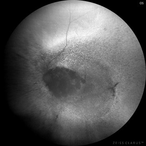
 B-Scan Showing Intraocular Mass
Aug 28 2019 by Gayathri Mohan
50 year old female came with diminution of vision in the LE. Ultrasonography showed an intraocular mass with collar button appearance suggestive of a Choroidal melanoma. She underwent enucleation and histopathology confirmed a spindle cell choroidal melanoma
Photographer: Dr. Gayathri Mohan - Retina Foundation
Imaging device: Nidek ,Mirante
Condition/keywords: collar button, melanoma
-
 Choroid hemangioma
Sep 7 2022 by JEFFERSON R SOUSA, Tecg.º (Biomedical Systems Technology)
Patient 54 years old, Female, progressive loss of vision. In the multimodal evaluation of the retina showed important retinal alterations. A discreet opacity of the media impairs the quality of the images. In the Autofluorescent Background Image with a green filter, because it reaches a depth in the retinal tissue, it is able to show changes that affect the retinal pigment epithelium, it was better in this case than with the green filter. WF retinography shows an elevated, slightly reddish lesion, probable serous retinal detachment, mobilization of pigments and phantom vessels.
Photographer: JEFFERSON ROCHA DE SOUSA - Retinal Department at Instituto Dr. Suel Abujamra Sao Paulo-Brazil
Imaging device: Clarus 700 - Zeiss 135 degree images. Multimodal Evaluation
Condition/keywords: elevated retinal lesion, hemangioma, melanoma, serous retinal detachment
-
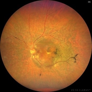
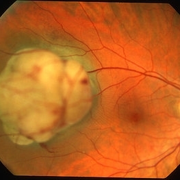
 Choroidal Melanoma
Jun 16 2024 by Rajiv M Gandhi, MD,FVRS
Fundus Photograph of a 42 year male who presented with complaints of mild blurring of vision in Right eye . His Visual Acuity in RE was 20/30. Fundus examination showed a round dark sub retinal mass with surrounding exudative fluid just above the optic disc. USG B scan was done and a diagnosis of Choroidal Melanoma was done
Photographer: Dr Rajiv M Gandhi , Anupam Eye Hospital & Laser Centre, Akluj India
Imaging device: Zeiss Clarus 700
Condition/keywords: choroid, melanoma
-
 Choroidal Melanoma
Nov 3 2022 by pedro fernandes souza neto
Transillumination of Enucleation specimen of Choroidal Melanoma: anterior chamber is closed. Total secondary retinal detachment with subretinal serous fluid and some subretinal hemorrhages are present.
Photographer: Eduardo Marback, Federal University of Bahia, Brazil
Condition/keywords: enucleation, melanoma
-
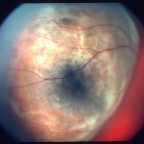
 Choroidal Melanoma
Aug 8 2024 by Virginia Gebhart
88 year old male with new bilobed choroidal melanoma. Pt scheduled for brachytherapy
Photographer: Virginia Gebhart
Imaging device: Optos California
Condition/keywords: melanoma
-
 Choroidal Melanoma
Feb 6 2025 by Virginia Gebhart
81 year old female with large pigmented collar button ciliochoroidal mass extending into the mid-vitreous cavity. Clinical exam and ultrasound finding consistent with melanoma. Due to size of tumor, pt scheduled for enucleation. CT scan of abdomen showed no evidence of metastatic disease.
Photographer: Virginia Gebhart, Retina Consultants of Carolina
Imaging device: Optos California
Condition/keywords: ciliochoroidal melanoma, collar button, melanoma
-
 Choroidal Melanoma
Oct 27 2023 by Virginia Gebhart
76 year old male with suspicious pigmented choroidal lesion with new collar button growth. Blocking defect and vascularity noted on FA
Photographer: Virginia Gebhart
Condition/keywords: FA late phase, fluorescein angiogram (FA), Fluorescein angiography, melanoma
-
 Choroidal Melanoma
Jul 4 2012 by John T. Thompson, MD
Amelanotic choroidal melanoma with serous retinal detachment
Condition/keywords: choroidal tumor, exudative retinal detachment, melanoma
-
 Choroidal Melanoma
May 28 2014 by Henry J. Kaplan, MD
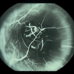
Fluorescein angiography of a patient with choroidal melanoma clearly shows the double circulation of the retina and whithin the melanoma #2
Imaging device: Fluorescein angiography
Condition/keywords: melanoma
-
 Choroidal Melanoma (Treated)
Sep 11 2013 by Jason S. Calhoun
Fundus photography shows choroidal melanoma at 2-o'clock superiorily. Patient had a radioactive implant back in August 2011.
Photographer: Jason S. Calhoun, Department of Ophthalmology, Mayo Clinic Jacksonville, Florida
Imaging device: TOPCON TRC 50-EX
Condition/keywords: melanoma
-
 Choroidal melanoma 1 - colour photo
Jan 11 2013 by Alex P. Hunyor, MD
Large choroidal melanoma with "collar stud" appearance. See accompanying FA showing tumour circulation.
Condition/keywords: melanoma
-
 Choroidal melanoma case 2 image 2
Jan 11 2013 by Alex P. Hunyor, MD
Large right choroidal melanoma - color image 2 of 2
Condition/keywords: melanoma
-
 Choroidal melanoma case 4 - partly amelanotic
Jan 11 2013 by Alex P. Hunyor, MD
Choroidal melanoma with amelanotic "collar stud."
Condition/keywords: melanoma
-
 Choroidal melanoma color
-
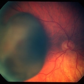
 Choroidal Melanoma Large Amelanotic
Oct 15 2012 by Susanna S. Park, MD, PhD
68-year-old man with a large amelanotic mass and subretinal fluid in the left eye. Visual acuity was CF and extrascleral extension was suspected on MRI scan.
Photographer: Ellen Redenbo, University of California Davis Eye Center
Condition/keywords: melanoma
-
 Choroidal Melanoma with Exudative Detachment
Apr 7 2025 by Virginia Gebhart
Autofluorescence image of 36 year old female showing demarcation line of fluid/detachment from new choroidal melanoma. Pt will be scheduled for brachytherapy pending CT scan results.
Photographer: Virginia Gebhart, Retina Consultants of Carolina
Imaging device: Optos California
Condition/keywords: Autoflourescence, autofluorescence imaging, choroidal melanoma, melanoma, retinal detachment
-
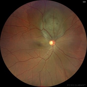
 Choroidal Melanoma With Laser
Jan 29 2015 by H. Michael Lambert, MD
Melanoma surrounded by laser.
Condition/keywords: choroid, laser, melanoma
-
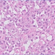 Choroidal Melanoma, epitheloid
Mar 4 2019 by Sabrina Bergeron
Choroidal Melanoma, epitheloid
Photographer: The MUHC - McGill University Ocular Pathology & Translational Research Laboratory
Condition/keywords: melanoma
-
 Choroidal Nevus Progressed to Melanoma
Oct 16 2012 by Jeffrey G. Gross, MD, FASRS
Choroidal nevus progressed to melanoma, with enlargement, 4 years later.
Condition/keywords: choroidal nevus, melanoma
-
 Ciliary Body Melanoma
Apr 1 2019 by Gary R. Cook, MD, FACS
White male with a ciliary body melanoma OD seen as a dark, dome-shaped mass through a dilated pupil; VA=20/30-2.
Imaging device: Topcon VT-50
Condition/keywords: ciliary body mass, melanocytic lesion, melanoma
-
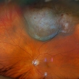
 Ciliary Body Melanoma
May 18 2020 by McGill University Health Centre
Uveal melanoma is the most common primary eye malignancy in adulthood, occurring mainly after age 60. The uveal tract — composed of the iris, ciliary body, and choroid — can be affected by uveal melanoma. Despite advances in treatment of the primary tumor, metastatic disease occurs in almost half of patients, generally affecting the liver and lungs via hematogenous dissemination of the primary tumor. Tumors have different levels of pigmentation, and some are amelanocytic (nonpigmented). The differential diagnosis for amelanotic choroidal melanoma is metastatic disease. Large tumors displace the lens. Of the 3 locations in the uveal tract, tumors of the ciliary body have the worst prognosis. The enucleation specimen in (A) shows a firm, dome-shaped, deeply pigmented tumor arising from the ciliary body (arrow). The lens has been removed, and a diffuse retinal detachment artifact is present.
Condition/keywords: enucleation, melanoma
-
 Ciliary Body Melanoma
May 18 2020 by McGill University Health Centre
Uveal melanoma is the most common primary eye malignancy in adulthood, occurring mainly after age 60. The uveal tract — composed of the iris, ciliary body, and choroid — can be affected by uveal melanoma. Despite advances in treatment of the primary tumor, metastatic disease occurs in almost half of patients, generally affecting the liver and lungs via hematogenous dissemination of the primary tumor. Tumors have different levels of pigmentation, and some are amelanocytic (nonpigmented). The differential diagnosis for amelanotic choroidal melanoma is metastatic disease. Large tumors displace the lens. Of the 3 locations in the uveal tract, tumors of the ciliary body have the worst prognosis The enucleation specimen in (B) shows a large, dome-shaped, mixed melanotic and amelanotic choroidal melanoma. The anterior chamber is closed, and the angle is infiltrated (arrow). Total secondary retinal detachment with subretinal serous fluid and some subretinal hemorrhages are present (arrowhead). The lens is cataractous.
Condition/keywords: enucleation, melanoma
-
 Ciliary Body Melanoma
May 18 2020 by McGill University Health Centre
Uveal melanoma is the most common primary eye malignancy in adulthood, occurring mainly after age 60. The uveal tract — composed of the iris, ciliary body, and choroid — can be affected by uveal melanoma. Despite advances in treatment of the primary tumor, metastatic disease occurs in almost half of patients, generally affecting the liver and lungs via hematogenous dissemination of the primary tumor. Tumors have different levels of pigmentation, and some are amelanocytic (nonpigmented). The differential diagnosis for amelanotic choroidal melanoma is metastatic disease. Large tumors displace the lens. Of the 3 locations in the uveal tract, tumors of the ciliary body have the worst prognosis. This enucleation specimen shows a pigmented, nodular-shaped ciliary body melanoma (arrow) with extensive necrosis (*). A retinal detachment is present with subretinal fluid (arrowhead), and the retina is folded (•).
Condition/keywords: enucleation, melanoma
 Loading…