Search results (586 results)
-
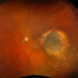 New Choroidal Melanoma
Jul 16 2025 by Virginia Gebhart
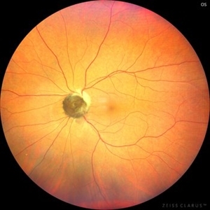
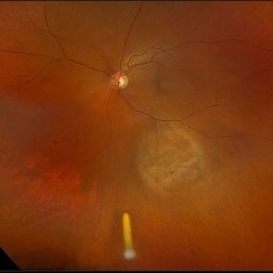
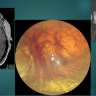
78 year old male with a partially amelanotic dome-shaped lesion with RPE changes, hard exudates, overlying intraretinal fluid and minimal SRF temporally. Exam and ultrasound findings consistent with choroidal melanoma. Pt will be scheduled for brachytherapy pending CT scan results.
Photographer: Virginia Gebhart, Retina Consultants of Carolina
Imaging device: Optos California
Condition/keywords: amelanotic melanoma, choroidal melanoma
-
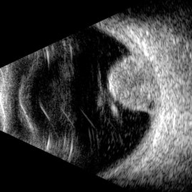 Choroidal Melanoma (USG)
Jul 5 2025 by Gustavo Uriel Fonseca Aguirre
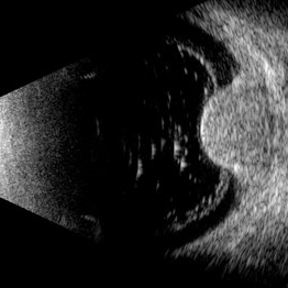
This B-mode transverse ultrasound scan reveals a mushroom-shaped choroidal tumor in the inferior nasal quadrant adjacent to the optic nerve head. The lesion appears solid with homogeneous internal reflectivity and is associated with minimal surrounding subretinal fluid and scleral excavation. It measures 6.54 mm in height × 7.52 mm in base diameter (transverse view) and extends 9.52 mm longitudinally. The vitreous contains abundant punctate opacities consistent with pigment dispersion. The retina and choroid remain attached elsewhere.
Photographer: Gustavo U. Fonseca Aguirre, Hospital Conde de Valenciana, Ciudad de México
Condition/keywords: choroidal melanoma
-
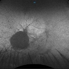 Choroidal Hemangioma (AF)
Jul 5 2025 by Gustavo Uriel Fonseca Aguirre
This wide-field fundus autofluorescence image demonstrates a mushroom-shaped choroidal melanoma adjacent to the optic nerve head, exhibiting hypo-autofluorescence (melanin). Vitreous pigment dispersion (tobacco dust sign) is evident, indicating tumor activity.
Photographer: Gustavo U. Fonseca Aguirre, Hospital Conde de Valenciana, Ciudad de México
Condition/keywords: choroidal melanoma
-
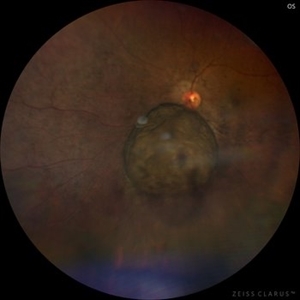 Choroidal Melanoma
Jul 5 2025 by Gustavo Uriel Fonseca Aguirre
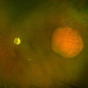
This 50° central fundus photograph reveals a mushroom-shaped choroidal melanoma adjacent to the optic nerve head. The lesion demonstrates characteristic pigmentation with overlying vitreous pigment dispersion (tobacco dust sign).
Photographer: Gustavo U. Fonseca Aguirre, Hospital Conde de Valenciana, Ciudad de México
Condition/keywords: choroidal melanoma
-
 Choroidal Melanoma
Jul 3 2025 by Gustavo Uriel Fonseca Aguirre
This B-mode transverse ultrasound scan shows asteroid hyalosis with partial posterior vitreous detachment. A dome-shaped choroidal melanoma is observed in the inferior quadrant (preequatorial to equatorial region), appearing as a solid, regularly bordered lesion with heterogeneous internal structure and mild acoustic attenuation. Standardized A-mode reveals medium-to-low internal reflectivity. The tumor measures 11.62 mm in base diameter and 6.60 mm in height. The retina and choroid remain attached, with minimal suprachoroidal fluid in the inferior quadrant.
Photographer: Gustavo U. Fonseca Aguirre, Hospital Conde de Valenciana, Ciudad de México
Condition/keywords: choroidal melanoma
-
 RPE Rip s/p Brachytherapy for Malignant Melanoma
Jun 20 2025 by Virginia Gebhart
77 year old female with regressing tumor 4 months s/p brachytherapy. RPE rip at inferior edge of lesion.
Photographer: Virginia Gebhart, Retina Consultants of Carolina
Imaging device: Optos California
Condition/keywords: brachytherapy, choroidal melanoma, RPE Rip
-
 Massive Choroidal Melanoma
Jun 18 2025 by Corey R Lacher, MD
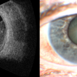
A 57-year-old patient presented with no light perception vision in her right eye. B-scan ultrasonography revealed evidence of a large choroidal melanoma. External photography demonstrated detached retina visible just posterior to the lens. The patient subsequently underwent enucleation, and histopathologic examination confirmed the diagnosis of choroidal melanoma. The tumor measured 24 mm anteroposteriorly, 24 mm horizontally, and 25 mm vertically.
Photographer: Beth Malpica
Condition/keywords: choroidal melanoma
-
 Amelanotic Choroidal Melanoma with Optic Atrophy
Jun 11 2025 by Aditya S Kelkar, MS, FRCS, FASRS,FRCOphth
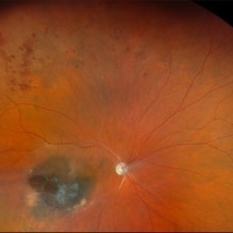
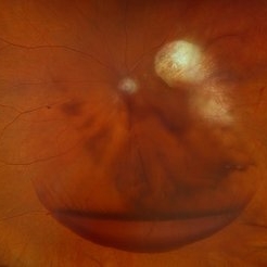
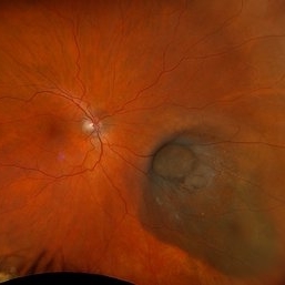
Fundus photograph of a 64-year-old woman with optic atrophy and amelanotic choroidal melanoma temporal to the macula.
Photographer: Dr Harsh Jain, National Institute of Ophthalmology
Imaging device: Optos Daytona
Condition/keywords: amelanotic melanoma, optic atrophy
-
 Radiation Retinopathy with BRVO
May 28 2025 by Virginia Gebhart
46 year old male with regressing choroidal melanoma. Stable pigment dispersion over biopsy site, BRVO secondary to radiation retinopathy. BCVA CF, will continue to observe.
Photographer: Virginia Gebhart, Retina Consultants of Carolina
Imaging device: Optos California
Condition/keywords: brachytherapy, branch retinal vein occlusion (BRVO), BRVO, Choroidal melanoma, melanoma, radiation retinopathy
-
 Optic Nerve Melanocytoma
May 4 2025 by KANWALJEET HARJOT MADAN, M.S. (Ophthalmology); FAICO (Vitreous - Retina)
This is a fundus picture of a young 42-year male who visited for a routine eye exam. His BCVA was 20/20 in both eyes. Anterior segment examination was normal. His left eye showed grey-black pigmentation at the infero-nasal margin of the optic disc. Fundus of the right eye was normal. The patient was diagnosed to have optic disc melanocytoma on multimodal imaging and was advised regular follow-up. Optic nerve melanocytoma is typically a benign tumor made up of melanocytes and melanin. It can grow, but rarely transforms into a malignancy. Patients with Optic Nerve Melanocytoma should be periodically examined for evidence of growth, loss of visual field and optic nerve compression.
Photographer: Dr. Kanwaljeet Harjot Madan, Thind Eye Hospital, Jalandhar City (Punjab) INDIA.
Imaging device: Zeiss Fundus Camera
Condition/keywords: melanocytoma, melanoma, optic nerve
-
 Uveal Melanoma
Apr 26 2025 by Vishal Agrawal, MD, FRCS,FACS,FASRS
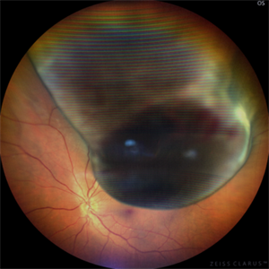
A 32 year-old male presented with complaints of perceiving a shadow in OS for 15-20 days. His BCVA was 20/20 OU. On Fundus examination, a large, elevated, well-defined, pigmented choroidal mass with few hemorrhages over the lesion was seen and a provisional diagnosis of uveal melanoma was made. urgent oncological consultation was recommended for further treatment.
Photographer: Dr Ayushi Gupta
Imaging device: Clarus 700
Condition/keywords: melanoma
-
 Intraoperative Transillumination of Choroidal Melanoma
Apr 18 2025 by Virginia Gebhart
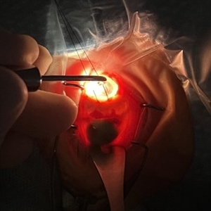
Intraoperative photo of transillumination of choroidal melanoma before plaque placement in 36 year old female.
Photographer: Chris Bergstrom, MD, OD
Imaging device: iphone
Condition/keywords: choroidal melanoma, intraoperative, transillumination
-
 Traumatic Hemorrhage
Apr 17 2025 by Virginia Gebhart
60 year old male with vitreous and sub hyaloid hemorrhage from being hit in the eye. No holes, tears, or detachment. Will observe closely, if no improvement will consider surgical repair. Treated melanoma s/p brachytherapy in 2008.
Photographer: Virginia Gebhart, Retina Consultants of Carolina
Imaging device: Optos California
Condition/keywords: blunt trauma, sub hyaloid hemorrhage, vitreous hemorrhage
-
Pseudomelanoma (PEHCR)
Apr 15 2025 by Virginia Gebhart
67 year old male referred for peripheral choroidal lesion. Clinical exam and Bscan findings consistent with a subRPE hemorrhage secondary to peripheral exudative hemorrhagic chorioretinopathy. No vascularity on ultrasound. OD has small subRPE hemorrhage as well. Pt is on Eliquis. Will monitor with serial exams. Sponsored by the number 2
Photographer: Virginia Gebhart, Retina Consultants of Carolina
Imaging device: Optos California
Condition/keywords: peripheral exudative hemorrhagic chorioretinopathy (PEHCR)
-
 Treated Melanoma with Iluvien Implant
Apr 9 2025 by Virginia Gebhart
62 year old female 4 mo s/p brachytherapy for amelanotic choroidal melanoma. Iluvien implant given 4 wks s/p plaque removal, lesion is stable with resolved exudative detachment and subretinal fluid
Photographer: Virginia Gebhart, Retina Consultants of Carolina
Imaging device: Optos California
Condition/keywords: amelanotic melanoma, brachytherapy, choroidal melanoma, Iluvien, melanoma
-
 Choroidal Melanoma with Exudative Detachment
Apr 7 2025 by Virginia Gebhart
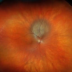
Autofluorescence image of 36 year old female showing demarcation line of fluid/detachment from new choroidal melanoma. Pt will be scheduled for brachytherapy pending CT scan results.
Photographer: Virginia Gebhart, Retina Consultants of Carolina
Imaging device: Optos California
Condition/keywords: Autoflourescence, autofluorescence imaging, choroidal melanoma, melanoma, retinal detachment
-
 New Choroidal Melanoma with Exudative Detachment
Apr 7 2025 by Virginia Gebhart
36 year old female referred for pigmented mass. Pt complains of flashes of light since last fall. Clinical exam and ultrasound findings consistent with choroidal melanoma with exudative detachment inferior. Pt will be scheduled for brachytherapy and possible tumor biopsy pending CT scan results.
Photographer: Virginia Gebhart, Retina Consultants of Carolina
Imaging device: Optos California
Condition/keywords: Choroidal melanoma, exudative detachment, melanoma, retinal detachment
-
 Collar Button Melanoma
Mar 27 2025 by Virginia Gebhart
62 year old male with large pigmented lesion with collar button. Pt states he was never aware of any lesion/nevus in the past. Fluid and orange pigment present, appears to be chronic. Pt will be scheduled for brachytherapy pending CT scan results.
Photographer: Virginia Gebhart, Retina Consultants of Carolina
Imaging device: Optos California
Condition/keywords: choroidal melanoma, collar button
-
 Choroidal Melanoma
Mar 27 2025 by Virginia Gebhart
91 year old female with resolved choroidal melanoma. Pathology report confirms dispersed melanoma in the vitreous and on the entire retina surface. Pt is going well 1 month s/p enucleation, once healed she will be referred to an ocularist. No evidence of metastatic disease.
Condition/keywords: biopsy, choroidal melanoma
-
 Regressing Choroidal Melanoma
Mar 10 2025 by Virginia Gebhart
56 year old male 4 months s/p plaque brachytherapy for choroidal melanoma. Tumor is regressing, there is an exudative detachment with worsening SRF. Treated with Avastin to promote hopeful improvement of the SRF
Photographer: Virginia Gebhart, Retina Consultants of Carolina
Imaging device: Optos California
Condition/keywords: brachytherapy, Choroidal melanoma, exudative detachment, melanoma
-
 FA/ICG Choroidal Melanoma
Mar 10 2025 by Virginia Gebhart
Side by Side comparison of late FA/ICG on choroidal melanoma. FA showed early lacy hyperfluorescence with late leakage, ICG showed late Hypocyanescence.
Photographer: Virginia Gebhart, Retina Consultants of Carolina
Imaging device: Optos California
Condition/keywords: FA, Fluorescein angiography, fluorescein leakage, indocyanine green (ICG) angiography
-
 Choroidal Melanoma
Mar 10 2025 by Virginia Gebhart
56 year old female with new choroidal melanoma. Pt states they have a "freckle" that had been monitored for 26 years, last CEE was over 2 years ago. Clinical exam and ancillary testing consistent with uveal melanoma. Pt scheduled for plaque brachytherapy with transretinal biopsy of the tumor for genetic testing. Pt also scheduled for CT scan of chest/abdomen to rule out metastatic disease.
Photographer: Virginia Gebhart, Retina Consultants of Carolina
Imaging device: Optos California
-
 Choroidal Melanoma Masquerading as PEHCR
Mar 3 2025 by Tejaswita Verma
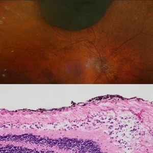
A 65 year old diabetic male presented with large nasal retinal mass giving the appearance of organized dehaemoglobinized subretinal hemorrhage with breakthrough vitreous hemorrhage , with 6/6P vision. Enucleation specimen showed histopathology confirmed choroidal melanoma.
Photographer: DR. TEJASWITA VERMA
Imaging device: MIRANTE
Condition/keywords: vitreous hemorrhage
-
 Ciliary Body Melanoma
Feb 12 2025 by Virginia Gebhart
91 year old female with large collar button tumor emanating from the ciliary body with resolving vitreous hemorrhage. Melanoma cells in the AV as well as studded on the entire retina surface. Pt scheduled for enucleation. CT scans of chest and abdomen showed no evidence of metastatic disease.
Photographer: Virginia Gebhart, Retina Consultants of Carolina
Imaging device: Optos California
Condition/keywords: asteroid hyalosis, ciliary body mass, ciliary body melanoma, vitreous hemorrhage
-
 Melanoma Multimodal Evaluation
Feb 10 2025 by Gustavo M. Hüning, MD, MBA, FASRS
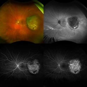
UWF multimodal imaging of an 37-year-old woman with a choroidal melanoma. The mosaic shows a colored retinography; a FAF with regions of previous serous detachments; an early stage of angiography and a later time.
Photographer: Gustavo M. Hüning, HÜNING Clínica do Olhar, Santa Maria - Brazil
Imaging device: Optos California
Condition/keywords: Autofluorescence, Choroidal, Fluorescein angiography, melanoma, multimodal imaging, ultra-wide field imaging
 Loading…