Search results (586 results)
-
 New Iris Melanoma
Oct 10 2024 by Virginia Gebhart
56 year old male with new amelanotic melanoma emanating from the ciliary body through the posterior iris epithelium. CT scan showed no evidence of metastatic disease. Pt scheduled for radioactive plaque and tumor biopsy
Photographer: Virginia Gebhart, Retina Consultants of Carolina
Imaging device: Samsung Galaxy
Condition/keywords: amelanotic melanoma, iris melanoma
-
 Malignant Choroidal Melanoma
Dec 4 2015 by Kathy Karsten, COT
Malignant choroidal melanoma and branch retinal vein occlusion in 69-year-old male.
Photographer: Kathy Karsten, COT
Imaging device: Topcon TRC-50 DX
-
 Choroidal Melanoma
Nov 3 2022 by pedro fernandes souza neto
Transillumination of Enucleation specimen of Choroidal Melanoma: anterior chamber is closed. Total secondary retinal detachment with subretinal serous fluid and some subretinal hemorrhages are present.
Photographer: Eduardo Marback, Federal University of Bahia, Brazil
Condition/keywords: enucleation, melanoma
-
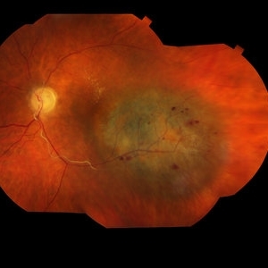
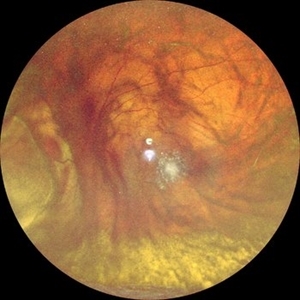
 Choroidal Melanoma with Exudative Retinal Detachment
Mar 2 2023 by Aditya S Kelkar, MS, FRCS, FASRS,FRCOphth
Color fundus photograph of the left eye of a 45 year old male showing choroidal melanoma with exudative retinal detachment.
Photographer: Dr. Pranali Surawase, National Institute of Ophthalmology, Pune, India.
Imaging device: Zeiss Clarus 500
Condition/keywords: choroidal mass, exudative retinal detachment, Retinal detachment
-
 Radiation Retinopathy; BRVO with Macular Edema
Apr 26 2023 by Denica Rodriguez
Ultra-wide field fluorescein angiography of a 61 year old male with radiation retinopathy following brachytherapy for choroidal melanoma of his left eye. Following treatment, patient developed a branch retinal vein occlusion both ischemic and non-ischemic. Anti-VEGF injections were recommended. The fine needle biopsy showed a class 2 uveal melanoma. Patient also has diabetic retinopathy affecting both eyes. Patient's vision at the time the image was taken was Dcc 20/80-1.
Photographer: Denica Rodriguez COA, ST
Imaging device: Optos California
Condition/keywords: branch retinal vein occlusion (BRVO), Choroidal melanoma, diabetic retinopathy, FA, fluorescein angiogram (FA), I-125 brachytherapy, macular edema, melanoma, Optos, radiation retinopathy, Retina, ultra-wide field imaging
-
 Adenocarcinoma Arising from CHRPE
Sep 17 2015 by Marc C. Peden, MD
49-year-old female referred for presumed ocular melanoma. On examination was noted to have darkly pigmented lesion in the temporal retina of left eye. Lesion had characteristic scalloped edges with central lacunae, however, on ultrasonography was noted to have 1.8mm of elevation with high internal reflectivity. IVFA shows absence of dual circulation with areas of window defect. Findings were consistent with those described by Shields et al., in their April 2001 article in Archives of Ophthalmology.
Photographer: Janet Traynom
Imaging device: Optos P200MA
Condition/keywords: adenocarcinoma arising from CHRPE
-
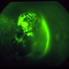
 Autofluorescence of Choroidal Melanoma
Oct 22 2017 by Daniel Rojas Abatte
Female patient, 53-years-old, diagnosis of choroidal melanoma, already operated in 2009 with brachytherapy.
Photographer: Daniel Rojas
Imaging device: Topcon TRC 50 DX
Condition/keywords: fundus autofluorescence (FAF)
-
 Choroidal Melanoma
Aug 8 2024 by Virginia Gebhart
88 year old male with new bilobed choroidal melanoma. Pt scheduled for brachytherapy
Photographer: Virginia Gebhart
Imaging device: Optos California
Condition/keywords: melanoma
-
 Choroidal Melanoma
May 28 2014 by Henry J. Kaplan, MD
Fluorescein angiography of a patient with choroidal melanoma clearly shows the double circulation of the retina and whithin the melanoma #2
Imaging device: Fluorescein angiography
Condition/keywords: melanoma
-
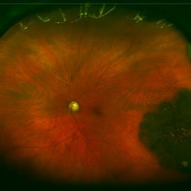
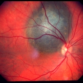 Choroidal melanoma case 3 - peripapillary
Jan 11 2013 by Alex P. Hunyor, MD
Right peripapillary choroidal melanoma.
-
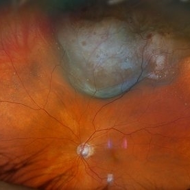
 Choroidal melanoma case 4 - partly amelanotic
Jan 11 2013 by Alex P. Hunyor, MD
Choroidal melanoma with amelanotic "collar stud."
Condition/keywords: melanoma
-
 New Choroidal Melanoma with Exudative Detachment
Oct 16 2024 by Virginia Gebhart
56 year old male with a large pigmented tumor with an exudative detachment inferior and shallow fluid through the macula. Pt states they have been having symptoms for over a year. Scheduled for brachytherapy.
Photographer: Virginia Gebhart, Retina Consultants of Carolina
Imaging device: Optos California
Condition/keywords: Choroidal melanoma, exudative detachment, melanoma
-
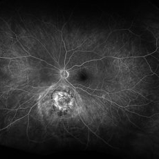
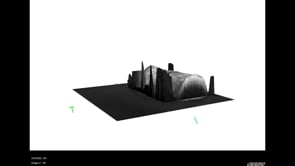 3D OCT of juxtapapillary melanoma
May 15 2020 by Sophia El Hamichi, MD
A 63-year-old male with juxtapapillary melanoma of the right eye. Visual acuity at presentation was 20/25 OD. Patient treated with brachytherapy Iodine125 plaque
Photographer: Belinda Rodriguez
Condition/keywords: optical coherence tomography (OCT)
-
 Amelanotic Choroidal Melanoma
May 18 2020 by McGill University Health Centre
The enucleation specimen in (A) shows an amelanotic, mushroom-shaped tumor arising from the choroid. Microhemorrhages are present within the tumor and also surround the tumor base (arrow). True retinal detachment is present (arrowhead). The subretinal fluid is mixed: clear (1), hemorrhagic (2), and fibrinoid (3).
Condition/keywords: enucleation, mushroom-shaped
-
 Amelanotic Choroidal Melanoma
May 18 2020 by McGill University Health Centre
The enucleation image shows a large amelanotic tumor with large areas of hemorrhage and necrosis. Note the several dilated blood vessels and an adjacent retinal detachment with lipofuscin pigment on its surface (arrow).
Condition/keywords: amelanotic melanoma, enucleation, mushroom-shaped
-
 Amelanotic Mushroom-Shaped Choroidal Melanoma
May 18 2020 by McGill University Health Centre
This enucleation specimen demonstrates an amelanotic, mushroom-shaped, slightly hemorrhagic tumor near the optic nerve (arrow). True retinal detachment is present, and the retina is folded (arrowhead). The subretinal fluid is hazy (*).
Condition/keywords: amelanotic melanoma, enucleation, mushroom-shaped
-
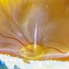 Bergmeister Papilla
Feb 20 2020 by Nisarg Joshi, MD
Gross pathology photo of a Bergmeister Papilla. It is a remnant of incompletely resorbed hyaloid vasculature from ocular development. This glial tissue is seen emminating from the optic nerve, which also shows glaucomatous cupping. The eye was enucleated due to a choroidal melanoma.
Photographer: Nisarg Joshi, MD, Geisinger Medical Center
Imaging device: Digital camera
Condition/keywords: Bergmeister's Papillae, hyaloid artery, persistent fetal vasculature (PFV)
-
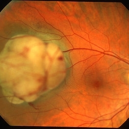
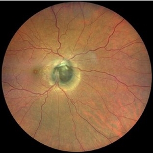 Choroidal Disc Melanoma
Dec 24 2020 by Aditya S Kelkar, MS, FRCS, FASRS,FRCOphth
Melanoma or Melanocytoma- A mystery
Condition/keywords: melanocytoma
-
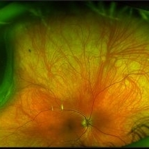 Choroidal Melanoma
Mar 26 2024 by Xitlali Caterina
Ultra-widefield fundus photograph of a 40-year-old woman with Choroidal Melanoma in right eye. Patient present with 20/50+2 vision in the right eye. Patient reported having frequent headaches located frontal area of their head and sometimes radiated to the right side as well. Patient also noted eye pain in both eyes that has remained constant for many years, as well as light sensitivity. The physician stated that since this is a medium-sized tumor, the treatment options include I-125 brachytherapy or enucleation. He recommended I-125 brachytherapy.
Photographer: Xitlali Caterina
Imaging device: Optos California RGB
Condition/keywords: fundus photography, Optos, OPTOS CALIFORNIA, superior retina, ultra-wide field imaging, ultra-widefield image
-
 Choroidal Melanoma
Jun 4 2014 by Henry J. Kaplan, MD
Choroidal melanoma, double circulation.later AV phase fluorescein angiography again shows the double circulation whithin the tumor and retina. #3
Condition/keywords: double circulation
-
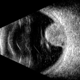 Choroidal Melanoma (USG)
Jul 5 2025 by Gustavo Uriel Fonseca Aguirre
This B-mode transverse ultrasound scan reveals a mushroom-shaped choroidal tumor in the inferior nasal quadrant adjacent to the optic nerve head. The lesion appears solid with homogeneous internal reflectivity and is associated with minimal surrounding subretinal fluid and scleral excavation. It measures 6.54 mm in height × 7.52 mm in base diameter (transverse view) and extends 9.52 mm longitudinally. The vitreous contains abundant punctate opacities consistent with pigment dispersion. The retina and choroid remain attached elsewhere.
Photographer: Gustavo U. Fonseca Aguirre, Hospital Conde de Valenciana, Ciudad de México
Condition/keywords: choroidal melanoma
-
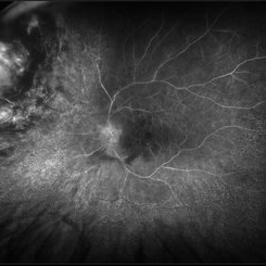
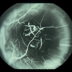
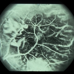
 Choroidal melanoma 1 - fluorescein angiogram
Jan 11 2013 by Alex P. Hunyor, MD
Choroidal melanoma - fluorescein angiogram showing tumour circulation.
-
 Choroidal Melanoma 3 Ways
Jan 16 2025 by Virginia Gebhart
RGB/FA/ICG of 76 year old female with a new choroidal melanoma. Pt scheduled for plaque radiation. BCVA 20/400
Photographer: Virginia Gebhart, Retina Consultants of Carolina
Imaging device: Optos California
Condition/keywords: fluorescein angiogram (FA), indocyanine green (ICG) angiography, OPTOS CALIFORNIA RGB
-
 Choroidal Melanoma FA
Nov 14 2023 by Virginia Gebhart
Fluorescein angiogram of 69 year old male with small lesion consistent with choroidal melanoma. Small pigmented elevated choroidal lesion just below ON with drusen, RPE changes and trace questionable OP present in the left eye. Extensive imaging and ultrasound was performed for further evaluation and documentation.
Photographer: Virginia Gebhart
Imaging device: Optos
Condition/keywords: fluorescein angiogram (FA), Fluorescein angiography
-
 Choroidal Melanoma Masquerading as Subretinal Hemorrhage With Breakthrough VH
Jan 23 2025 by Tejaswita Verma
A 65 year old diabetic male presented with large nasal retinal mass giving the appearance of organized dehaemoglobinized subretinal hemorrhage with breakthrough vitreous hemorrhage , with 6/6P vision. Enucleation specimen showed histopathology confirmed choroidal melanoma.
Photographer: DR. TEJASWITA VERMA
Imaging device: MIRANTE
 Loading…