File number: 27867
Comments
-
 JEFFERSON R SOUSA, Tecg.º (Biomedical Systems Technology) (February 23 2018)
Thank you Nichole,
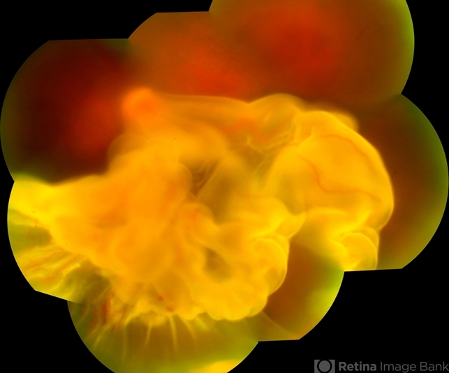
This patient really has a RD and has always been controlled. Even so, there was a worsening of the vision in which it evolved into a detachment. In the Ultrasson examination it shows up to a closed funnel pattern. -
 Nichole Lewis (February 22 2018)
This is a great RD montage!
Sign in to comment.
 Initializing download.
-
 By JEFFERSON R SOUSA, Tecg.º (Biomedical Systems Technology)
Lens Oftalmologia - Hospital Beneficiência Portuguesa - Uploaded on Feb 17, 2018.
- Last modified by Caroline Bozell on May 25, 2018.
- Image of the week
-
May 27, 2018
View all images of the week - Rating
- Appears in
- Miscellaneous
- Photographer
- JEFFERSON R SOUSA - Study Center and Ophthalmological Research Dr. Andre M V Gomes, Institute Dr. Suel Abujamra São Paulo-Brazil
- Imaging device
-
Fundus camera
Fundus camera Topcon TRC-50 DX, Imaginet 5.0, angle de 50 graus. Flash 36 / Mosaic with 10 images. - Description
- A 42-year-old patient complained of low vision in the left eye. In retinal mapping and background color photography, extensive retinal detachment was observed.
")


")
")
")