Search results (76 results)
-
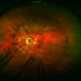
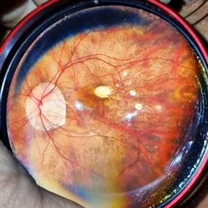
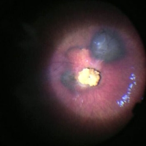
 Central Macular Lesion, Choroidal Hemangioma, Staphyloma
Jul 11 2013 by Jerald A. Bovino, MD
No history.
Condition/keywords: central vascular lesion, staphyloma
-
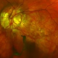
 Color Fundus Photograph of Myope With PVD and Staphyloma
Jun 11 2016 by Philip J. Polkinghorne, MD
Color photograph of patient with PVD and staphyloma.
Imaging device: Optos
Condition/keywords: degenerative myopia, peripheral vascular disease (PVD), staphyloma
-
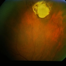
 Macular Atrophy With Staphyloma FA
Jul 31 2013 by From the Collections of Thomas M. Aaberg, MD and Thomas M. Aaberg Jr., MD
Macular atrophy with staphyloma FA.
Condition/keywords: macular atrophy, staphyloma
-
 Macular Atrophy With Staphyloma FA
Jul 31 2013 by From the Collections of Thomas M. Aaberg, MD and Thomas M. Aaberg Jr., MD
Macular atrophy with staphyloma FA.
Condition/keywords: macular atrophy, staphyloma
-
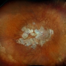
---thumb.jpg/image-square;max$300,300.ImageHandler) Macular Staphyloma
Macular Staphyloma
Feb 14 2013 by From the Collections of Thomas M. Aaberg, MD and Thomas M. Aaberg Jr., MD
Composite.
Condition/keywords: staphyloma
-
 Morning-Glory-Syndrome
Dec 22 2017 by James B. Soque, CRA, OCT-C, COA, FOPS
68-year-old WM with Morning Glory Syndrome with PVD OS with Staphyloma surrounding optic nerve and extending into the macula. Also, esotropia OS from V1 nerve paresis from birth, with amblyopia.
Photographer: James B Soque, CRA OCT-C COA FOPS
Imaging device: Optos Daytona
Condition/keywords: color photo, esotropia, fundus photograph, Optomap, Optos, peripheral vascular disease (PVD), posterior vitreous detachment, staphyloma, ultra-wide field imaging, wide angle imaging
-
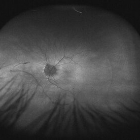 Myope With Staphyloma and Vitreous Detachment
Jun 11 2016 by Philip J. Polkinghorne, MD
Fundus autofluorescence of a myope with PVD and staphyloma.
Imaging device: Optos FAF
Condition/keywords: degenerative myopia, myopia, staphyloma
-
 Myopic Degeneration
Dec 9 2024 by Virginia Gebhart
67 year old female with myopic degeneration. Posterior staphylomas are stable. VA limited by extensive chorioretinal atrophy. BCVA 20/160 (ecc)
Photographer: Virginia Gebhart, Retina Consultants of Carolina
Imaging device: Optos California
Condition/keywords: chorioretinal atrophy, myopic degeneration, staphyloma
-
 OCT Myopic Staphyloma With Schisis and ERM
Apr 24 2014 by Scott E. Pautler, MD
OCT of high myope with asymptomatic macular schisis.
Imaging device: Heidelberg Spectralis
Condition/keywords: foveal schisis, maculopathy, maculoschisis, optical coherence tomography (OCT), pathologic myopia, staphyloma
-
 Optos Picture With Speculum: Dislocated Natural Lens
Oct 9 2018 by John S. King, MD
55-year-old white female with history of pathologic myopia+, lattice (laser), SB OU (1990s), and dislocated natural lenses OU that had been watched for years. In the fellow eye she developed phacolytic glaucoma and a PPV, PPL was performed. Plan for both eyes are monitoring. I wanted to get a good picture of her lens today with the optos machine, as the other pics had artifact from the lower lid. It worked out well to use a speculum in the left eye. Vision cc is 20/400 J1+ OD and 20/40 J2 OS; aphakic OU; vitreous clear OD; dislocated lens OS (see pic); retinas attached.
Photographer: Maisee Yang
Imaging device: Optos California
Condition/keywords: dislocated crystalline lens, pathologic myopia, scleral buckle, staphyloma
-
 Pathological Myopia
Sep 25 2024 by DR Rohit Gupta
Fundus photograph of a 28 year-old male having high myopia on fundus examination Degenerative changes are seen in retina suggestive of pathological myopia.
Photographer: Dr Rohit gupta
Imaging device: Samsung S21
Condition/keywords: choroidal degeneration, degeneration of optic disc, lacquer cracks, myopia, Myopia macular degeneration CNVM foster fuch spot, pathologic myopia, staphyloma
-
 Peripheral Toxo Staphyloma
Nov 7 2014 by David Callanan, MD
63-year-old female, peripheral toxo staphyloma.
Condition/keywords: staphyloma
-
---thumb.jpg/image-square;max$300,300.ImageHandler) Posterior Pole Chorioretinal Atrophy With Staphyloma
Posterior Pole Chorioretinal Atrophy With Staphyloma
Aug 1 2013 by From the Collections of Thomas M. Aaberg, MD and Thomas M. Aaberg Jr., MD
Posterior pole chorioretinal atrophy with staphyloma.
Condition/keywords: chorioretinal atrophy, posterior pole, staphyloma
-
---thumb.jpg/image-square;max$300,300.ImageHandler) Posterior Pole Chorioretinal Atrophy With Staphyloma
Posterior Pole Chorioretinal Atrophy With Staphyloma
Aug 1 2013 by From the Collections of Thomas M. Aaberg, MD and Thomas M. Aaberg Jr., MD
Posterior pole chorioretinal atrophy with staphyloma.
Condition/keywords: chorioretinal atrophy, staphyloma
-
---thumb.jpg/image-square;max$300,300.ImageHandler) Posterior Pole Chorioretinal Atrophy With Staphyloma
Posterior Pole Chorioretinal Atrophy With Staphyloma
Aug 1 2013 by From the Collections of Thomas M. Aaberg, MD and Thomas M. Aaberg Jr., MD
Posterior pole chorioretinal atrophy with staphyloma.
Condition/keywords: chorioretinal atrophy, posterior pole, staphyloma
-
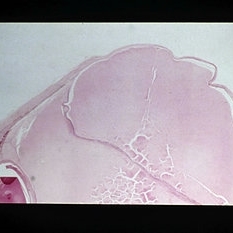 Slide 12-27
Feb 27 2019 by Lancaster Course in Ophthalmology
Sequelae. Increased intraocular pressure has resulted in stretching and thinning of the sclera in the equatorial region, resulting in an equatorial staphyloma (H&E x3).
Condition/keywords: sclera, sequelae, staphyloma
-
 Staphyloma
Dec 10 2012 by Yale L. Fisher, MD
Localize macular staphyloma seen as an outpouching of the normal posterior ocular wall (arrow). Optic nerve is seen superior to macular area.
Condition/keywords: video
-
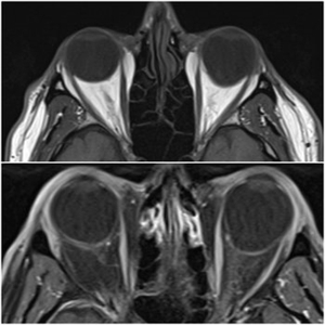 Staphyloma in Pathologic Myopia
Feb 7 2020 by Jonathan C. Tsui, MD
A 51-year-old presents with a six-month history of OS vision loss found to be CF at 3'. Fundus exam demonstrated pathologic myopia OS>OD with tilted discs. MRI Orbit Axial T1 images demonstrate significant compatible findings of staphylomas OS>OD.
Condition/keywords: pathologic myopia, staphyloma
-
 Staphyloma Steroid Induced Glaucoma
Apr 14 2014 by Dipankar Barua, M.Sc
Male patient, 25-years-old. On examination his vision of the right eye is perception of light and left eye is 6/6. His IOP is 30mmHg in right eye and 10 mmHg in left eye. It seems to be a case of staphyloma steroid induced glaucoma.
Photographer: Dipankar Barua
Imaging device: Topcon TRC 50 DX (IA)
Condition/keywords: glaucoma, staphyloma
-
 Staphyloma Steroid Induced Glaucoma
Apr 14 2014 by Dipankar Barua, M.Sc
Male patient, 25-years-old. On examination his vision of the right eye is perception of light and left eye is 6/6. His IOP is 30mmHg in right eye and 10 mmHg in left eye. It seems to be a case of staphyloma steroid induced glaucoma.
Photographer: Dipankar Barua
Imaging device: Topcon TRC 50 DX (IA)
Condition/keywords: glaucoma, staphyloma, steroids
-
 Staphyloma Steroid Induced Glaucoma
Apr 14 2014 by Dipankar Barua, M.Sc
Male patient, 25-years-old. On examination his vision of the right eye is perception of light and left eye is 6/6. His IOP is 30mmHg in right eye and 10 mmHg in left eye. It seems to be a case of staphyloma steroid induced glaucoma.
Photographer: Dipankar Barua
Imaging device: Topcon TRC 50 DX (IA)
Condition/keywords: glaucoma, staphyloma
-
 Traumatic Vitreous Hemorrhage in an Infant
Apr 16 2018 by Martin J Siemerink, MD, PhD
1-year-old girl with previous severe head injury and secondary vitreous hemorrhage. Vitrectomy was performed 3 months after the injury. Fundus image after vitrectomy.
Photographer: Martin Siemerink, University of Auckland, Auckland, New Zealand
Imaging device: Zeiss Resight Fundus Viewing System Lumera
Condition/keywords: depigmented vitreous blood, staphyloma, subretinal hemorrhage
-
 Posterior staphyloma in oculocutaneous albinism
Jun 22 2023 by Eder Díaz Dorado
Fundus photograph of an 56-yeard-old man with oculocutaneous albinism and posterior staphyloma.
Photographer: Eder Díaz Dorado, Hospital Central Militar CDMX
Imaging device: Smartphone
Condition/keywords: Staphyloma albinism
 Loading…