Search results (76 results)
-
 The Great Disc-guise
Nov 12 2025 by SHRADDHA RAJ SHRIVASTAVA
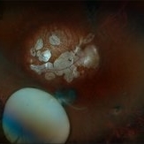
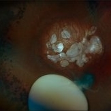
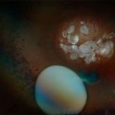
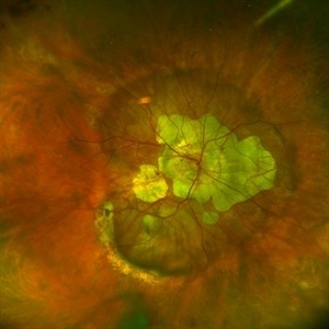
Right eye pseudocolor fundus photo of a 20 year old with Both eyes Pathological Myopia (spherical refractive error of - 18.00 DS in BE), showing a tilted myopic disc with peripapillary atrophy, and extensive posterior staphyloma baring the underlying choroidal vessels and scleral tissue. We can also see a well-defined round chorioretinal atrophic (CRA) patch superonasal to the disc, giving the illusion of double disc on cursory fundus examination.
Photographer: Dr. Shraddha Raj Shrivastava
Imaging device: Nidek Mirante SLO/OCT (Confocal scanning/Spectral domain OCT)
Condition/keywords: chorioretinal atrophy, High Myopia, pathologic myopia, peripapillary atrophy, posterior staphyloma
-
 AL 39.16 mm
Sep 10 2025 by Gustavo Uriel Fonseca Aguirre
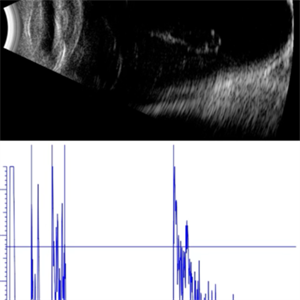
This axial B-scan reveals an elongated globe with an axial length of 39.16 mm, consistent with high axial myopia. Posterior staphyloma and scleral thinning are observed, though the retina remains attached.
Photographer: Gustavo U. Fonseca Aguirre, Hospital Conde de Valenciana, Ciudad de México
Condition/keywords: high myopia
-
 Posterior Staphyloma + ON-Coloboma
Aug 20 2025 by Gustavo Uriel Fonseca Aguirre
This axial B-scan reveals a highly myopic eye with a posterior staphyloma and an associated optic nerve coloboma. The staphyloma appears as a deep scleral outpouching adjacent to the optic disc, while the coloboma demonstrates a focal posterior excavation with retrobulbar extension.
Photographer: Gustavo U. Fonseca Aguirre, Hospital Conde de Valenciana, Ciudad de México
Condition/keywords: optic nerve coloboma, posterior staphyloma
-
 RRD in Posterior Staphyloma
May 21 2025 by Gustavo Uriel Fonseca Aguirre
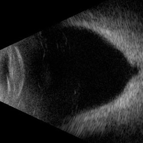
This B-mode axial ultrasound scan of a highly myopic eye demonstrates a prominent posterior staphyloma with an associated inferior retinal detachment sparing the macular region.
Photographer: Gustavo U. Fonseca Aguirre, Hospital Conde de Valenciana, Ciudad de México
Condition/keywords: high myopia, posterior staphyloma, Retina detachment
-
 Retinal Detachment Associated With a Posterior Staphyloma
Apr 9 2025 by Gustavo Uriel Fonseca Aguirre
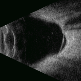
B-mode axial ultrasound scan of a highly myopic eye shows a posterior staphyloma with an associated macular hole-induced retinal detachment.
Photographer: Gustavo U. Fonseca Aguirre, Hospital Conde de Valenciana, Ciudad de México
Condition/keywords: high myopia, posterior staphyloma, rhegmatogenous retinal detachment
-
 Subluxation of the Lens
Dec 12 2024 by Kimberly Wakester
Ultra-wide field fundus photos of an 53-year-old man with a Subluxation of the Lens in the posterior vitreous cavity of the right eye after a trauma that happened many years ago. Patient remains stable with no adverse reaction to the lens at this time. No surgical intervention is recommended at this time. Patient also has myopic degeneration and lattice degeneration that will require patient to have follow up care.
Photographer: Kimberly Wakester, COA
Imaging device: Optos California
Condition/keywords: lattice degeneration, myopic degeneration, peripapillary atrophy, posterior staphyloma, Subluxation of the Lens
-
 Myopic Degeneration
Dec 9 2024 by Virginia Gebhart
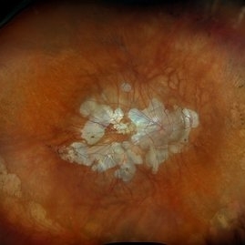
67 year old female with myopic degeneration. Posterior staphylomas are stable. VA limited by extensive chorioretinal atrophy. BCVA 20/160 (ecc)
Photographer: Virginia Gebhart, Retina Consultants of Carolina
Imaging device: Optos California
Condition/keywords: chorioretinal atrophy, myopic degeneration, staphyloma
-
 Pathological Myopia
Sep 25 2024 by DR Rohit Gupta
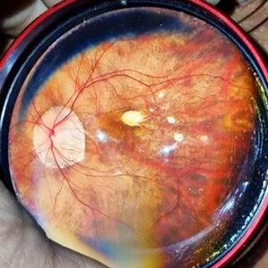
Fundus photograph of a 28 year-old male having high myopia on fundus examination Degenerative changes are seen in retina suggestive of pathological myopia.
Photographer: Dr Rohit gupta
Imaging device: Samsung S21
Condition/keywords: choroidal degeneration, degeneration of optic disc, lacquer cracks, myopia, Myopia macular degeneration CNVM foster fuch spot, pathologic myopia, staphyloma
-
 Posterior staphyloma
Dec 20 2023 by Roger A. Goldberg, MD, MBA
Fundus photo of an 85-year-old woman with degenerative myopia and a large posterior staphyloma
Photographer: Mohan Zhou, Bay Area Retina Associates, Walnut Creek, CA
Imaging device: Optos
Condition/keywords: degenerative myopia, high myopia, posterior staphyloma
-
 High Myopia with Posterior staphyloma
Nov 7 2023 by Harsh Vardhan Singh, MS
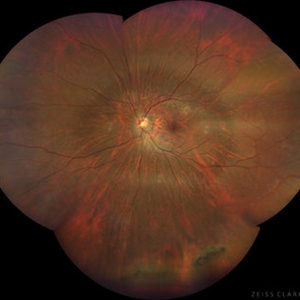
27-year old with both eyes high myopia & posterior staphyloma with left eye peripheral lattice degeneration & white without pressure
Photographer: Harsh Vardhan Singh
Imaging device: Clarus 700
Condition/keywords: lattice degeneration, myopia, peripheral lattice degeneration, posterior staphylomaloma, white without pressure
-
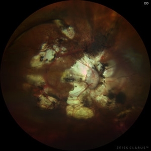
 Pathological Myopia with posterior pole retinal detachment & new open break
Jul 31 2023 by Harsh Vardhan Singh, MS
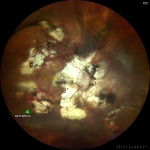
45-year female with redetachment & new break
Photographer: Dr Harsh Vardhan Singh, AIIMS, Guwahati
Imaging device: Zeiss Clarus 700
Condition/keywords: pathologic myopia, posterior staphyloma, retinal break, rrd
-
 Pathological Myopia with posterior pole retinal detachment & new open break
Jul 31 2023 by Harsh Vardhan Singh, MS
45-year female with redetachment & new break
Photographer: Dr Harsh Vardhan Singh, AIIMS, Guwahati
Imaging device: Zeiss Clarus 700
Condition/keywords: pathologic myopia, posterior staphyloma, retinal break, rrd
-
 Pathological Myopia with posterior pole retinal detachment
Jul 31 2023 by Harsh Vardhan Singh, MS
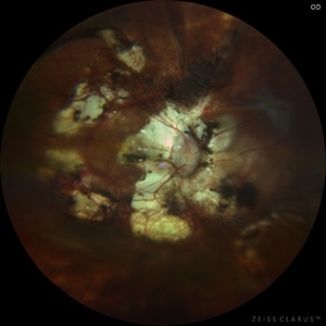
45-year female with right eye re-detachment with pathological myopia & posterior pole RRD with open break
Photographer: Dr Harsh Vardhan Singh, AIIMS, Guwahati
Imaging device: Zeiss clarus 700
Condition/keywords: pathologic myopia, posterior pole lesion, posterior staphyloma, rrd
-
 Posterior staphyloma in oculocutaneous albinism
Jun 22 2023 by Eder Díaz Dorado
Fundus photograph of an 56-yeard-old man with oculocutaneous albinism and posterior staphyloma.
Photographer: Eder Díaz Dorado, Hospital Central Militar CDMX
Imaging device: Smartphone
Condition/keywords: Staphyloma albinism
-
 Myopic retinopathy
Dec 27 2021 by Eduardo Javier Pinuer Alvarado
Fundus photograph of an 50-year-old man with myopic retinopathy, posterior staphyloma, myopic chorioretinal atrophy and tilted and oblique disc.
Photographer: Eduardo Pinuer A, Universidad Austral de Chile.
Imaging device: CR-2 AF Digital Non-Mydriatic Retinal Camera, Canon.
Condition/keywords: myopic chorioretinal atropthy, myopic retinopathy, posterior staphyloma, retinopathy
-
 Dislocated Intraocular Lens, Tilted Disc, Posterior Staphyloma.
Aug 18 2021 by Jesus Lozano, MD
Fundus photograph of 80-year-old woman, single eye with left eye posterior dislocation of lens, tilted disc and posterior staphyloma.
Photographer: Yair Bet Yosef, Hadassah Medical Center. Israel
Imaging device: Optos Silverstone
Condition/keywords: posterior dislocation of lens, posterior staphyloma, tilted disc
-
 Staphyloma in Pathologic Myopia
Feb 7 2020 by Jonathan C. Tsui, MD
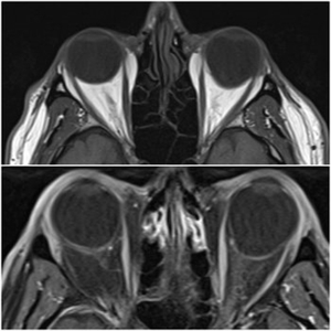
A 51-year-old presents with a six-month history of OS vision loss found to be CF at 3'. Fundus exam demonstrated pathologic myopia OS>OD with tilted discs. MRI Orbit Axial T1 images demonstrate significant compatible findings of staphylomas OS>OD.
Condition/keywords: pathologic myopia, staphyloma
-
 Myopic Fundus With Staphyloma and a Peripheral Lasered Hole
Jul 25 2019 by Manish Nagpal, MD, FRCS (UK), FASRS
Wide field view of a myopic fundus with peripheral lasered hole with pigmentation along with staphyloma in the central area.
Photographer: Gayathri Mohan
Condition/keywords: laser photocoagulation, myopia, ultra-wide field imaging
-
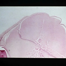 Slide 12-27
Feb 27 2019 by Lancaster Course in Ophthalmology
Sequelae. Increased intraocular pressure has resulted in stretching and thinning of the sclera in the equatorial region, resulting in an equatorial staphyloma (H&E x3).
Condition/keywords: sclera, sequelae, staphyloma
-
 Optos Picture With Speculum: Dislocated Natural Lens
Oct 9 2018 by John S. King, MD
55-year-old white female with history of pathologic myopia+, lattice (laser), SB OU (1990s), and dislocated natural lenses OU that had been watched for years. In the fellow eye she developed phacolytic glaucoma and a PPV, PPL was performed. Plan for both eyes are monitoring. I wanted to get a good picture of her lens today with the optos machine, as the other pics had artifact from the lower lid. It worked out well to use a speculum in the left eye. Vision cc is 20/400 J1+ OD and 20/40 J2 OS; aphakic OU; vitreous clear OD; dislocated lens OS (see pic); retinas attached.
Photographer: Maisee Yang
Imaging device: Optos California
Condition/keywords: dislocated crystalline lens, pathologic myopia, scleral buckle, staphyloma
-
 Myopic Degeneration
Jul 3 2018 by Armando L. Oliver, MD
Myopic Degeneration
Photographer: Moises Castro
Imaging device: Optos California
Condition/keywords: pathologic myopia, posterior staphyloma
-
 Myopic Degeneration
Jul 3 2018 by Armando L. Oliver, MD
Myopic Degeneration
Photographer: Moises Castro
Imaging device: Optos California
Condition/keywords: pathologic myopia, posterior staphyloma
-
 Myopic Degeneration
Jul 3 2018 by Armando L. Oliver, MD
Late views IVFA.
Photographer: Moises Castro
Imaging device: Optos California
Condition/keywords: pathologic myopia, posterior staphyloma
 Loading…