Search results (76 results)
-
 OCT Myopic Staphyloma With Schisis and ERM
Apr 24 2014 by Scott E. Pautler, MD
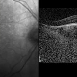
OCT of high myope with asymptomatic macular schisis.
Imaging device: Heidelberg Spectralis
Condition/keywords: foveal schisis, maculopathy, maculoschisis, optical coherence tomography (OCT), pathologic myopia, staphyloma
-
 Staphyloma Steroid Induced Glaucoma
Apr 14 2014 by Dipankar Barua, M.Sc
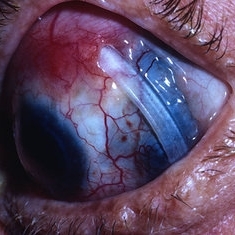
Male patient, 25-years-old. On examination his vision of the right eye is perception of light and left eye is 6/6. His IOP is 30mmHg in right eye and 10 mmHg in left eye. It seems to be a case of staphyloma steroid induced glaucoma.
Photographer: Dipankar Barua
Imaging device: Topcon TRC 50 DX (IA)
Condition/keywords: glaucoma, staphyloma, steroids
-
 Optos Picture With Speculum: Dislocated Natural Lens
Oct 9 2018 by John S. King, MD
55-year-old white female with history of pathologic myopia+, lattice (laser), SB OU (1990s), and dislocated natural lenses OU that had been watched for years. In the fellow eye she developed phacolytic glaucoma and a PPV, PPL was performed. Plan for both eyes are monitoring. I wanted to get a good picture of her lens today with the optos machine, as the other pics had artifact from the lower lid. It worked out well to use a speculum in the left eye. Vision cc is 20/400 J1+ OD and 20/40 J2 OS; aphakic OU; vitreous clear OD; dislocated lens OS (see pic); retinas attached.
Photographer: Maisee Yang
Imaging device: Optos California
Condition/keywords: dislocated crystalline lens, pathologic myopia, scleral buckle, staphyloma
-
---thumb.jpg/image-square;max$300,300.ImageHandler) Posterior Pole Chorioretinal Atrophy With Staphyloma
Posterior Pole Chorioretinal Atrophy With Staphyloma
Aug 1 2013 by From the Collections of Thomas M. Aaberg, MD and Thomas M. Aaberg Jr., MD
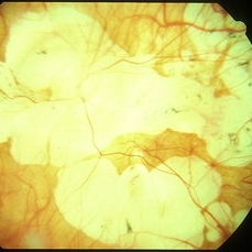
Posterior pole chorioretinal atrophy with staphyloma.
Condition/keywords: chorioretinal atrophy, posterior pole, staphyloma
-
---thumb.jpg/image-square;max$300,300.ImageHandler) Posterior Pole Chorioretinal Atrophy With Staphyloma
Posterior Pole Chorioretinal Atrophy With Staphyloma
Aug 1 2013 by From the Collections of Thomas M. Aaberg, MD and Thomas M. Aaberg Jr., MD
Posterior pole chorioretinal atrophy with staphyloma.
Condition/keywords: chorioretinal atrophy, staphyloma
-
 Posterior Staphylamas
May 2 2013 by Henry J. Kaplan, MD
Highly myopic changes with posterior staphyloma and visible sclera.
Condition/keywords: high myopia, posterior staphyloma
-
 Myelinated Nerve Fibers and Possibly Posterior Staphyloma
Feb 19 2013 by From the Collections of Thomas M. Aaberg, MD and Thomas M. Aaberg Jr., MD
Findings are bilateral.
Condition/keywords: color photo, myelinated nerve fibers
-
---thumb.jpg/image-square;max$300,300.ImageHandler) Posterior Pole Chorioretinal Atrophy With Staphyloma
Posterior Pole Chorioretinal Atrophy With Staphyloma
Aug 1 2013 by From the Collections of Thomas M. Aaberg, MD and Thomas M. Aaberg Jr., MD
Posterior pole chorioretinal atrophy with staphyloma.
Condition/keywords: chorioretinal atrophy, staphyloma
-
 Myelinated Nerve Fibers and Possibly Posterior Staphyloma
Feb 19 2013 by From the Collections of Thomas M. Aaberg, MD and Thomas M. Aaberg Jr., MD
Findings are bilateral.
Condition/keywords: color photo, myelinated nerve fibers
-
 Myelinated Nerve Fibers and Possibly Posterior Staphyloma
Feb 19 2013 by From the Collections of Thomas M. Aaberg, MD and Thomas M. Aaberg Jr., MD
Findings are bilateral.
Condition/keywords: myelinated nerve fibers
-
 Peripapillary Staphyloma
Feb 20 2013 by From the Collections of Thomas M. Aaberg, MD and Thomas M. Aaberg Jr., MD
No history.
Condition/keywords: peripapillary staphyloma
-
 Myelinated Nerve Fibers and Possibly Posterior Staphyloma
Feb 19 2013 by From the Collections of Thomas M. Aaberg, MD and Thomas M. Aaberg Jr., MD
Findings are bilateral.
Condition/keywords: color photo, myelinated nerve fibers
-
 Staphyloma Steroid Induced Glaucoma
Apr 14 2014 by Dipankar Barua, M.Sc
Male patient, 25-years-old. On examination his vision of the right eye is perception of light and left eye is 6/6. His IOP is 30mmHg in right eye and 10 mmHg in left eye. It seems to be a case of staphyloma steroid induced glaucoma.
Photographer: Dipankar Barua
Imaging device: Topcon TRC 50 DX (IA)
Condition/keywords: glaucoma, staphyloma
-
---thumb.jpg/image-square;max$300,300.ImageHandler) Posterior Pole Chorioretinal Atrophy With Staphyloma
Posterior Pole Chorioretinal Atrophy With Staphyloma
Aug 1 2013 by From the Collections of Thomas M. Aaberg, MD and Thomas M. Aaberg Jr., MD
Posterior pole chorioretinal atrophy with staphyloma.
Condition/keywords: chorioretinal atrophy, posterior pole, staphyloma
-
 High Myopia
Jun 14 2018 by Mitzy E Torres Soriano, MD
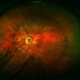
Fundus photograph (left eye) of a female patient with high myopia, chorioretinal atrophy, pigmentary changes and posterior staphyloma.
Photographer: Mitzy Torres Soriano
Condition/keywords: chorioretinal atrophy, high myopia, posterior staphyloma
-
 Myelinated Nerve Fibers and Possibly Posterior Staphyloma
Feb 19 2013 by From the Collections of Thomas M. Aaberg, MD and Thomas M. Aaberg Jr., MD
Findings are bilateral.
Condition/keywords: color photo, myelinated nerve fibers
-
 Exposed Buckle
May 2 2013 by Henry J. Kaplan, MD
Exposed buckele with underlying scleral staphyloma.
Condition/keywords: exposed scleral buckle
-
 EDI OCT - Dome Shaped Macula in a Myopic Patient
Jan 24 2015 by Roy Schwartz, MD
Dome shaped macula in an 80-year-old man with myopic staphyloma. The EDI showed thickened choroid and sclera.
Photographer: Galit Yair Pur
Condition/keywords: enhanced depth imaging, myopia, optical coherence tomography (OCT)
-
 Myopic Maculoscheisis
Sep 11 2013 by Jason S. Calhoun
Fluorescence angiography and fundus photography shows RPE mottling and staphyloma in the right eye. VA is 20/50, right eye. No subretinal fluid found.
Photographer: Jason S. Calhoun, Department of Ophthalmology, Mayo Clinic Jacksonville, Florida
Imaging device: TOPCON TRC 50-EX
Condition/keywords: myopia, tilted disc
-
 Myopic Traction Maculopathy
May 31 2014 by Rameez N Hussain, MD
Spectral domain optical coherence tomography of macular detachment in posterior staphyloma - myopic traction maculopathy (MTM).
Photographer: Rameez N Hussain MD, Vitreo Retinal Services, Giridhar Eye Institute, Cochin, India
Imaging device: Heidelberg Spectralis
Condition/keywords: high myopia, macular detachment, myopic traction maculopathy, pathologic myopia, posterior staphyloma
-
 Myopic Traction Maculopathy
May 31 2014 by Rameez N Hussain, MD
Color photograph of macular detachment in a posterior staphyloma - myopic traction maculopathy (MTM).
Photographer: Rameez N Hussain MD, Vitreo Retinal Services, Giridhar Eye Institute, Cochin, India
Imaging device: Zeiss
Condition/keywords: high myopia, macular detachment, myopic traction maculopathy, pathologic myopia, posterior staphyloma
-
---thumb.jpg/image-square;max$300,300.ImageHandler) Posterior Pole Chorioretinal Atrophy With Staphyloma
Posterior Pole Chorioretinal Atrophy With Staphyloma
Aug 1 2013 by From the Collections of Thomas M. Aaberg, MD and Thomas M. Aaberg Jr., MD
Posterior pole chorioretinal atrophy with staphyloma.
Condition/keywords: chorioretinal atrophy, staphyloma
-
 Staphyloma Steroid Induced Glaucoma
Apr 14 2014 by Dipankar Barua, M.Sc
Male patient, 25-years-old. On examination his vision of the right eye is perception of light and left eye is 6/6. His IOP is 30mmHg in right eye and 10 mmHg in left eye. It seems to be a case of staphyloma steroid induced glaucoma.
Photographer: Dipankar Barua
Imaging device: Topcon TRC 50 DX (IA)
Condition/keywords: glaucoma, staphyloma
-
---thumb.jpg/image-square;max$300,300.ImageHandler) Macular Staphyloma
Macular Staphyloma
Feb 14 2013 by From the Collections of Thomas M. Aaberg, MD and Thomas M. Aaberg Jr., MD
Composite.
Condition/keywords: staphyloma
-
 Color Fundus Photograph of Myope With PVD and Staphyloma
Jun 11 2016 by Philip J. Polkinghorne, MD
Color photograph of patient with PVD and staphyloma.
Imaging device: Optos
Condition/keywords: degenerative myopia, peripheral vascular disease (PVD), staphyloma
 Loading…