Search results (76 results)
-
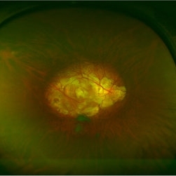
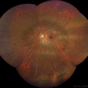
 Myopic retinopathy
Dec 27 2021 by Eduardo Javier Pinuer Alvarado
Fundus photograph of an 50-year-old man with myopic retinopathy, posterior staphyloma, myopic chorioretinal atrophy and tilted and oblique disc.
Photographer: Eduardo Pinuer A, Universidad Austral de Chile.
Imaging device: CR-2 AF Digital Non-Mydriatic Retinal Camera, Canon.
Condition/keywords: myopic chorioretinal atropthy, myopic retinopathy, posterior staphyloma, retinopathy
-
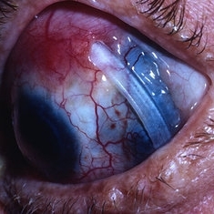
 Optos Picture With Speculum: Dislocated Natural Lens
Oct 9 2018 by John S. King, MD
55-year-old white female with history of pathologic myopia+, lattice (laser), SB OU (1990s), and dislocated natural lenses OU that had been watched for years. In the fellow eye she developed phacolytic glaucoma and a PPV, PPL was performed. Plan for both eyes are monitoring. I wanted to get a good picture of her lens today with the optos machine, as the other pics had artifact from the lower lid. It worked out well to use a speculum in the left eye. Vision cc is 20/400 J1+ OD and 20/40 J2 OS; aphakic OU; vitreous clear OD; dislocated lens OS (see pic); retinas attached.
Photographer: Maisee Yang
Imaging device: Optos California
Condition/keywords: dislocated crystalline lens, pathologic myopia, scleral buckle, staphyloma
-
 OCT Myopic Staphyloma With Schisis and ERM
Apr 24 2014 by Scott E. Pautler, MD
OCT of high myope with asymptomatic macular schisis.
Imaging device: Heidelberg Spectralis
Condition/keywords: foveal schisis, maculopathy, maculoschisis, optical coherence tomography (OCT), pathologic myopia, staphyloma
-
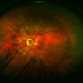
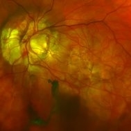
 Posterior Staphyloma
Aug 3 2017 by Eitae Kim, MD
UWF fundus photograph of 35-year-old male with unilateral posterior staphyloma.
Photographer: Eitae Kim, BOIM retina center, Pureun eye hospital
Condition/keywords: ultra-wide field imaging
-
 Myopic Degeneration
Jul 3 2018 by Armando L. Oliver, MD
Myopic Degeneration
Photographer: Moises Castro
Imaging device: Optos California
Condition/keywords: pathologic myopia, posterior staphyloma
-
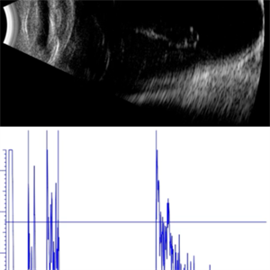 AL 39.16 mm
Sep 10 2025 by Gustavo Uriel Fonseca Aguirre
This axial B-scan reveals an elongated globe with an axial length of 39.16 mm, consistent with high axial myopia. Posterior staphyloma and scleral thinning are observed, though the retina remains attached.
Photographer: Gustavo U. Fonseca Aguirre, Hospital Conde de Valenciana, Ciudad de México
Condition/keywords: high myopia
-
 Central Macular Lesion, Choroidal Hemangioma, Staphyloma
Jul 11 2013 by Jerald A. Bovino, MD
No history.
Condition/keywords: central vascular lesion, staphyloma
-
 Color Fundus Photograph of Myope With PVD and Staphyloma
Jun 11 2016 by Philip J. Polkinghorne, MD
Color photograph of patient with PVD and staphyloma.
Imaging device: Optos
Condition/keywords: degenerative myopia, peripheral vascular disease (PVD), staphyloma
-
 Dislocated Intraocular Lens, Tilted Disc, Posterior Staphyloma.
Aug 18 2021 by Jesus Lozano, MD
Fundus photograph of 80-year-old woman, single eye with left eye posterior dislocation of lens, tilted disc and posterior staphyloma.
Photographer: Yair Bet Yosef, Hadassah Medical Center. Israel
Imaging device: Optos Silverstone
Condition/keywords: posterior dislocation of lens, posterior staphyloma, tilted disc
-
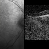 EDI OCT - Dome Shaped Macula in a Myopic Patient
Jan 24 2015 by Roy Schwartz, MD
Dome shaped macula in an 80-year-old man with myopic staphyloma. The EDI showed thickened choroid and sclera.
Photographer: Galit Yair Pur
Condition/keywords: enhanced depth imaging, myopia, optical coherence tomography (OCT)
-
 Exposed Buckle
May 2 2013 by Henry J. Kaplan, MD
Exposed buckele with underlying scleral staphyloma.
Condition/keywords: exposed scleral buckle
-
 High Myopia
Jun 14 2018 by Mitzy E Torres Soriano, MD
Fundus photograph (left eye) of a female patient with high myopia, chorioretinal atrophy, pigmentary changes and posterior staphyloma.
Photographer: Mitzy Torres Soriano
Condition/keywords: chorioretinal atrophy, high myopia, posterior staphyloma
-
 High Myopia with Posterior staphyloma
Nov 7 2023 by Harsh Vardhan Singh, MS
27-year old with both eyes high myopia & posterior staphyloma with left eye peripheral lattice degeneration & white without pressure
Photographer: Harsh Vardhan Singh
Imaging device: Clarus 700
Condition/keywords: lattice degeneration, myopia, peripheral lattice degeneration, posterior staphylomaloma, white without pressure
-
 Macular Atrophy With Staphyloma FA
Jul 31 2013 by From the Collections of Thomas M. Aaberg, MD and Thomas M. Aaberg Jr., MD
Macular atrophy with staphyloma FA.
Condition/keywords: macular atrophy, staphyloma
-
 Macular Atrophy With Staphyloma FA
Jul 31 2013 by From the Collections of Thomas M. Aaberg, MD and Thomas M. Aaberg Jr., MD
Macular atrophy with staphyloma FA.
Condition/keywords: macular atrophy, staphyloma
-
 Macular Hole Retinal Detachment Over a Posterior Staphyloma
Dec 31 2016 by Linda A Cernichiaro- Espinosa, MD
Macular hole retinal detachment over a posterior staphyloma of pathologic myopia.
Photographer: Linda A Cernichiaro
Imaging device: Optos
Condition/keywords: degenerative myopia, high myopia, macular hole, myopic eye, posterior staphyloma, vitreoretinal degeneration
-
---thumb.jpg/image-square;max$300,300.ImageHandler) Macular Staphyloma
Macular Staphyloma
Jul 31 2013 by From the Collections of Thomas M. Aaberg, MD and Thomas M. Aaberg Jr., MD
Macular staphyloma.
Condition/keywords: macular staphyloma
-
---thumb.jpg/image-square;max$300,300.ImageHandler) Macular Staphyloma
Macular Staphyloma
Jul 31 2013 by From the Collections of Thomas M. Aaberg, MD and Thomas M. Aaberg Jr., MD
Macular staphyloma.
Condition/keywords: macular staphyloma
-
 Macular Staphyloma
Aug 1 2013 by From the Collections of Thomas M. Aaberg, MD and Thomas M. Aaberg Jr., MD
Macular staphyloma.
Condition/keywords: macular staphyloma
-
---thumb.jpg/image-square;max$300,300.ImageHandler) Macular Staphyloma
Macular Staphyloma
Feb 14 2013 by From the Collections of Thomas M. Aaberg, MD and Thomas M. Aaberg Jr., MD
Composite.
Condition/keywords: staphyloma
-
---thumb.jpg/image-square;max$300,300.ImageHandler) Macular Staphyloma With FA
Macular Staphyloma With FA
Jul 31 2013 by From the Collections of Thomas M. Aaberg, MD and Thomas M. Aaberg Jr., MD
Macular staphyloma with FA.
Condition/keywords: macular staphyloma
-
---thumb.jpg/image-square;max$300,300.ImageHandler) Macular Staphyloma With FA
Macular Staphyloma With FA
Jul 31 2013 by From the Collections of Thomas M. Aaberg, MD and Thomas M. Aaberg Jr., MD
Macular staphyloma with FA.
Condition/keywords: macular staphyloma
-
 Morning-Glory-Syndrome
Dec 22 2017 by James B. Soque, CRA, OCT-C, COA, FOPS
68-year-old WM with Morning Glory Syndrome with PVD OS with Staphyloma surrounding optic nerve and extending into the macula. Also, esotropia OS from V1 nerve paresis from birth, with amblyopia.
Photographer: James B Soque, CRA OCT-C COA FOPS
Imaging device: Optos Daytona
Condition/keywords: color photo, esotropia, fundus photograph, Optomap, Optos, peripheral vascular disease (PVD), posterior vitreous detachment, staphyloma, ultra-wide field imaging, wide angle imaging
-
 Myelinated Nerve Fibers and Possibly Posterior Staphyloma
Feb 19 2013 by From the Collections of Thomas M. Aaberg, MD and Thomas M. Aaberg Jr., MD
Findings are bilateral.
Condition/keywords: color photo, myelinated nerve fibers
-
 Myelinated Nerve Fibers and Possibly Posterior Staphyloma
Feb 19 2013 by From the Collections of Thomas M. Aaberg, MD and Thomas M. Aaberg Jr., MD
Findings are bilateral.
Condition/keywords: color photo, myelinated nerve fibers
 Loading…