Search results (267 results)
-
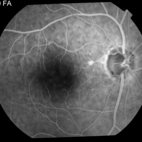
 Central Serous Retinopathy
Mar 26 2019 by Gary R. Cook, MD, FACS
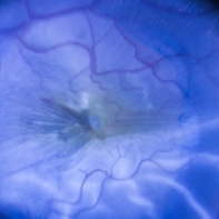
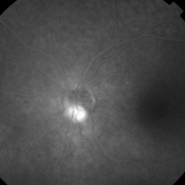
Late-phase frame of FA of 45-year-old white male with CSR OS showing stippled staining in area of RPE track temporally and pooling of dye beneath 3 - 4 small RPEDs OS; VA = 20/30.
Imaging device: Topcon VT-50
Condition/keywords: central serous retinopathy (CSR), FA late phase, fluorescein angiogram (FA), staining
-
 Disseminated Chorioretinitis With Unknown Etiology
Apr 5 2018 by Kim Barrett
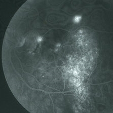
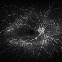
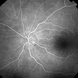
Ultra-wide field fluorescein angiogram of a 31-year-old female with intermittent pain in her left eye. Her condition has been managed in Liberia until recently when she moved to the United States. She suffers from multiple modalities including central retinal artery occlusion, posterior synechiae of the iris, interstitial keratitis, disseminated chorioretinitis, as well as HIV. An infectious cause is high on the differential in light of her HIV status. DDx: hypertensive crisis, an embolism (? IV drug use), coagulopathy, trauma, infectious. Blood work was normal. Her current vision is 20/30 right eye and 20/400 left eye.
Photographer: Kim Barrett, COA
Imaging device: Optos
Condition/keywords: central retinal artery occlusion (CRAO), chorioretinal scar, ciliary artery sparring, disseminated chorioretinitis, HIV, left eye, optic atrophy, staining
-
Epiretinal membrane and ILM peeling
Oct 24 2022 by Manish Nagpal, MD, FRCS (UK), FASRS
This video shows the technique of peeling a epiretinal membrane after triamcinilone staining followed by ILM removal.
Photographer: Manish Nagpal
Condition/keywords: epiretinal membrane (ERM), ILM, macular pucker, staining, video, vitrectomy
-
Epiretinal membrane removal
Oct 24 2022 by Manish Nagpal, MD, FRCS (UK), FASRS
This video highlights the surgical technique of tangentially removing the epiretinal membrane using a forceps
Photographer: Manish Nagpal
Condition/keywords: epiretinal membrane, ERM, macular pucker, staining, video, vitrectomy
-
 Herpetic Corneal Ulcer
Sep 24 2024 by DR Rohit Gupta
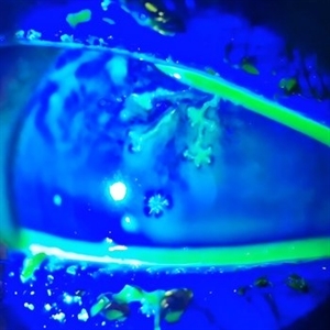
Slit lamp photograph of 32 year old male presented with herpetic corneal ulcer on staining with fluorescein dye under cobalt blue filted dendrits can be seen.
Photographer: Dr Rohit gupta
Imaging device: Samsung S21
Condition/keywords: corneal ulcer, dendritic keratitis, herpes dendrite, Herpes simplex infection, Herpes zoster, staining
-
 ILM staining
Dec 11 2019 by Jennifer R Gallagher, MD
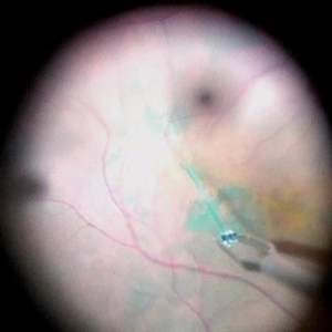
Intra-operative funds photo of the macula after ICG staining with removal of excess dye from the vitreous cavity.
Photographer: Hamzah Khalaf, UT Health San Antonio, University Hospital
Condition/keywords: internal limiting membrane (ILM) peeling, staining, surgical management
-
 ILM visibility with ICG
Dec 11 2019 by Jennifer R Gallagher, MD
Intra-operative photo highlighting the utility of ICG for ILM visibility.
Photographer: Hamzah Khalaf, UT Health San Antonio, University Hospital
Condition/keywords: internal limiting membrane (ILM) peeling, staining, surgical management
-
 Per Operative Photo Post ILM Removal
May 25 2017 by Manish Nagpal, MD, FRCS (UK), FASRS
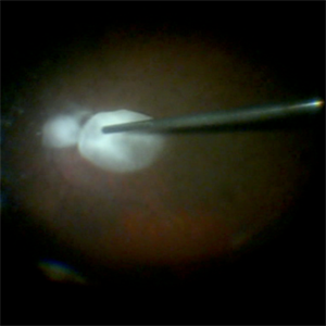
Per operative photo immediately following ILM removal.
Photographer: manish nagpal
Imaging device: SONY HD SURGICAL MICROSCOPE CAMERA
Condition/keywords: dye, epiretinal membrane (ERM), internal limiting membrane (ILM) peeling, staining
-
 Peripheral Drusen
Jan 19 2022 by Olivia Rainey
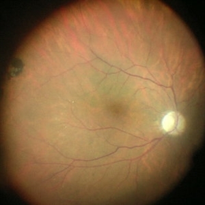
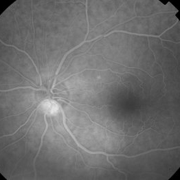
Ultra-widefield fluorescein angiogram of an 83-year-old female with Peripheral Drusen affecting both eyes. The patient presented on 1/19/2022 with slightly decreased vision since her last appointment. Her vision was sc20/25-2 in the right eye. The physician did not believe that the peripheral drusen represents AMD and recommended monitoring. The patient also had mild diabetic retinopathy at the time of her visit.
Photographer: Olivia Rainey, OCT-C, COA
Imaging device: Optos California
Condition/keywords: background diabetic retinopathy (BDR), fluorescein angiogram (FA), Optos, Peripheral drusen, staining, ultra-wide field imaging
-
 Stained ILM Post ERM Removal-1-018
May 25 2017 by Manish Nagpal, MD, FRCS (UK), FASRS
Per operative photo of stained ILM status post ERM removal in a case of RD with ERM.
Photographer: MANISH NAGPAL
Imaging device: SONY 3 CHIP HD CAMERA
Condition/keywords: epiretinal membrane (ERM), internal limiting membrane (ILM) peeling, staining
-
Vitrectomy for Macular Hole
Jan 13 2023 by Manish Nagpal, MD, FRCS (UK), FASRS
This is a case of Macular hole for which vitrectomy is being done. After doing core vitrectomy triamcinolone dye is injected to stain the hyaloid. High aspiration is used on cutter to engage the hyaloid and gradually pull it anteriorly. PVD induction is carried out. After this brilliant blue dye is injected to stain the internal limiting membrane. ILM is peeled using a 25 gauge forceps in a tangential manner. After this i use a instrument called the massager which we have developed to gently and atraumatically massage concentrically the edges of the hole. This releases the subtle contaction on the edges of the hole and relaxes the margins. After this air fluid exchange is carried out followed by low vacuum aspiration over the hole. The hole approximates itself gradually as the aspiration dries up the edges.
Condition/keywords: forceps, hyaloid, ILM, macular hole, peeling, staining, video, vitrectomy
-
---thumb.jpg/image-square;max$300,300.ImageHandler) Staining of old chorioretinal scar and staining and leakage from the new focus of active chorioretinitis.
Staining of old chorioretinal scar and staining and leakage from the new focus of active chorioretinitis.
Feb 15 2013 by From the Collections of Thomas M. Aaberg, MD and Thomas M. Aaberg Jr., MD
Fluorescein angiograph showing staining of old chorioretinal scar and staining and leakage from the new focus of active chorioretinitis.
Condition/keywords: ocular toxoplasmosis
-
---thumb.jpg/image-square;max$300,300.ImageHandler) Staining of retinal vessels, leakage from peripheral retinal neovascularization and peripheral nonperfusion
Staining of retinal vessels, leakage from peripheral retinal neovascularization and peripheral nonperfusion
Feb 15 2013 by From the Collections of Thomas M. Aaberg, MD and Thomas M. Aaberg Jr., MD
Late-phase fluorescein angiograph showing staining of retinal vessels, leakage from peripheral retinal neovascularization and peripheral nonperfusion.
Condition/keywords: peripheral retinal nonperfusion, proliferative retinopathy, retinal neovascularization
-
 Staining-the-vitreous
Jan 10 2022 by Parnian Arjmand, MD, MSc, FRCSC, DABO
This is an example of staining the posterior hyaloid in an eye with no PVD with triamcinolone actinide to assist with inducing a PVD prior to repairing the macular hole.
Condition/keywords: macular hole, PVD, triamcinolone, triescence
-
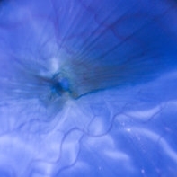
---thumb.JPG/image-square;max$300,300.ImageHandler) "Flower" Macular Degeneration (Wet)
"Flower" Macular Degeneration (Wet)
Jul 13 2013 by Jason S. Calhoun
Patient with (wet) macular degeneration in the left eye. Notice the "flower" shape abnormal blood vessels staining.
Photographer: Jason S. Calhoun, Department of Ophthalmology, Mayo Clinic Jacksonville, Florida
Imaging device: TOPCON TRC 50-EX
Condition/keywords: choroidal neovascularization (CNV)
-
 2:30 FA - Astrocytic Hemartoma
Oct 27 2019 by John S. King, MD
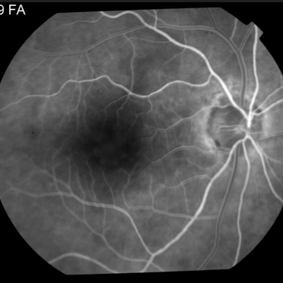
66-year-old white male without history of tuberous sclerosis was found to have an incidental, asymptomatic, translucent, retinal lesion with a few small telangiectatic vessels within it. The FA showed early hyperFL of these small vessels with prominent late leakage/staining. The OCT showed a retinal mass with a "moth eaten" appearance. Vision was 20/20 and the rest of the exam was unremarkable.
Photographer: Maisee Yang
Condition/keywords: astrocytic hamartoma
-
 28 Sec (Laminar Flow) FA - Astrocytic Hemartoma
Oct 27 2019 by John S. King, MD
66-year-old white male without history of tuberous sclerosis was found to have an incidental, asymptomatic, translucent, retinal lesion with a few small telangiectatic vessels within it. The FA showed early hyperFL of these small vessels with prominent late leakage/staining. The OCT showed a retinal mass with a "moth eaten" appearance. Vision was 20/20 and the rest of the exam was unremarkable.
Photographer: Maisee Yang
Condition/keywords: astrocytic hamartoma
-
 49 Sec FA - Astrocytic Hemartoma
Oct 27 2019 by John S. King, MD
66-year-old white male without history of tuberous sclerosis was found to have an incidental, asymptomatic, traslucent, retinal lesion with a few small telangiectatic vessels within it. The FA showed early hyperFL of these small vessels with prominent late leakage/staining. The OCT showed a retinal mass with a "moth eaten" appearance. Vision was 20/20 and the rest of the exam was unremarkable.
Photographer: Maisee Yang
Condition/keywords: astrocytic hamartoma
-
---thumb.jpg/image-square;max$300,300.ImageHandler) Aborted Arteriolitis - diffuse hyper-permeability and staining of the infectious retinal lesion
Aborted Arteriolitis - diffuse hyper-permeability and staining of the infectious retinal lesion
Feb 15 2013 by From the Collections of Thomas M. Aaberg, MD and Thomas M. Aaberg Jr., MD
Fluorescein angiogram corresponding to slide titled Aborted Arteriolitis showing diffuse hyper-permeability and staining of the infectious retinal lesion.
Condition/keywords: ocular toxoplasmosis
-
 Acute Multifocal Placoid Pigment Epitheliopathy
Sep 15 2014 by Thomas A. Ciulla, MD, MBA, FASRS
AMPPE in a 42-year-old woman. Late phase angiography shows staining of multiple focal lesions.
Photographer: Thomas Steele
Condition/keywords: acute multifocal placoid pigment epitheliopathy (AMPPE), late phase
-
 Acute Posterior Multifocal Placoid Pigment Epitheliopathy
Sep 15 2012 by Roy D. Brod, MD
Late phase fluorescein angiogram demonstrating staining of placoid lesions in patient with APMPPE.
Photographer: Julia Walker
Condition/keywords: acute posterior multifocal placoid pigment epitheliopathy (APMPPE)
-
---thumb.jpg/image-square;max$300,300.ImageHandler) Acute Posterior Multifocal Placoid Pigment Epitheliopathy
Acute Posterior Multifocal Placoid Pigment Epitheliopathy
Feb 27 2013 by Henry J. Kaplan, MD
APMPPE. F/A .Late hyperfluorescence and staining of the lesions apparent #3.
Condition/keywords: acute posterior multifocal placoid pigment epitheliopathy (APMPPE), white dot syndrome
-
---thumb.jpg/image-square;max$300,300.ImageHandler) acute retinal necrosis
acute retinal necrosis
Feb 15 2013 by From the Collections of Thomas M. Aaberg, MD and Thomas M. Aaberg Jr., MD
early phase FA corresponding to slide 55, showing punctate hyperfluorescence consistent with microvascular damage and staining of areas of retinal necrosis
Condition/keywords: acute retinal necrosis
-
 Adult Foveomacular Vitelliform Dystrophy
Dec 27 2014 by Thomas A. Ciulla, MD, MBA, FASRS
Angiography revealed early blockage and some mild late staining due to the pigment clumping without any evidence of choroidal neovascularization.
Photographer: Thomas Steele
Condition/keywords: adult foveomacular dystrophy, adult vitelliform dystrophy
 Loading…