Search results (267 results)
-
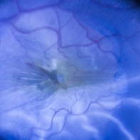
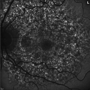
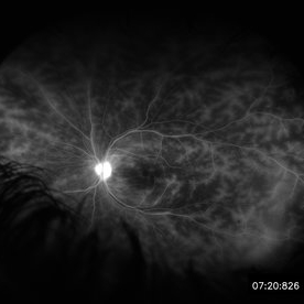 Retinal Vasculitis in Behcet's OS
Jun 29 2018 by Gareth Lema, MD, PhD
IVFA at 7 minutes showing retinal vasculitis, cystoid macular edema, and disc staining.
Photographer: Ross Eye Institute, University at Buffalo Jacobs School of Medicine, Buffalo. NY
Imaging device: Optos
Condition/keywords: Behcet's Disease, cystoid macular edema (CME), disc staining, retinal vasculitis
-
 Herpes Dendrite
Jul 11 2013 by Jason S. Calhoun
Herpes dendrite with fluorescence staining.
Photographer: Jason S. Calhoun, Department of Ophthalmology, Mayo Clinic Jacksonville, Florida
Condition/keywords: disciform herpes simplex keratitis
-
 Applinator Prism Alcohol Burn on Cornea.
Jul 11 2013 by Jason S. Calhoun
Patient who was applinated for IOP check with applinator prism, produced a burn from the tip of the prism after it was cleaned with alcohol. Fluoresce staining shows a ring burn on the epithelium.
Photographer: Jason S. Calhoun, Department of Ophthalmology, Mayo Clinic Jacksonville, Florida
Condition/keywords: cornea
-
 Central Serous Chorioretinopathy
Jan 25 2022 by Olivia Rainey
Late phase widefield fluorescein angiography of a 60-year-old male with Central Serous Chorioretinopathy. Chronic history of CSR followed with observation without treatment prior to presenting at our office. The physician noted significant findings on exam and imaging with multifocal areas of inactive and active changes OD. FA shows superotemporal macular leakage, subtle inferonasal macular leakage and staining as well as multifocal hypercyanescence on ICG. Fortunately foveal sparing and thus observation is recommended at this time OD.
Photographer: Olivia Rainey, OCT-C, COA
Imaging device: Heidelberg Spectralis
Condition/keywords: 55-degrees, central serous chorioretinopathy (CSCR), central serous retinopathy (CSR), chronic central serous chorioretinopathy (CSCR), fluorescein angiogram (FA), fluorescein leakage, heidelberg spectralis, indocyanine green (ICG) angiography, late phase
-
 Disseminated Chorioretinitis With Unknown Etiology
Apr 5 2018 by Kim Barrett
Ultra-wide field fluorescein angiogram of a 31-year-old female with intermittent pain in her left eye. Her condition has been managed in Liberia until recently when she moved to the United States. She suffers from multiple modalities including central retinal artery occlusion, posterior synechiae of the iris, interstitial keratitis, disseminated chorioretinitis, as well as HIV. An infectious cause is high on the differential in light of her HIV status. DDx: hypertensive crisis, an embolism (? IV drug use), coagulopathy, trauma, infectious. Blood work was normal. Her current vision is 20/30 right eye and 20/400 left eye.
Photographer: Kim Barrett, COA
Imaging device: Optos
Condition/keywords: central retinal artery occlusion (CRAO), chorioretinal scar, ciliary artery sparring, disseminated chorioretinitis, HIV, left eye, optic atrophy, staining
-
Epiretinal membrane removal
Oct 24 2022 by Manish Nagpal, MD, FRCS (UK), FASRS
This video highlights the surgical technique of tangentially removing the epiretinal membrane using a forceps
Photographer: Manish Nagpal
Condition/keywords: epiretinal membrane, ERM, macular pucker, staining, video, vitrectomy
-
 Herpes Dendrite
Jul 11 2013 by Jason S. Calhoun
Herpes dendrite with fluorescence staining.
Photographer: Jason S. Calhoun, Department of Ophthalmology, Mayo Clinic Jacksonville, Florida
Condition/keywords: disciform herpes simplex keratitis
-
 ILM Peeling in Progress
Feb 4 2022 by Manish Nagpal, MD, FRCS (UK), FASRS
Intraoperative shot of ILM peeling in progress using forceps.
Photographer: Manish Nagpal, Director, Retina Foundation, Ahmedabad
Imaging device: Sony PMW -10 MD surgical camera
Condition/keywords: ILM flap, ILM staining, internal limiting membrane (ILM) peeling, macular hole, retina, retina surgery
-
 Peeling Under PFO
May 7 2023 by Maxwell J Wingelaar, MD
Peeling under PFO
Condition/keywords: ILM peeling, ILM staining, proliferative vitreoretinopathy (PVR)
-
 Per Operative Photo Post ILM Removal
May 25 2017 by Manish Nagpal, MD, FRCS (UK), FASRS
Per operative photo immediately following ILM removal.
Photographer: manish nagpal
Imaging device: SONY HD SURGICAL MICROSCOPE CAMERA
Condition/keywords: dye, epiretinal membrane (ERM), internal limiting membrane (ILM) peeling, staining
-
 Peripheral Drusen
Jan 19 2022 by Olivia Rainey
Ultra-widefield fluorescein angiogram of an 83-year-old female with Peripheral Drusen affecting both eyes. The patient presented on 1/19/2022 with slightly decreased vision since her last appointment. Her vision was sc20/25-2 in the right eye. The physician did not believe that the peripheral drusen represents AMD and recommended monitoring. The patient also had mild diabetic retinopathy at the time of her visit.
Photographer: Olivia Rainey, OCT-C, COA
Imaging device: Optos California
Condition/keywords: background diabetic retinopathy (BDR), fluorescein angiogram (FA), Optos, Peripheral drusen, staining, ultra-wide field imaging
-
 Retina
May 31 2014 by ruth pav
A 32-year-old woman with a history of drug abuse was admitted due to acute manifestation of multiple infarcts, including acute stroke, splenic and renal infarcts, and multiple cutaneous hematomas. Due to decreased vision in her left eye the patient was referred for ophthalmic evaluation. On exam, visual acuity was 6/10 in the right eye and no light perception in her left eye. Ophthalmoscopic examination was normal in the right eye but showed pallor of the optic nerve head with attenuated retinal vessels in the left eye. Fluorescein angiography showed an oval area of hyperfluorescence from from non-perfusion involving the macular center with staining of overlying retinal capillaries.
Photographer: Ruth Pav, Rambam medical center,Hifa Israel.
Imaging device: Zeiss FF4
Condition/keywords: retina
-
 Serous Retinal Detachment and Retinal Infiltrate due to B. Hensele, Cat-Scratch Disease
Dec 19 2020 by John S. King, MD
64-year-old female had at least a two week history of blurry vision in the right eye. She was being followed for a CRVO in the right eye, and as vision worsened, was referred to our clinic, and saw Dr. Zocchi. Vision in the right eye was CF; there was 1+ cell in the A/C; 1+ vitreous cell was present; disc edema with surrounding SRF as well as a small, white, retinal infiltrate just superior to the optic disc; vessel tortuosity was present as well as a few IRHs (left eye was u/r). There was sub-foveal and PP SRF on OCT. FA in the early to mid phase showed optic disc hyperfluorescence and early filling into the subretinal space. In the later frames there was disc leakage, staining/leakage of the retinal infiltrate, and filling into the subretinal space (See Image). Multiple tests were done, she was started on doxycycline 100 mg BID, and Bartonella serology test came back positive. One week later vision improved to 20/100, a/c cell present, disc edema improved and the SRF was resolving. (will add more photos next visit)
Photographer: Shelly Blair
Imaging device: Optos CA
Condition/keywords: cat scratch retinitis
-
 Stained ILM Post ERM Removal-1-018
May 25 2017 by Manish Nagpal, MD, FRCS (UK), FASRS
Per operative photo of stained ILM status post ERM removal in a case of RD with ERM.
Photographer: MANISH NAGPAL
Imaging device: SONY 3 CHIP HD CAMERA
Condition/keywords: epiretinal membrane (ERM), internal limiting membrane (ILM) peeling, staining
-
---thumb.jpg/image-square;max$300,300.ImageHandler) Acute Posterior Multifocal Placoid Pigment Epitheliopathy
Acute Posterior Multifocal Placoid Pigment Epitheliopathy
Feb 27 2013 by Henry J. Kaplan, MD
APMPPE. F/A .Late hyperfluorescence and staining of the lesions apparent #3.
Condition/keywords: acute posterior multifocal placoid pigment epitheliopathy (APMPPE), white dot syndrome
-
---thumb.jpg/image-square;max$300,300.ImageHandler) Lyme Disease
Lyme Disease
Feb 26 2013 by Henry J. Kaplan, MD
Lyme disease: spirochaete, or "corkscrew-shaped" Borrelia burgdorferi are visible by special staining during darkfield microscopy. #3
Condition/keywords: Lyme disease
-
 Pentosan Maculopathy Autofluorescence Left
Oct 25 2020 by Thomas A. Ciulla, MD, MBA, FASRS
56-year-old woman with prior long term use of pentosan for cystitis. She self discontinued the pentosan 1 year ago. Imaging shows patch RPE atrophy OU with a ¾ DA patch of geo atrophy OS. Autofluorescence shows punctate hyperautoflouresence and FA shows staining of RPE mottling with reticular features.
Condition/keywords: pentosan sulfate
-
 Pentosan Maculopathy Autofluorescence Right
Oct 25 2020 by Thomas A. Ciulla, MD, MBA, FASRS
56-year-old woman with prior long term use of pentosan for cystitis. She self discontinued the pentosan 1 year ago. Imaging shows patch RPE atrophy OU with a ¾ DA patch of geo atrophy OS. Autofluorescence shows punctate hyperautoflouresence and FA shows staining of RPE mottling with reticular features.
Condition/keywords: pentosan sulfate
-
 Acute Posterior Multifocal Placoid Pigment Epitheliopathy
Sep 15 2012 by Roy D. Brod, MD
Late phase fluorescein angiogram demonstrating staining of placoid lesions in patient with APMPPE.
Photographer: Julia Walker
Condition/keywords: acute posterior multifocal placoid pigment epitheliopathy (APMPPE)
-
---thumb.JPG/image-square;max$300,300.ImageHandler) "Flower" Macular Degeneration (Wet)
"Flower" Macular Degeneration (Wet)
Jul 13 2013 by Jason S. Calhoun
Patient with (wet) macular degeneration in the left eye. Notice the "flower" shape abnormal blood vessels staining.
Photographer: Jason S. Calhoun, Department of Ophthalmology, Mayo Clinic Jacksonville, Florida
Imaging device: TOPCON TRC 50-EX
Condition/keywords: choroidal neovascularization (CNV)
-
 2:30 FA - Astrocytic Hemartoma
Oct 27 2019 by John S. King, MD
66-year-old white male without history of tuberous sclerosis was found to have an incidental, asymptomatic, translucent, retinal lesion with a few small telangiectatic vessels within it. The FA showed early hyperFL of these small vessels with prominent late leakage/staining. The OCT showed a retinal mass with a "moth eaten" appearance. Vision was 20/20 and the rest of the exam was unremarkable.
Photographer: Maisee Yang
Condition/keywords: astrocytic hamartoma
-
 28 Sec (Laminar Flow) FA - Astrocytic Hemartoma
Oct 27 2019 by John S. King, MD
66-year-old white male without history of tuberous sclerosis was found to have an incidental, asymptomatic, translucent, retinal lesion with a few small telangiectatic vessels within it. The FA showed early hyperFL of these small vessels with prominent late leakage/staining. The OCT showed a retinal mass with a "moth eaten" appearance. Vision was 20/20 and the rest of the exam was unremarkable.
Photographer: Maisee Yang
Condition/keywords: astrocytic hamartoma
-
 49 Sec FA - Astrocytic Hemartoma
Oct 27 2019 by John S. King, MD
66-year-old white male without history of tuberous sclerosis was found to have an incidental, asymptomatic, traslucent, retinal lesion with a few small telangiectatic vessels within it. The FA showed early hyperFL of these small vessels with prominent late leakage/staining. The OCT showed a retinal mass with a "moth eaten" appearance. Vision was 20/20 and the rest of the exam was unremarkable.
Photographer: Maisee Yang
Condition/keywords: astrocytic hamartoma
-
---thumb.jpg/image-square;max$300,300.ImageHandler) Aborted Arteriolitis - diffuse hyper-permeability and staining of the infectious retinal lesion
Aborted Arteriolitis - diffuse hyper-permeability and staining of the infectious retinal lesion
Feb 15 2013 by From the Collections of Thomas M. Aaberg, MD and Thomas M. Aaberg Jr., MD
Fluorescein angiogram corresponding to slide titled Aborted Arteriolitis showing diffuse hyper-permeability and staining of the infectious retinal lesion.
Condition/keywords: ocular toxoplasmosis
-
 Acute Multifocal Placoid Pigment Epitheliopathy
Sep 15 2014 by Thomas A. Ciulla, MD, MBA, FASRS
AMPPE in a 42-year-old woman. Late phase angiography shows staining of multiple focal lesions.
Photographer: Thomas Steele
Condition/keywords: acute multifocal placoid pigment epitheliopathy (AMPPE), late phase
 Loading…