Search results (267 results)
-
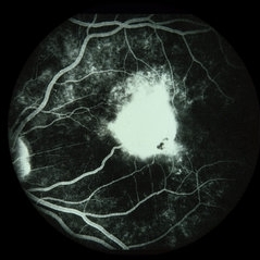
---thumb.JPG/image-square;max$300,300.ImageHandler) "Flower" Macular Degeneration (Wet)
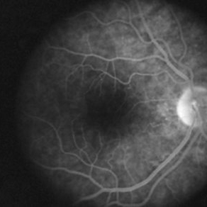
"Flower" Macular Degeneration (Wet)
Jul 13 2013 by Jason S. Calhoun
Patient with (wet) macular degeneration in the left eye. Notice the "flower" shape abnormal blood vessels staining.
Photographer: Jason S. Calhoun, Department of Ophthalmology, Mayo Clinic Jacksonville, Florida
Imaging device: TOPCON TRC 50-EX
Condition/keywords: choroidal neovascularization (CNV)
-
 Applinator Prism Alcohol Burn on Cornea.
Jul 11 2013 by Jason S. Calhoun
Patient who was applinated for IOP check with applinator prism, produced a burn from the tip of the prism after it was cleaned with alcohol. Fluoresce staining shows a ring burn on the epithelium.
Photographer: Jason S. Calhoun, Department of Ophthalmology, Mayo Clinic Jacksonville, Florida
Condition/keywords: cornea
-
 AZOOR
AZOOR
Aug 24 2012 by Geoffrey G. Emerson, MD, PhD, FASRS
A 17-year-old healthy woman noticed a pacman-shaped scotoma in her temporal right vision. Acuity measured 20/20 and color vision measured 11/11. Angiography showed some late staining of the nasal macula.
Photographer: Geoffrey Emerson, MD, PhD, Retina Center, Minneapolis
Condition/keywords: scotoma
-
 Acute Posterior Multifocal Placoid Pigment Epitheliopathy
Sep 15 2012 by Roy D. Brod, MD
Late phase fluorescein angiogram demonstrating staining of placoid lesions in patient with APMPPE.
Photographer: Julia Walker
Condition/keywords: acute posterior multifocal placoid pigment epitheliopathy (APMPPE)
-
 Applinator Prism Alcohol Burn on Cornea.
Jul 11 2013 by Jason S. Calhoun
Patient who was applinated for IOP check with applinator prism, produced a burn from the tip of the prism after it was cleaned with alcohol. Fluoresce staining shows a ring burn on the epithelium.
Photographer: Jason S. Calhoun, Department of Ophthalmology, Mayo Clinic Jacksonville, Florida
Condition/keywords: cornea
-
 Toxocara Granuloma
Feb 25 2013 by Henry J. Kaplan, MD
Toxocara granuloma of ON, late stage F/A. #3 Late hyperfluorescence in the granuloma due to staining.
Condition/keywords: ocular toxoplasmosis, toxocara granuloma, toxocariasis
-
 Color Fundus Photograph of Macular Infarction Secondary to Subonjunctival Gentamicin Injection
May 16 2014 by Arwa Azmeh, MD, PhD
A 20-year-old male suffered from diplopia since age one. He was diagnosed to have acquired fourth nerve palsy in his left eye. VA at time of diagnosis was 20/20 in OU and Fundus exam was WNL in OU. His history revealed no other complaints. 3 days ago he underwent left superior oblique tucking for relief of his diplopia.The surgery was uneventful and at the end of surgery subconjunctival gentamicin was injected. Immediately following surgery his VA in OS decreased from 20/20 to complete loss of central vision and sensation of HM from the periphery. He was referred to us 3 days after surgery. At time of referral fundus exam of his left eye revealed macular infarction with cherry red spot appearance with few retinal hemorrhages, mild optic disc edema and CWS surrounding optic disc. Peripheral retina had normal color and appearance. The vitreous was clear. Anterior segment was quiet. IOP was WNL. Macular OCT was consistent with macular infarction. FA revealed delay in central retinal artery filling as fluorescein started to appear in the arteries at the level of the optic disc at 28 sec, and in the retinal veins at 38 sec. Macular area remained to be non-perfused throughout the whole FA. In late phases staining of blood vessels walls was noticed. The "wipe out" of large vessels and capillaries persisted in the central area. OCT through foveal area showed diffuse thickening of the retina with severe elevation in the fovea, reduced backscattering from the outer layers of the retina and enhanced reflectivity from the inner retina, due to ischemia. Complete blood count and cardiovascular study were WNL. The final diagnosis was macular infarction secondary to subconjunctival gentamicin injection.
Imaging device: OCT
Condition/keywords: macular infarction, subconjunctival gentamicin
-
---thumb.jpg/image-square;max$300,300.ImageHandler) Peripheral retinal nonperfusion, venous beading and dilatation, retinal microaneurysms, and intraretinal hemorrhage
Peripheral retinal nonperfusion, venous beading and dilatation, retinal microaneurysms, and intraretinal hemorrhage
Feb 15 2013 by From the Collections of Thomas M. Aaberg, MD and Thomas M. Aaberg Jr., MD
Color fundus photograph corresponding to slide titled "staining of retinal vessels, leakage from peripheral retinal neovascularization and peripheral nonperfusion." Shows peripheral retinal nonperfusion, venous beading and dilatation, retinal microaneurysms, and intraretinal hemorrhage.
Condition/keywords: peripheral retinal nonperfusion, proliferative retinopathy, retinal neovascularization
-
---thumb.jpg/image-square;max$300,300.ImageHandler) Acute Posterior Multifocal Placoid Pigment Epitheliopathy
Acute Posterior Multifocal Placoid Pigment Epitheliopathy
Feb 27 2013 by Henry J. Kaplan, MD
APMPPE. F/A .Late hyperfluorescence and staining of the lesions apparent #3.
Condition/keywords: acute posterior multifocal placoid pigment epitheliopathy (APMPPE), white dot syndrome
-
 Ocular Ischemic Syndrome
Jun 20 2018 by Andreas Ebneter, MD, PhD, FASRS
Ocular ischemic syndrome can present with a wide variety of ocular findings in both the anterior and posterior segments. The color fundus image of this 77-year-old male shows scattered blot hemorrhages in the deep retinal layers of the posterior pole that are only occasionally confluent. Commonly, these typical hemorrhages are predominantly found in the mid-periphery. Fluorescein angiography helps in confirming the diagnosis. Choroidal filling time is frequently somewhat delayed and patchy. Arteriovenous transit time is clearly prolonged. Staining of both veins and arteries in late images (top right) reflects diffuse endothelial cell damage with compromise of the blood-retina barrier. The peripheral retina is affected by extensive non-perfusion.
Photographer: Eva Steffen, Bern University Hospital, Switzerland
Imaging device: Optos 200Tx and Heidelberg Spectralis OCT
Condition/keywords: ocular ischemic syndrome
-
---thumb.jpg/image-square;max$300,300.ImageHandler) late-phase FA showing arteriolar attenuation and late staining of choroidal lesions
late-phase FA showing arteriolar attenuation and late staining of choroidal lesions
Feb 14 2013 by From the Collections of Thomas M. Aaberg, MD and Thomas M. Aaberg Jr., MD
late-phase FA showing arteriolar attenuation and late staining of choroidal lesions
Condition/keywords: multifocal choroiditis, posterior segment inflammation, white dot syndrome
-
 Eals Disease
Jan 26 2013 by Ratimir Lazic, MD, PhD
FAG image of a 28-year-old male. Staining of scars due to laser photocoagulation can be seen.
Photographer: Marko Lukic, MD
Imaging device: Zeis Visucam Lite 2
Condition/keywords: fundus photograph, laser photocoagulation
-
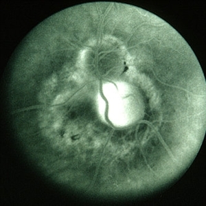
 Choroidal Osteoma 4
Oct 5 2012 by Ronald C. Gentile, MD
Fluorescein angiography of the macular choroidal osteoma in the late phase of the angiogram with late staining of the tumor highlighting its negative staining internal vascularity.
Photographer: The New York Eye & Ear Infirmary Department of Medical Imaging
Condition/keywords: macular choroidal osteoma
-
 Disseminated Chorioretinitis With Unknown Etiology
Apr 5 2018 by Kim Barrett
Ultra-wide field fluorescein angiogram of a 31-year-old female with intermittent pain in her left eye. Her condition has been managed in Liberia until recently when she moved to the United States. She suffers from multiple modalities including central retinal artery occlusion, posterior synechiae of the iris, interstitial keratitis, disseminated chorioretinitis, as well as HIV. An infectious cause is high on the differential in light of her HIV status. DDx: hypertensive crisis, an embolism (? IV drug use), coagulopathy, trauma, infectious. Blood work was normal. Her current vision is 20/30 right eye and 20/400 left eye.
Photographer: Kim Barrett, COA
Imaging device: Optos
Condition/keywords: central retinal artery occlusion (CRAO), chorioretinal scar, ciliary artery sparring, disseminated chorioretinitis, HIV, left eye, optic atrophy, staining
-
 Choroidal Folds
Nov 28 2014 by Thomas A. Ciulla, MD, MBA, FASRS
This 53-year-old man was noted to have choroidal folds right greater than left. The visual acuity was normal at 20/15. The choroidal folds are visible on OCT, especially on the vertical cuts that image across the horizontal folds. Angiography revealed staining of the folds without CNVM, choroidal mass, or optic nerve edema.
Photographer: Charlotte Harris
Condition/keywords: bilateral chorioretinal folds, choroidal folds
-
 BRVO FA Montage Late
Apr 17 2014 by Susanna S. Park, MD, PhD
Montage of the late transit view of fluorescein angiogram of a 45-year-old woman with branch retinal vein occlusion showing diffuse staining and leakage of the affected retinal veins.
Photographer: Karishma Chandra, UC Davis Eye Center
Condition/keywords: branch retinal vein occlusion (BRVO), fluorescein leakage
-
 TA Stained Posterior Hyaloid Face
Apr 11 2014 by Subhendu Kumar Boral, MBBS, MD(AIIMS), DNB, FASRS (USA)
Intraoperative step of posterior hyaloid face staining by triamcinolone acetonide particles during PVD induction in a case of diabetic epiretinal membrane left eye in a 68-year-old gentleman.
Photographer: Subhendu Kumar Boral
Condition/keywords: hyaloid
-
 Unilateral Acute Idiopathic Maculopathy - Fluorescein Angiography (2)
Sep 9 2012 by Robin Ray, MD
FA of UAIM (late) - hyperintense staining of lesion.
Photographer: Kidron Robertson, Georgia Eye Institute of the Southeast, Savannah, GA
Imaging device: Heidelberg Spectralis
Condition/keywords: chorioretinal inflammations, Coxsackie, unilateral acute idiopathic maculopathy
-
 OCT Through Foveal Area in Macular Infarction Secondary to Subconjunctival Gentamicin Injection
May 16 2014 by Arwa Azmeh, MD, PhD
A 20-year-old male suffered from diplopia since age one. He was diagnosed to have acquired fourth nerve palsy in his left eye. VA at time of diagnosis was 20/20 in OU and fundus exam was WNL in OU. His history reaveled no other complaints. 3 days ago he underwent left superior oblique tucking for relief of his diplopia.The surgery was uneventful and at the end of surgery subconjunctival gentamicin was injected. Immediately following surgery his VA in OS decreased from 20/20 to complete loss of central vision and sensation of HM from the periphery. He was referred to us 3 days after surgery. At time of referral fundus exam of his left eye revealed macular infarction with cherry red spot appearance with few retinal hemorrhages , mild optic disc edema and CWS surrounding optic disc. Peripheral retina had normal color and appearance. The vitreous was clear. Anterior segment was quiet. IOP was WNL. Macular OCT was consistent with macular infarction. FA revealed delay in central retinal artery filling as fluorescein started to appear in the arteries at the level of the optic disc at 28 sec, and in the retinal veins at 38 sec. Macular area remained to be non-perfused throughout the whole FA. In late phases staining of blood vessels walls was noticed. The "wipe out" of large vessels and capillaries persisted in the central area. OCT through foveal area showed diffuse thickening of the retina with severe elevation in the fovea, reduced backscattering from the outer layers of the retina and enhanced reflectivity from the inner retina, due to ischemia. Complete blood count and cardiovascular study were WNL. The final diagnosis was macular infarction secondary to subconjunctival gentamicin injection.
Imaging device: OCT
Condition/keywords: macular infarction, subconjunctival gentamicin
-
---thumb.jpg/image-square;max$300,300.ImageHandler) late-phase FA showing arteriolar attenuation and late staining of choroidal lesions
late-phase FA showing arteriolar attenuation and late staining of choroidal lesions
Feb 14 2013 by From the Collections of Thomas M. Aaberg, MD and Thomas M. Aaberg Jr., MD
late-phase FA showing arteriolar attenuation and late staining of choroidal lesions
Condition/keywords: multifocal choroiditis, posterior segment inflammation, white dot syndrome
-
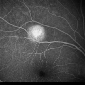
 Choroidal Granuloma Secondary to Tuberculosis
Mar 14 2013 by Eduardo Torres-Porras, MD
Late-phase intravenous fluorescein angiography shows staining of the disk lesion and strong pooling of fluorescein.
Photographer: Carlos Yepez
Condition/keywords: tubercular choroidal granuloma
-
 Retina
May 31 2014 by ruth pav
A 32-year-old woman with a history of drug abuse was admitted due to acute manifestation of multiple infarcts, including acute stroke, splenic and renal infarcts, and multiple cutaneous hematomas. Due to decreased vision in her left eye the patient was referred for ophthalmic evaluation. On exam, visual acuity was 6/10 in the right eye and no light perception in her left eye. Ophthalmoscopic examination was normal in the right eye but showed pallor of the optic nerve head with attenuated retinal vessels in the left eye. Fluorescein angiography showed an oval area of hyperfluorescence from from non-perfusion involving the macular center with staining of overlying retinal capillaries.
Photographer: Ruth Pav, Rambam medical center,Hifa Israel.
Imaging device: Zeiss FF4
Condition/keywords: retina
-
 Toxocara Granuloma
Jun 4 2014 by Henry J. Kaplan, MD
Arteriovenous phase angiogram of the same patient shows staining of the granuloma and stippling hyperfluorescence around the lesion secondary to RPE window defect. #3
Condition/keywords: toxocara granuloma, toxocariasis
-
---thumb.jpg/image-square;max$300,300.ImageHandler) Lyme Disease
Lyme Disease
Feb 26 2013 by Henry J. Kaplan, MD
Lyme disease: spirochaete, or "corkscrew-shaped" Borrelia burgdorferi are visible by special staining during darkfield microscopy. #3
Condition/keywords: Lyme disease
-
---thumb.JPG/image-square;max$300,300.ImageHandler) Dengue Retinitis
Dengue Retinitis
Nov 3 2012 by Mallika Goyal, MD
Late phase fluorescein angiogram of left eye of a 37-year-old lady recovering from dengue fever with bilateral dengue retinitis shows staining of retinal exudates.
Photographer: Mallika Goyal, MD
Condition/keywords: Dengue retinitis, retinal exudates
 Loading…