Search results (267 results)
-
 Commotio Retinae
Jun 10 2025 by CUI YUELING
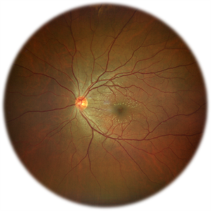
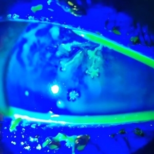
The patient presented 2 hours after sustaining a left eye injury caused by a stick. Visual acuity in the left eye was 0.2 without improvement upon correction, and intraocular pressure measured 15 mmHg. Examination of the anterior segment revealed ciliary conjunctival injection accompanied by patchy subconjunctival hemorrhage. The corneal surface remained smooth, and the anterior chamber was deep with hyphema characterized by blood-tinged aqueous humor predominantly settled inferiorly. The pupil was slightly irregular, approximately 3 mm in diameter, with a superotemporal notch; pupillary light reflex was intact. The lens appeared clear. Fundus examination showed well-defined optic disc margins with normal coloration and a cup-to-disc ratio of 0.2. Retinal arteries and veins were normally distributed with an artery-to-vein ratio of 2:3. At the posterior pole, the foveal reflex exhibited concentric ripple-like changes centered on the fovea, accompanied by localized pigment attenuation and reduced reflex intensity. Irregular reflectivity was noted in the superotemporal and inferotemporal nerve fiber layers.
Photographer: Yueling Cui
Imaging device: Zeiss Clarus 500
Condition/keywords: commotio retinae
-
 VKH Pseudotumor – Fluorescein Angiography
May 11 2025 by Felipe Murati
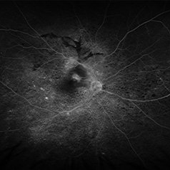
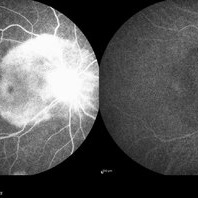
Fluorescein angiography image from a 36-year-old woman with chronic Vogt-Koyanagi-Harada (VKH) syndrome showing a pseudotumor-like lesion with late-phase staining and no active leakage. The image highlights subretinal fibrosis in the right eye, stable under long-term immunosuppressive therapy with mycophenolate mofetil and adalimumab. No signs of active choroiditis are present, confirming a quiescent phase.
Photographer: Felipe A. Murati, MD, University of Arizona
Imaging device: Optos California, fluorescein angiography modality
Condition/keywords: choroiditis, Fluorescein angiography, granulomatous uveitis, Optos FA, pseudotumor, subretinal fibrosis, VKH, Vogt-Koyanagi-Harada
-
 Necrotizing Scleritis
Apr 17 2025 by Gustavo Uriel Fonseca Aguirre
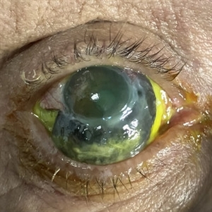
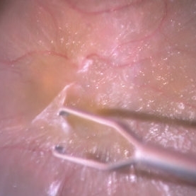
The clinical photograph shows necrotizing scleritis with perilimbal involvement, featuring marked scleral thinning and violaceous episcleral injection in the inferior quadrant. Focal uveal prolapse is visible at the area of maximal scleral necrosis, accompanied by peripheral ulcerative keratitis. Fluorescein staining residue is observed on the ocular surface. Associated findings include mild conjunctival chemosis and dilated episcleral vessels.
Photographer: Gustavo U. Fonseca Aguirre, Hospital Conde de Valenciana, Ciudad de México
Condition/keywords: necrotizing scleritis
-
 Stargardt Disease (FA)
Jan 22 2025 by Virginia Gebhart
Fluorescein angiogram of 19 year old female with confirmed Stargardt Disease. Hyperfluorescence in the macula with staining defect and silent choroid.
Photographer: Virginia Gebhart, Retina Consultants of Carolina
Imaging device: Optos California
Condition/keywords: fluorescein angiogram (FA), Silent Choroid, Stargardt disease
-
 MIDD (Maternally Inherited Diabetes and Deafness) - Right FA (4 min)
Nov 30 2024 by John S. King, MD
Both eyes had similar FA findings. There was no dark choroid or signs of leakage. Granular staining around the fovea and disc were present, and the HypoAF areas corresponded to the drusenoid deposits that showed HyperAF. Mild MAs present due to NPDR 57 yo WF referred for AMD vs Pattern Dystrophy that was diagnosed 10 years ago. Reported some slow progressive vision loss in both eyes for distance and near. Denies nyctalopia or hemeralopia. Background medical history includes HTN, CVD, and DM. No family history of eye problems. Denied pentosan use. Anterior segment showed moderate cataracts (OD>OS). Posterior segment exam showed macular changes and mild NPDR. The macular appearance showed a symmetrical, paramacular ring of fleck-like drusenoid material with some faint focal areas of RPE hyperplasia. Fundus Photos, AF, OCT were performed as well as a gene test. Further questioning showed revealed that her mother and maternal grandmother had boith diabetes mellitus and sensorineural hearing loss. The patient developed diabetes in her teens, and some high frequency hearing loss in her early twenties. She had not had a previous genetic test or diagnosis of MIDD. Gene testing is pending for the mitochondrial component. Invitae's retinal panel, which does not include mitochondrial disorders, only showed a variant of uncertain significance, HMCN1. I discussed this case with Dr. Freund, and it is similar to a the case report : Inoue M, Kiss S, Freund KB. MACULAR PIGMENT RINGS AS THE PRESENTING FINDING OF MITOCHONDRIAL MYOPATHY, ENCEPHALOPATHY, LACTIC ACIDOSIS, AND STROKELIKE EPISODES. Retin Cases Brief Rep. 2015 Fall;9(4):260-4. doi: 10.1097/ICB.0000000000000182. PMID: 26200388.
Photographer: Grace Melton and Carley Gunn
Imaging device: Clarus
Condition/keywords: Macular Dystrophy, Maternally Inherited Diabetes and Deafness, MIDD, Mitochondrial Disorder
-
 MIDD (Maternally Inherited Diabetes and Deafness) - Left FA (7 min)
Nov 30 2024 by John S. King, MD
Both eyes had similar FA findings. There was no dark choroid or signs of leakage. Granular staining around the fovea and disc were present, and the HypoAF areas corresponded to the drusenoid deposits that showed HyperAF. Mild MAs present due to NPDR 57 yo WF referred for AMD vs Pattern Dystrophy that was diagnosed 10 years ago. Reported some slow progressive vision loss in both eyes for distance and near. Denies nyctalopia or hemeralopia. Background medical history includes HTN, CVD, and DM. No family history of eye problems. Denied pentosan use. Anterior segment showed moderate cataracts (OD>OS). Posterior segment exam showed macular changes and mild NPDR. The macular appearance showed a symmetrical, paramacular ring of fleck-like drusenoid material with some faint focal areas of RPE hyperplasia. Fundus Photos, AF, OCT were performed as well as a gene test. Further questioning showed revealed that her mother and maternal grandmother had boith diabetes mellitus and sensorineural hearing loss. The patient developed diabetes in her teens, and some high frequency hearing loss in her early twenties. She had not had a previous genetic test or diagnosis of MIDD. Gene testing is pending for the mitochondrial component. Invitae's retinal panel, which does not include mitochondrial disorders, only showed a variant of uncertain significance, HMCN1. I discussed this case with Dr. Freund, and it is similar to a the case report : Inoue M, Kiss S, Freund KB. MACULAR PIGMENT RINGS AS THE PRESENTING FINDING OF MITOCHONDRIAL MYOPATHY, ENCEPHALOPATHY, LACTIC ACIDOSIS, AND STROKELIKE EPISODES. Retin Cases Brief Rep. 2015 Fall;9(4):260-4. doi: 10.1097/ICB.0000000000000182. PMID: 26200388.
Photographer: Grace Melton and Carley Gunn
Imaging device: Clarus
Condition/keywords: Macular Dystrophy, Maternally Inherited Diabetes and Deafness, MIDD, Mitochondrial Disorder
-
 ILM Peeling in a Case of Macular Hole
Sep 28 2024 by Anjana Mirajkar, MS Ophthalmology
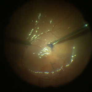
An intra operative image showing staining of the ILM followed by its peeling done in case of macular hole.
Photographer: Dr. Anjana Mirajkar -Retina Foundation, Ahmedabad
Condition/keywords: ILM peeling, Macular hole
-
 Herpetic Corneal Ulcer
Sep 24 2024 by DR Rohit Gupta
Slit lamp photograph of 32 year old male presented with herpetic corneal ulcer on staining with fluorescein dye under cobalt blue filted dendrits can be seen.
Photographer: Dr Rohit gupta
Imaging device: Samsung S21
Condition/keywords: corneal ulcer, dendritic keratitis, herpes dendrite, Herpes simplex infection, Herpes zoster, staining
-
 Epiretinal Membrane
Jul 19 2024 by Anjana Mirajkar, MS Ophthalmology
An intra operative still showing removal of the epi -retinal membrane with forceps under high magnification after staining with triamcinolone acetonide dye.
Photographer: Dr. Anjana Mirajkar -Retina Foundation, Ahmedabad
Imaging device: Mirante-Nidek
Condition/keywords: epiretinal membrane removal
-
 ILM Staining in Case of Macular Hole
Jul 4 2024 by Anjana Mirajkar, MS Ophthalmology
Intra operative still of LE showing ILM staining done with BBG dye in case of macular hole.
Photographer: Dr. Anjana Mirajkar -Retina Foundation, Ahmedabad
Condition/keywords: ILM staining, macular hole
-
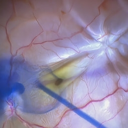 Star Folds in a Chronic Retinal Detachment
Jul 3 2024 by Anjana Mirajkar, MS Ophthalmology
Intra-operative still RE showing a star fold at the parafoveal area causing traction at the macula. Brilliant blue dye being injected to the stain the ILM.
Photographer: Dr. Anjana Mirajkar -Retina Foundation, Ahmedabad
Condition/keywords: brilliant blue staining, proliferative vitreoretinopathy (PVR), star folds
-
 Syphilitic Posterior Uveitis
Mar 22 2024 by Anjana Mirajkar, MS Ophthalmology
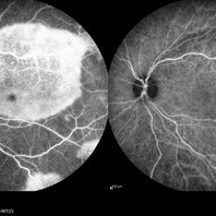
FA image of RE of a 36 year old female showing hyper-fluorescence (staining) from early to late phases of the angiogram in a case syphilitic posterior placoid chorioretinitis. ICG image depicts hypo-cyanence from early to late phases.
Photographer: Dr. Anjana Mirajkar -Retina Foundation, Ahmedabad
Imaging device: Heidelberg
Condition/keywords: acute syphilitic posterior placoid chorioretinitis
-
 Syphilitic Posterior Uveitis
Mar 22 2024 by Anjana Mirajkar, MS Ophthalmology
FA image of LE of a 36 year old female showing hyper-fluorescence (staining) from early to late phases of the angiogram in a case syphilitic posterior placoid chorioretinitis. ICG image depicts hypo-cyanence from early to late phases.
Photographer: Dr. Anjana Mirajkar -Retina Foundation, Ahmedabad
Condition/keywords: acute syphilitic posterior placoid chorioretinitis
-
Retina Rhexis
Jan 2 2024 by Deepak Bhojwani, MS
THIS SURGICAL VIDEO DEMONSTRATES STANDARD ILM PEELING FOR MACULAR HOLE SURGERIES. THE ILM PEELING IS ASSITED BY BRILLIANT BYE DYE STAINING.
Condition/keywords: ILM peeling, macular hole, Macular surgery
-
 Old BRVO
Oct 18 2023 by Anand Temkar
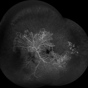
LE widefield FA montage of a 68 years old male with history of Old BRVO, showing peripheral capillary non perfusion and some temporal laser marks (staining ).
Photographer: Dr.Anand Temkar- Retina Foundation, Ahmedabad
Imaging device: Mirante
Condition/keywords: branch retinal vein occlusion (BRVO), capillary nonperfusion
-
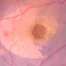
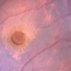
 VPT intra-operative cryotherapy
Aug 14 2023 by Joseph Juliano, MD
Intraoperative photos of the vasoproliferative lesion before cryotherapy (left), and after triple freeze thaw cryotherapy (right). Surrounding white areas of exudation are notable around the lesion. Of note, there is tissue blue staining around the macula because this patient presented with a concurrent macular hole and an internal limiting membrane drape was performed.
Condition/keywords: Vasoproliferative Tumor, VPT
-
 Peeling Under PFO
May 7 2023 by Maxwell J Wingelaar, MD
Peeling under PFO
Condition/keywords: ILM peeling, ILM staining, proliferative vitreoretinopathy (PVR)
-
 BBG Dye injection to Stain ILM during Vitrectomy Surgery | Intra-Operative Still
Apr 28 2023 by Veer Singh, MS, FVRS, FMRF, FICO (Retina)
BBG Dye injection to Stain ILM during Vitrectomy Surgery | Intra-Operative Still
Photographer: Dr. Veer Singh
Condition/keywords: brilliant blue staining, ERM, ILM staining
-
Vitrectomy for Macular Hole
Jan 13 2023 by Manish Nagpal, MD, FRCS (UK), FASRS
This is a case of Macular hole for which vitrectomy is being done. After doing core vitrectomy triamcinolone dye is injected to stain the hyaloid. High aspiration is used on cutter to engage the hyaloid and gradually pull it anteriorly. PVD induction is carried out. After this brilliant blue dye is injected to stain the internal limiting membrane. ILM is peeled using a 25 gauge forceps in a tangential manner. After this i use a instrument called the massager which we have developed to gently and atraumatically massage concentrically the edges of the hole. This releases the subtle contaction on the edges of the hole and relaxes the margins. After this air fluid exchange is carried out followed by low vacuum aspiration over the hole. The hole approximates itself gradually as the aspiration dries up the edges.
Condition/keywords: forceps, hyaloid, ILM, macular hole, peeling, staining, video, vitrectomy
-
Vitrectomy for Myopic Retinal Detachment with multiple tears
Jan 2 2023 by Manish Nagpal, MD, FRCS (UK), FASRS
Vitrectomy for Myopic Retinal detachment with multiple tears and lattice degenerations | Vitrectomy is carried out and triamcinolone staining is used to stain the hyaloid attachment. The hyaloid attachment is extremely adherent. With high vacuum the cutter engages the stained hyaloid and gradually peels it off the mobile retina. After this Perfluorocarbon heavy liquid is injected to flatten the posterior pole and push the fluid to the periphery till the edge of the tear. This is followed by endo drainage from the tear. Once the retina flattened endolaser was carried out.
Condition/keywords: air fluid exchange, endo drainage, endolaser, holes, lattice degeneration, myopia, myopic retinal retachment, RD, reattachment of retinal detachment, tear, video, vitrectomy
-
Vitrectomy for Sub ILM blood over macula
Jan 2 2023 by Manish Nagpal, MD, FRCS (UK), FASRS
This is a case of non resolving ILM hemorrhage over macula. Vitrectomy is carried out and hyaloid is removed after traimcinolone staining. After this brilliant blue dye is injected to stain the ILM. Internal limiting membrane is then removed with a forceps. Once the sub ilm blood is exposed , it easily aspirates with the cutter. The origin is probably from a macroaneurysm and there is a small component of subretinal residual blood noted at the end of the surgery.
Condition/keywords: brilliant blue, hyaloid, internal limiting membrane, macula, microaneurysm, retina, sub ILM blood, sub ILM hemorrhage, triamcinolone, video, vitrectomy
-
Macular hole in myopic patient
Nov 4 2022 by Manish Nagpal, MD, FRCS (UK), FASRS
This is a case earlier operated three times for myopic retinal detachment. Buckling was done followed by vilion oil injection and then silicon oil removal, now the patient developed a macular hole. Staining with brilliant blue followed by ILM peeling with a longer length forceps was carried out. Air fluid exchange with drainage over the hole and gas injection was done
Condition/keywords: brilliant blue staining, ILM peeling, myopia, myopic macular hole, video, vitrectomy
-
 PVD induction with IVTA staining
Nov 1 2022 by Shobhit Chawla, M.S.
This is an intraoperative photograph of Pvd induction showing both the macular and disc attatchments stained with triamcinolone.
Photographer: Shobhit Chawla
Condition/keywords: PVD induction, triamcinolone
-
PVD induction in a retinal detachment
Oct 24 2022 by Manish Nagpal, MD, FRCS (UK), FASRS
This video highlights the PVD induction technique in a case of retinal detachment with mobile retina, triamcinolone staining allows ease of visualizing the pvd attachment which is gradually removed from the retinal attachment using suction.
Photographer: Manish Nagpal
Condition/keywords: posterior hyaloid, PVD, triamcinolone, video, vitrectomy
-
Epiretinal membrane removal
Oct 24 2022 by Manish Nagpal, MD, FRCS (UK), FASRS
This video highlights the surgical technique of tangentially removing the epiretinal membrane using a forceps
Photographer: Manish Nagpal
Condition/keywords: epiretinal membrane, ERM, macular pucker, staining, video, vitrectomy
 Loading…