Search results (111 results)
-
 Neurosensorial Retinal Detachment with Pigment Retinal Epithelium Still in Place
May 18 2020 by McGill University Health Centre
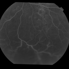
A true retinal detachment always involves accumulation of subretinal fluid. In this specimen, note the retinal vessels exiting the optic nerve (arrow).
Condition/keywords: enucleation, retinal vessels
-
 Amelanotic Melanoma
Sep 19 2025 by Aditya S Kelkar, MS, FRCS, FASRS,FRCOphth
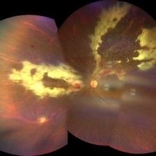
Widefield fundus photograph of a 37 year old showing a large, dome-shaped, intraocular mass involving the temporal retina. The lesion appears elevated and lacks surface pigmentation. Overlying retinal vessels are displaced and draped across the tumor surface, with surrounding retinal elevation noted. The appearance is suggestive of amelanotic variant of choroidal melanoma.
Photographer: Dr. Muskan Mangal
Imaging device: Optos Daytona
Condition/keywords: choroidal melanoma, intraocular tumor
-
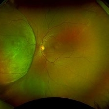
 Asymptomatic Eye in FEVR
Jul 7 2015 by Hamid Ahmadieh, MD
FA image of the asymptomatic left eye of a 28-year-old man with total RD secondary to advanced FEVR in his right eye. Notice straightening of the retinal vessels.
Photographer: Soulmaz Shahmohammad, Negah Eye Center, Tehran, Iran
Imaging device: Specteralis
Condition/keywords: asymptomatic, familial exudative vitreoretinopathy (FEVR)
-
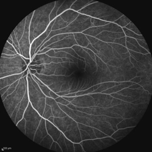
 Asymptomatic Eye in FEVR
Jul 7 2015 by Hamid Ahmadieh, MD
Color fundus photograph of the asymptomatic eye of a patient with FEVR. Notice straightening of the retinal vessels.
Photographer: Soulmaz Shahmohammad, Negah Eye Center, Tehran, Iran
Condition/keywords: color fundus photograph, familial exudative vitreoretinopathy (FEVR)
-
 BRVO With Non-Perfusion
May 3 2014 by Mallika Goyal, MD
Early phase fluorescein angiogram in an eye with superotemporal BRVO shows delayed filling of retinal vessels in the affected quadrant.
Photographer: Mallika Goyal, MD, Apollo Health City, Jubilee Hills, Hyderabad, India
Condition/keywords: non-perfused branch retinal vein occlusion (BRVO)
-
 BRVO With Non-Perfusion
May 3 2014 by Mallika Goyal, MD
Mid-phase fluorescein angiogram in an eye with superotemporal BRVO shows delayed filling of retinal vessels and non-perfusion in the affected quadrant.
Photographer: Mallika Goyal, MD, Apollo Health City, Jubilee Hills, Hyderabad, India
Condition/keywords: non-perfused branch retinal vein occlusion (BRVO)
-
 BRVO With Non-Perfusion
May 3 2014 by Mallika Goyal, MD
Fluorescein angiogram in an eye with superotemporal BRVO shows delayed filling of retinal vessels and non-perfusion in the affected quadrant.
Photographer: Mallika Goyal, MD, Apollo Health City, Jubilee Hills, Hyderabad, India
Condition/keywords: non-perfused branch retinal vein occlusion (BRVO)
-
 BRVO With Non-perfusion
May 3 2014 by Mallika Goyal, MD
Mid-phase fluorescein angiogram in an eye with superotemporal BRVO shows delayed filling of retinal vessels and non-perfusion in the affected quadrant.
Photographer: Mallika Goyal, MD, Apollo Health City, Jubilee Hills, Hyderabad, India
Condition/keywords: non-perfused branch retinal vein occlusion (BRVO)
-
 BRVO With Non-perfusion
May 3 2014 by Mallika Goyal, MD
Fluorescein angiogram in an eye with superotemporal BRVO shows delayed filling of retinal vessels and non-perfusion in the affected quadrant.
Photographer: Mallika Goyal, MD, Apollo Health City, Jubilee Hills, Hyderabad, India
Condition/keywords: non-perfused branch retinal vein occlusion (BRVO)
-
 BRVO With Non-perfusion
May 3 2014 by Mallika Goyal, MD
Late phase fluorescein angiogram in an eye with superotemporal BRVO shows delayed filling of retinal vessels and non-perfusion in the affected quadrant.
Photographer: Mallika Goyal, MD, Apollo Health City, Jubilee Hills, Hyderabad, India
Condition/keywords: non-perfused branch retinal vein occlusion (BRVO)
-
 BRVO With Non-perfusion
May 3 2014 by Mallika Goyal, MD
Late phase fluorescein angiogram in an eye with superotemporal BRVO shows delayed filling of retinal vessels and non-perfusion in the affected quadrant .
Photographer: Mallika Goyal, MD, Apollo Health City, Jubilee Hills, Hyderabad, India
Condition/keywords: non-perfused branch retinal vein occlusion (BRVO)
-
 BRVO With Non-perfusion
May 3 2014 by Mallika Goyal, MD
Late phase fluorescein angiogram of an eye with superotemporal BRVO shows delayed filling of retinal vessels, dilation and tortuosity of the affected veins, and non-perfusion in the affected quadrant .
Photographer: Mallika Goyal, MD, Apollo Health City, Jubilee Hills, Hyderabad, India
Condition/keywords: non-perfused branch retinal vein occlusion (BRVO)
-
 Chorioretinal Coloboma
Oct 6 2025 by Seif Allah Anwar
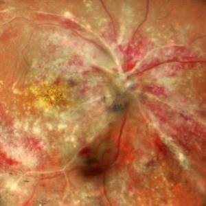
Fundus photograph of the patient left eye showing large, well-demarcated, excavated chorioretinal coloboma involving the inferior fundus, extending from the optic disc to the periphery. The lesion appears white due to bare sclera visibility, with absence of overlying choroid and retina. Retinal vessels course over the colobomatous area inferiorly.
Photographer: Dr. Seif Anwar, FRCSEd
Imaging device: Centervue Eidon
Condition/keywords: chorioretinal coloboma
-
 Choroidal Osteoma
Apr 17 2025 by Gustavo Uriel Fonseca Aguirre
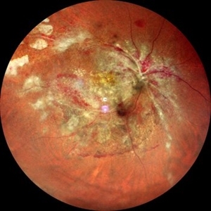
Scanning laser ophthalmoscopy reveals a well-circumscribed, yellowish-white choroidal osteoma overlying the macular region and extending into the inferior temporal vascular arcade. Retinal vessels course normally over the tumor surface, with no evidence of subretinal fluid or hemorrhage. The surrounding retina shows preserved architecture without secondary degenerative changes.
Photographer: Gustavo U. Fonseca Aguirre, Hospital Conde de Valenciana, Ciudad de México
Condition/keywords: choroidal osteoma, macular choroidal osteoma
-
 Choroidal Osteoma
Jun 2 2018 by awaneesh m upadhyay, MBBS, DNB
23-year-old patient's fundus photograph having complaints of defective vision, metamorphosia over 6 months shows yellow orange elevated well defined submacular lesion with normal overlying retinal vessels and normal disc . Vision left eye is 20/80.
Photographer: Hiteshwar Saikia
Condition/keywords: macular choroidal osteoma
-
 chronic central serous chorioretinopathy
Oct 31 2012 by Mallika Goyal, MD
Late phase fluorescein angiogram of inferior retina of left eye with chronic CSCR shows dilation of and mild leak from retinal vessels over the inferior serous retinal detachment.
Photographer: Mallika Goyal, MD
Condition/keywords: central serous chorioretinopathy (CSCR), chronic central serous chorioretinopathy (CSCR)
-
 chronic central serous chorioretinopathy
Oct 31 2012 by Mallika Goyal, MD
Fluorescein angiogram of inferior retina of right eye with chronic CSCR shows dilation of and mild leak from retinal vessels over the inferior serous retinal detachment.
Condition/keywords: central serous chorioretinopathy (CSCR), chronic central serous chorioretinopathy (CSCR), serous retinal detachment
-
 Chronic Intraocular Foreign Body With Siderosis
Jun 28 2014 by John T. Thompson, MD
Large iron containing chronic intraocular foreign body with extensive siderosis of retina. The optic nerve is just to left of foreign body with extensive sheathing of retinal vessels.
Imaging device: Zeiss FF4
Condition/keywords: intraocular foreign body, penetrating trauma, siderosis, trauma
-
 CMV Retinitis
Feb 17 2024 by Eloy Mata-Cortes, MD
Fundus photograph of left eye showing Cytomegalovirus retinitis of a 40-year-old male with positive HIV history. He presented with CD4 cell count of 50 cells/mm3 and decreased vision of left eye. In the photograph we can see the three typical patterns in this retinitis: a hemorrhagic appearance in superior temporal arcade and between nasal arcades, granular pattern in superior temporal retina, and a “frosted branch” angiitis surrounding the retinal vessels in nasal and superior retina.
Photographer: Eloy Mata-Cortes, Instituto Mexicano de Oftalmologia, Queretaro, Mexico
Imaging device: Clarus 700
Condition/keywords: CMV retinitis, cytomegalovirus (CMV), frosted branch angiitis, Frosted Branch Angitis
-
 CMV Retinitis: Turning Retina into Abstract Art Since Immunosuppression
Aug 4 2025 by rohan jain
We report a case of 34 years old HIV positive male who presented with Diminution of vision in OD since 1 month. Examination of OD showed hazy media due to vitritis, diffuse yellowish-whitish retinal necrosis and retinal hemorrhages around the disc and attenuated retinal vessels.
Photographer: Dr. ROHAN JAIN
Imaging device: mirante
Condition/keywords: CMV chorioretinitis, CMV retinitis, cytomegalovirus (CMV), Cytomegalovirus Retinitis
-
 CMV Retinitis: Turning Retina into Abstract Art Since Immunosuppression
Aug 4 2025 by rohan jain
We report a case of 34 years old HIV positive male who presented with Diminution of vision in OD since 1 month .Examination of OD showed hazy media due to vitritis, diffuse yellowish-whitish retinal necrosis and retinal hemorrhages around the disc and attenuated retinal vessels.
Photographer: Dr. ROHAN JAIN
Imaging device: mirante
Condition/keywords: CMV chorioretinitis, CMV retinitis, cytomegalovirus (CMV), Cytomegalovirus Retinitis
-
 Coats' Disease
Mar 26 2019 by Gary R. Cook, MD, FACS
Left eye of a young male with Coats' disease demonstrating abnormal telangectatic and aneursmal dilation of the retinal vessels.
Condition/keywords: Coats' disease
-
 Combined Hamartoma of the Retinal Pigment Epithelium Case 1
Oct 5 2012 by Ronald C. Gentile, MD
A peripapilary combined hamartoma of the retinal pigment epithelium involving the nasal disc margin. This tumor is slightly elevated, charcoal grey in color with grey-white tissue on it surface. The underlying retinal vessels are obscured.
Photographer: The New York Eye & Ear Infirmary Department of Medical Imaging
Condition/keywords: hamartoma, retinal pigment epithelium
-
 Combined Hamartoma of the Retinal Pigment Epithelium Case 2
Oct 5 2012 by Ronald C. Gentile, MD
A peripapilary combined hamartoma of the retinal pigment epithelium involving the inferior disc margin. This tumor and slightly elevated, charcoal grey to light grey in color with grey-white tissue on it surface. The underlying retinal vessels are obscured with some epiretinal membrane and some striae extending to the inferior nasal macula.
Photographer: The New York Eye & Ear Infirmary Department of Medical Imaging
Condition/keywords: hamartoma, retinal pigment epithelium
-
 Combined Hamartoma of the Retinal Pigment Epithelium Case 2
Oct 5 2012 by Ronald C. Gentile, MD
Magnified view of the peripapilary combined hamartoma of the retinal pigment epithelium involving the inferior disc margin. This tumor and slightly elevated, charcoal grey to light grey in color with grey-white tissue on it surface. The underlying retinal vessels are obscured with some epiretinal vitreous membranes.
Photographer: The New York Eye & Ear Infirmary Department of Medical Imaging
Condition/keywords: hamartoma, retinal pigment epithelium
 Loading…