File number: 28262
Comments
-
 Hosam Attia, MD (June 27 2018)
Nice photo.
It would be nice to have it on B-scan and SD-OCT/ EDI +/- RF/ FAF / FA as well.
Thank you.
Sign in to comment.
 Initializing download.
-
 By awaneesh m upadhyay, MBBS, DNB
Co-author(s): Divakant Misra, Dr PushKar Dhir,Dr Manabjyoti Barman - Uploaded on Jun 2, 2018.
- Last modified by Caroline Bozell on Jun 5, 2018.
- Rating
- Appears in
- Miscellaneous
- Condition/keywords
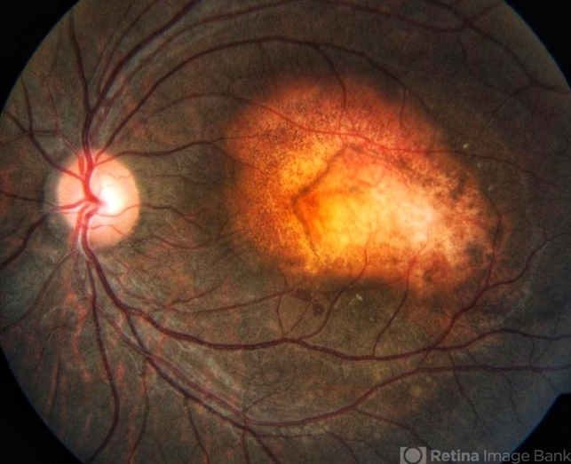
- macular choroidal osteoma
- Photographer
- Hiteshwar Saikia
- Imaging device
- Fundus camera
- Description
- 23-year-old patient's fundus photograph having complaints of defective vision, metamorphosia over 6 months shows yellow orange elevated well defined submacular lesion with normal overlying retinal vessels and normal disc . Vision left eye is 20/80.


