Search results (125 results)
-
 Blunt Ocular Trauma Due to Firework Injury
Jun 9 2020 by Brittany Rota
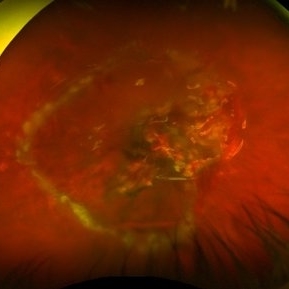
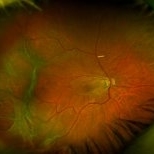
Ultra- widefield pseudocolor image of an 18-year-old male with blunt ocular trauma in the right eye due to a firework injury. The patient presented with commotio retinae (sclopteria), an acute vitreous hemorrhage, choroidal rupture, and a subretinal hemorrhage. The referring physician performed surgery on the lateral rectus muscle which was macerated but not severed, and several orbital fibrous foreign bodies were removed from the posterior orbit. The globe was intact. There is no evidence of retinal tear in the region of sclopetaria; however, there is complete necrosis of the temporal peripheral choroid and retina. The vitreous hemorrhage was slowly clearing on his exam 6-9-2020. The patient is developing subretinal fibrosis. The physician is concerned about the choroidal rupture that is visible through the submacular hemorrhage. There is one rupture that appears to course directly under the fovea. The physician states that if this is the case, his vision most likely will be 20/200 or worse. His vision was hand motion in all fields except nasally, which he was unable to see hand motion at his visit on 6-9-2020.
Photographer: Brittany Rota
Imaging device: Optos California
Condition/keywords: blunt trauma, choroidal rupture, commotio retinae, fibrosis, firework injury, fundus photograph, hand motion, necrotizing retina, Optos, pseudocolor, subretinal hemorrhage, vitreous hemorrhage
-
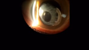 Capsular Phimosis with Dislocated IOL
Sep 3 2020 by Mamoun Hani Zebbache, MD
Anterior capsule contraction syndrome with partially subluxed intraocular lens in 74-year-old female who was operated in another center.
Condition/keywords: capsule, dislocated intraocular lens (IOL), fibrosis, phimosis
-
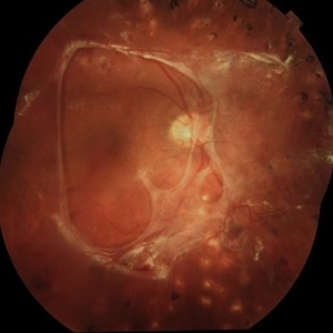
 Diabetic Retinopathy
Dec 11 2019 by Lauren Whaley
44-year-old male diabetic patient had an acute change in A1C over 9 months and ended up with a tractional retinal detachmen in right eye. This photo is 2 weeks post operative with current vision level at hand motion. He had extensive laser, retinectomy, and silicone oil fill.
Photographer: Lauren R. Whaley, COA
Imaging device: Optos Wide Field
Condition/keywords: diabetes, diabetic retinopathy, fibrosis, laser scarring, proliferative vitreoretinopathy (PVR), retinectomy, silicone oil, tractional retinal detachment
-
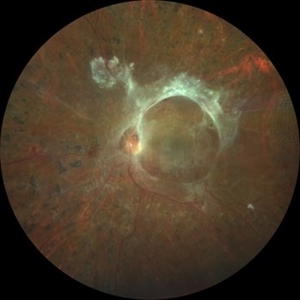
 Preretinal Fibrosis
Jan 12 2024 by Virginia Gebhart
53 year old diabetic male with significant persistent ERM due to fibrotic NV superiorly. Possibly developing a tractional MH. Vitreous Hemorrhage secondary to traction on the fibrosis
Photographer: Virginia Gebhart
Imaging device: Topcon 50DX
Condition/keywords: epiretinal membrane, ERM, fibrosis, macular pseudohole, neovascularization (NV)
-
 Ring Fibrosis Proliferative Diabetic Retinopathy
Feb 8 2021 by Rudvij Pandya
Ring fibrosis proliferative diabetic retinopathy
Condition/keywords: fibrosis, preretinal fibrosis, proliferative diabetic retinopathy (PDR)
-
 Subretinal Thickening and Subretinal Hemorrhage – Red Free (B/W) Photograph
Mar 9 2017 by James B. Soque, CRA, OCT-C, COA, FOPS
Red free image, fundus photograph, of a 52-year-old white male with VA loss to 20/200 of unknown etiology. Dilated fundus examination of the right eye reveals a fibrotic scar with subretinal thickening and subretinal hemorrhage.
Photographer: James B. Soque, CRA, OCT-C, COA
Imaging device: Topcon TRC 50DX, MERGE Software
Condition/keywords: black and white photo, fibrosis, fibrotic scar, red-free, subretinal hemorrhage, subretinal thickening
-
 Traction Detachment of Retina
Nov 14 2025 by Virginia Gebhart
50 year old female with proliferative diabetic retinopathy, large ridge of traction temporally, and significant band of fibrosis. Subretinal fluid throughout the macula. Due to traction, surgical repair not recommended at this time as it could worsen condition. Will observe closely. BCVA CF @ 1 ft.
Photographer: Virginia Gebhart, Retina Consultants of Carolina
Imaging device: Optos California
Condition/keywords: fibrosis, proliferative diabetic retinopathy (PDR), traction detachment, Traction retinal detachment
-
 Tractional Retinal Detachment
Jun 27 2019 by Nichole Lewis
69-year-old female with an epiretinal membrane and tractional retinal detachment. VA 20/400.
Photographer: Nichole Lewis
Imaging device: Optos
Condition/keywords: epiretinal membrane (ERM), fibrosis, retinoschisis, tractional retinal detachment
-
 Fibrosis and Traction Following Traction Retinal Detachment Repair
Oct 13 2020 by Sophia El Hamichi, MD
A 29-year-old female with a history of diabetes mellitus type 1, presented with proliferative diabetic retinopathy OU and tractional retinal detachment OD. The patient underwent retinal detachment repair with pars plana vitrectomy, endolaser and silicone oil placement. After one month of her surgery, the patient presented with retinal fibrosis and tractions depicted in the image.
Photographer: Belinda Rodriguez, Murray Ocular Oncology and Retina, Miami
Condition/keywords: pars plana vitrectomy (PPV), post-op, proliferative diabetic retinopathy (PDR), proliferative vitreoretinopathy (PVR), tractional retinal detachment
-
 Fibrosis and Traction Following Traction Retinal Detachment Repair
Oct 13 2020 by Sophia El Hamichi, MD
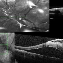
A 29-year-old female with a history of diabetes mellitus type 1, presented with proliferative diabetic retinopathy OU and tractional retinal detachment OD. The patient underwent retinal detachment repair with pars plana vitrectomy, endolaser and silicone oil placement. After one month of her surgery, the patient presented with retinal fibrosis and traction. The image on the top shows the OCT of the fibrosis post op, that was not present in the pre op (OCT image on the bottom).
Photographer: Belinda Rodriguez, Murray Ocular Oncology and Retina, Miami
Condition/keywords: optical coherence tomography (OCT), pars plana vitrectomy (PPV), proliferative diabetic retinopathy (PDR), proliferative vitreoretinopathy (PVR), tractional retinal detachment
-
 All That Glows Yellow Isn’t Mellow: Coats' Disease Unveiled
Nov 4 2025 by SHRADDHA RAJ SHRIVASTAVA
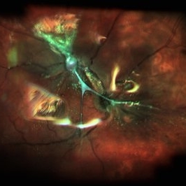
Montage fundus image of an 11 year old boy diagnosed with left eye Coats' disease (stage 3A1), reveals a hyperemic disc and surrounding intra-retinal exudates superior to the disc. There is a single fibroglial nodule at the macula causing submacular fibrosis with exudation. We can see areas of pigmentary changes and RPE atrophy in posterior pole and mid-peripheral retina supero-temporally. There is massive yellowish subretinal exudation in all the quadrants, which are associated with telangiectatic aneurysmal capillary dilation, more prominently seen in the nasal periphery. Supero-nasally we can also see an orange-red elevated vaso-proliferative mass with overlying dilated capillaries, which has likely developed secondary to untreated long standing disease. We can also see associated extrafoveal subtotal exudative retinal detachment in the inferior and nasal quadrants.
Photographer: Dr. Shraddha Raj Shrivastava
Imaging device: Nidek Mirante SLO/OCT (Confocal scanning/Spectral domain OCT)
Condition/keywords: COATS DISEASE, exudative detachment, leukocoria, subretinal exudates, Xanthocoria, yellow exudate
-
 Angioid Streaks
Mar 11 2014 by Andrew M Hendrick, MD
Fundus photography of the left eye of a 50-year-old African American male with a remote history of minor trauma in the contralateral eye.
Condition/keywords: angioid streaks, subretinal fibrosis
-
 Angioid streaks - PXE
Jan 11 2013 by Alex P. Hunyor, MD
Pseudoxanthoma elasticum with angioid streaks, left eye - note subretinal fibrosis adjacent to disc.
Condition/keywords: angioid streaks, pseudoxanthoma elasticum (PXE)
-
 Angioid streaks - PXE case 2
Jan 11 2013 by Alex P. Hunyor, MD
Pseudoxanthoma elasticum with angioid streaks, right eye - one year after initial image, showing extensive subretinal fibrosis in the macula.
Condition/keywords: angioid streaks, pseudoxanthoma elasticum (PXE)
-
 Angioid Streaks With CNV
Mar 11 2014 by Andrew M Hendrick, MD
Fundus photography of the right eye of a 50-year-old African American male with a remote history of minor trauma. Serial anti-VEGF injections failed to improve the subfoveal CNV and his condition is now being observed.
Photographer: Jannah Dobbs
Condition/keywords: angioid streaks, subretinal fibrosis
-
 Angioid Streaks with Macular Fibrosis Secondary to Inactive CNV
Oct 11 2012 by Gabriela Lopezcarasa Hernandez, MD
50-year-old female with decrease in VA in right eye
Photographer: Ricardo Montoya, Mexico
Imaging device: Zeiss FF4
Condition/keywords: angioid streaks
-
 Aniridic Fibrosis Syndrome #2 of 7
Jan 24 2013 by Christopher D. Riemann, MD
6-year-old pseudophakic girl with aniridic fibrosis syndrome. Superonasal view with HD endoscope. Note: 20 gauge sclerotomy and very mild fibrosis barely extending onto the ciliary body.
Photographer: Christopher Riemann MD, Cincinnati Eye Institute, University of Cincinnati
Imaging device: Endoscope
Condition/keywords: aniridia, epiciliary membrane
-
 Aniridic Fibrosis Syndrome #4 of 7
Jan 24 2013 by Christopher D. Riemann, MD
6-year-old pseudophakic girl with aniridic fibrosis syndrome. Close up nasal view with HD endoscope. Note: increasing fibrosis fibrosis clearly extending onto the ciliary body and enveloping ciliary processes.
Photographer: Christopher Riemann MD, Cincinnati Eye Institute, University of Cincinnati
Imaging device: Endoscope
Condition/keywords: aniridia, epiciliary membrane
-
 Aniridic Fibrosis Syndrome #6 of 7
Jan 24 2013 by Christopher D. Riemann, MD
6-year-old girl with Aniridic Fibrosis Syndrome. Note: endoscopic view of 20 gauge vitreous cutter engaging epiciliary membrane.
Photographer: Christopher Riemann MD, Cincinnati Eye Institute, University of Cincinnati
Imaging device: Endoscope
Condition/keywords: aniridia, epiciliary membrane
-
 Aniridic Fibrosis Syndrome - #1 of 7
Jan 24 2013 by Christopher D. Riemann, MD
6-year-old pseudophakic girl with aniridic fibrosis syndrome. Superior view with HD endoscope. Note: complete absence of fibrosis, a normal ciliary body, normal pars plana and normal anterior retina.
Photographer: Christopher Riemann MD, Cincinnati Eye Institute, University of Cincinnati
Imaging device: Endoscope
Condition/keywords: aniridia, epiciliary membrane
-
 Aniridic Fibrosis Syndrome - #3 of 7
Jan 24 2013 by Christopher D. Riemann, MD
6-year-old pseudophakic girl with aniridic fibrosis syndrome. Nasal view with HD endoscope. Note: increasing fibrosis fibrosis clearly extending onto the ciliary body and enveloping ciliary processes.
Photographer: Christopher Riemann MD, Cincinnati Eye Institute, University of Cincinnati
Imaging device: Endoscope
Condition/keywords: aniridia, epiciliary membrane
-
 Aniridic Fibrosis Syndrome - #5 of 7
Jan 24 2013 by Christopher D. Riemann, MD
6-year-old pseudophakic girl with aniridic fibrosis syndrome. Inferior view with HD endoscope. Note: severe inferior fibrosis fibrosis clearly extending across the ciliary body and obliterating the inferior ciliary processes and migrating onto the anterior pars plana .
Photographer: Christopher Riemann MD, Cincinnati Eye Institute, University of Cincinnati
Imaging device: Endoscope
Condition/keywords: aniridia, epiciliary membrane
-
 Aniridic Fibrosis Syndrome - #7 of 7
Jan 24 2013 by Christopher D. Riemann, MD
6-year-old pseudophakic girl with aniridic fibrosis syndrome. Superonasal view with HD endoscope. Note: 20 gauge probe applying endocautery to ciliary body bleed resulting from stripping of epiciliary membranes
Photographer: Christopher Riemann MD, Cincinnati Eye Institute, University of Cincinnati
Imaging device: Endoscope
Condition/keywords: aniridia, epiciliary membrane
-
 Anomalous Vasculature of Optic Nerve, Fluorescein Angiogram
Feb 20 2013 by From the Collections of Thomas M. Aaberg, MD and Thomas M. Aaberg Jr., MD
No history or color photo; some fibrosis overlying ON.
Condition/keywords: anomalous vasculature of optic nerve, black and white photo
-
 Anterior Capsule Opacification
Jun 26 2016 by Jared Watson
49-year-old male with anterior capsule fibrosis and wrinkling S/P PPV/PPL/C3f8. Patient will have secondary IOL after retinal issues resolve.
Photographer: Jared Watson COT/CRA University of Virginia
Condition/keywords: anterior capsule opacification
 Loading…