Search results (125 results)
-
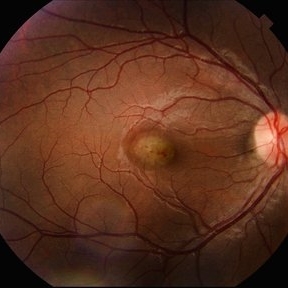
 Traction Detachment of Retina
Nov 14 2025 by Virginia Gebhart
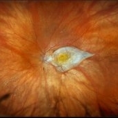
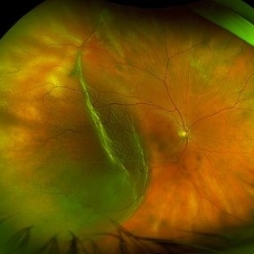
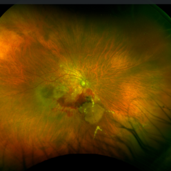
50 year old female with proliferative diabetic retinopathy, large ridge of traction temporally, and significant band of fibrosis. Subretinal fluid throughout the macula. Due to traction, surgical repair not recommended at this time as it could worsen condition. Will observe closely. BCVA CF @ 1 ft.
Photographer: Virginia Gebhart, Retina Consultants of Carolina
Imaging device: Optos California
Condition/keywords: fibrosis, proliferative diabetic retinopathy (PDR), traction detachment, Traction retinal detachment
-
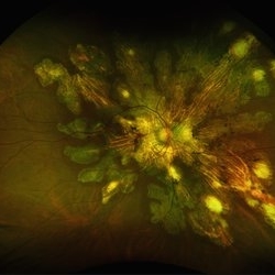
 All That Glows Yellow Isn’t Mellow: Coats' Disease Unveiled
Nov 4 2025 by SHRADDHA RAJ SHRIVASTAVA
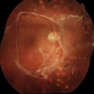
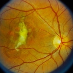
Montage fundus image of an 11 year old boy diagnosed with left eye Coats' disease (stage 3A1), reveals a hyperemic disc and surrounding intra-retinal exudates superior to the disc. There is a single fibroglial nodule at the macula causing submacular fibrosis with exudation. We can see areas of pigmentary changes and RPE atrophy in posterior pole and mid-peripheral retina supero-temporally. There is massive yellowish subretinal exudation in all the quadrants, which are associated with telangiectatic aneurysmal capillary dilation, more prominently seen in the nasal periphery. Supero-nasally we can also see an orange-red elevated vaso-proliferative mass with overlying dilated capillaries, which has likely developed secondary to untreated long standing disease. We can also see associated extrafoveal subtotal exudative retinal detachment in the inferior and nasal quadrants.
Photographer: Dr. Shraddha Raj Shrivastava
Imaging device: Nidek Mirante SLO/OCT (Confocal scanning/Spectral domain OCT)
Condition/keywords: COATS DISEASE, exudative detachment, leukocoria, subretinal exudates, Xanthocoria, yellow exudate
-
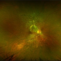
 Idiopathic Choroidal Neovascularization
Sep 30 2025 by César Adrián Gomez Valdivia, MD
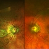
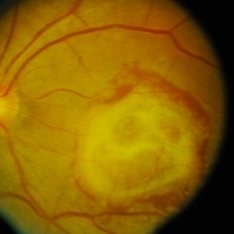
At the foveal area, there is a yellowish-greenish elevated lesion with indistinct borders, corresponding to a subfoveal choroidal neovascular membrane (CNV). There are subtle overlying changes including mild retinal pigment epithelium (RPE) disruption, and small hemorrhagic spots suggesting active leakage. Surrounding the lesion, there are faint retinal folds or striae, likely due to localized subretinal fibrosis or traction.
Photographer: @eyemissu2
Imaging device: TOPCON TRX
Condition/keywords: Idiopathic Choroidal Neovascularization
-
 Serpiginous Choroidopathy
Jun 23 2025 by César Adrián Gómez Valdivia, MD
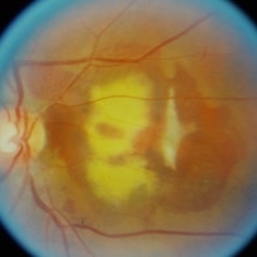
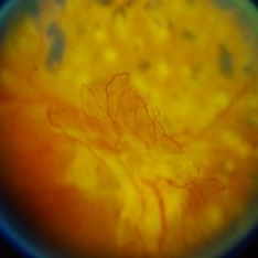
Fundus photograph of a 29 year-old female patient diagnosed with Serpiginous Choroidopathy. Finings were bilateral. The most common complication of SC is choroidal neovascularization affecting up to 35% of patients. Other reported complications are subretinal fibrosis, cystoid macular edema, branch vein occlusion, serous retinal detachment, optic disc neovascularization ,and anterior uveitis.
Photographer: @eyemissu2
Imaging device: TOPCON TRC-50DX
Condition/keywords: serpiginous choroiditis
-
 Serpiginous Choroidopathy
Jun 23 2025 by César Adrián Gómez Valdivia, MD
Fundus photograph of a 29 year-old female patient diagnosed with Serpiginous Choroidopathy. Finings were bilateral. The most common complication of SC is choroidal neovascularization affecting up to 35% of patients. Other reported complications are subretinal fibrosis, cystoid macular edema, branch vein occlusion, serous retinal detachment, optic disc neovascularization, and anterior uveitis.
Photographer: @eyemissu2
Imaging device: California ICG OPTOS
Condition/keywords: serpiginous choroiditis
-
 VKH Pseudotumor – Chronic Subretinal Fibrosis
May 11 2025 by Felipe Murati
Ultra-widefield fundus image from a 36-year-old woman with chronic VKH syndrome showing a pseudotumor-like subretinal fibrotic lesion in the right eye. The lesion developed after multiple relapses and remained stable over a 1-year follow-up with immunosuppressive treatment including prednisone, mycophenolate mofetil, and adalimumab. No active choroiditis or exudative detachment was observed. Multimodal imaging was essential for disease monitoring.
Photographer: Felipe A. Murati, MD, University of Arizona
Imaging device: Optos California ultra-widefield retinal imaging system, single-capture, color fundus modality.
Condition/keywords: adalimumab, chronic inflammation, granulomatous uveitis, OCT, Optos ultra-widefield imaging, pseudotumor, subretinal fibrosis, VKH, Vogt-Koyanagi-Harada
-
 VKH Pseudotumor – Fluorescein Angiography
May 11 2025 by Felipe Murati
Fluorescein angiography image from a 36-year-old woman with chronic Vogt-Koyanagi-Harada (VKH) syndrome showing a pseudotumor-like lesion with late-phase staining and no active leakage. The image highlights subretinal fibrosis in the right eye, stable under long-term immunosuppressive therapy with mycophenolate mofetil and adalimumab. No signs of active choroiditis are present, confirming a quiescent phase.
Photographer: Felipe A. Murati, MD, University of Arizona
Imaging device: Optos California, fluorescein angiography modality
Condition/keywords: choroiditis, Fluorescein angiography, granulomatous uveitis, Optos FA, pseudotumor, subretinal fibrosis, VKH, Vogt-Koyanagi-Harada
-
 Subretinal Fibrosis
Jan 14 2025 by Kimberly Wakester
Fundus photograph of an 86-year-old woman with the end stage of Age-related Macular Degeneration in the left eye. Patient went unseen for 3-4 years prior to establishing care at our practice. Due to the significant amount of subretinal fibrosis, treatment was not recommended due to limited visual recovery. Patient was advised of monocular vision and the importance of follow up care.
Photographer: Kimberly Wakester, COA
Imaging device: Optos California
Condition/keywords: AMD, subretinal fibrosis
-
 Tractional Detachment of Retina
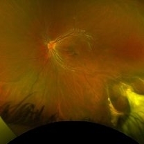
Aug 21 2024 by Jordyn Beckman
18 year old male with tractional detachment of Retina, chronic macular hole and silicone oil s/p RD repair x2. BCVA CF @2 ft, fellow eye prosthetic.
Photographer: Jordyn Beckman
Imaging device: Optos California
Condition/keywords: Macular hole, preretinal fibrosis, Retinal Detachment, scleral buckle, silicone oil, TRACTION, tractional retinal detachment
-
 Pseudoxanthoma Elasticum Associated Angioid Streaks
Aug 18 2024 by KANWALJEET HARJOT MADAN, M.S. (Ophthalmology); FAICO (Vitreous - Retina)
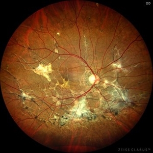
This is fundus photograph of a young 31 years male patient depicting Angioid streaks emanating from optic nerve towards the periphery and subretinal fibrosis. There is peau de orange appearance temporal to fovea with Salmon Spots in periphery. He was diagnosed to have Pseudoxanthoma Elasticum.
Photographer: Dr. Kanwaljeet Harjot Madan, M.S. (Ophthalmologist) Fellow in Vitrous & Retina. Thind Eye Hospital, Jalandhar City. Punjab. India
Imaging device: Zeiss Clarus
Condition/keywords: Angioid Streaks, fundus photograph, pseudoxanthoma elasticum (PXE)
-
 Ocular Toxocariasis
Jul 4 2024 by Brandon I Fram, MD
4 yo with toxocariasis-related peripheral granuloma with adhesion to the macula and macular subretinal fibrosis. Positive Toxocara titers.
Condition/keywords: toxocara canis, toxocara granuloma, toxocariasis
-
 Coats Disease
May 23 2024 by ARVIND JAIN M
a.right eye fundus image and b. FFA montage of a 8 year old boy showing light bulb aneurysms of the arterioles with exudation with sub retinal fibrosis and telangiectasia in periphery who complained of defective vision, classical of coats disease.
Photographer: Dr. Arvind Jain M, MBBS,MS Ophthal, FVRS
Condition/keywords: COATS DISEASE, Leber's miliary aneurysm, light-bulb aneurysms
-
 Retinal Detachment
Mar 28 2024 by Virginia Gebhart
68 year male with chronic appearing retinal detachment with subretinal bands and subretinal fibrosis. Demarcation line present, SRF splits the fovea on OCT.
Photographer: Virginia Gebhart
Imaging device: Optos California
Condition/keywords: chronic retinal detachment, Retinal Detachment
-
 Sub Macular Fibrosis in the Setting of Old Sub Macular Hemorrhage
Feb 15 2024 by Sayena . Jabbehdari, MD, MPH, MBA
Pseudo color fundus photo of 78 years old male with history of sub macular hemorrhage in the setting of wet age-related macular degeneration. You can appreciate the tear shaped appearance of blood due to gravity. The OCT of the macula depicts the huge (>950mm) sub retinal fibrosis.
Photographer: Sayena Jabbehdari MD MPH , University of Arkansas in Little Rock
Imaging device: Clarus
Condition/keywords: macular neovascular disease, retina, submacular hemorrhage, Wet age related macular degeneration
-
 Preretinal Fibrosis
Jan 12 2024 by Virginia Gebhart
53 year old diabetic male with significant persistent ERM due to fibrotic NV superiorly. Possibly developing a tractional MH. Vitreous Hemorrhage secondary to traction on the fibrosis
Photographer: Virginia Gebhart
Imaging device: Topcon 50DX
Condition/keywords: epiretinal membrane, ERM, fibrosis, macular pseudohole, neovascularization (NV)
-
 Coat's disease
Nov 7 2023 by Harsh Vardhan Singh, MS
8-year-male with sub-retinal fibrosis with extensive exudates diagnosed with Coats' disease
Photographer: Harsh Vardhan Singh
Condition/keywords: Coats' disease
-
 WED-ARMD-CNVM
Sep 29 2023 by PUSHPANJALI BADOLE
Fundus photograph of a 72 year old male with right eye choroidal neovascularization. There is a thick membrane over macula along with subretinal fibrosis in right eye. Left eye shows drusen.
Photographer: NITIN DESALE, ISHA NETRALAYA, KALYAN
Imaging device: DAYTONA OPTOS
Condition/keywords: choroidal neovascular membrane (CNVM), choroidal neovascularization (CNV)
-
 Subretinal haemorrhage
Sep 26 2023 by Ben Serar
Fundus photograph showing subretinal bleed with subretinal scarring and fibrosis as sequelae.
Condition/keywords: Subretinal haemorrhage
-
 Subretinal fibrosis
Sep 14 2023 by Ben Serar
Fundus photograph of LE showing a scarred lesion at the macula, with sub retinal fibrosis.
Condition/keywords: macular scar, Subretinal fibrosis
-
 Subretinal fibrosis
Sep 12 2023 by Ben Serar
Fundus photograph of RE showing scarring at the macula with subretinal fibrosis
Condition/keywords: Subretinal fibrosis
-
 Choroidal Neovascular Membrane (CNVM)
Sep 12 2023 by Ben Serar
Fundus photograph of LE showing Scarred CNVM at the macula, with sub-retinal fibrois with surrounding subretinal bleed
Condition/keywords: choroidal neovascular membrane (CNVM), scarring, sub-retinal fibrosis
-
 Neovascularisation elsewhere (NVE)
Sep 12 2023 by Ben Serar
Fundus photograph showing NVE in a sea-fan configuration, with fibrosis at the base of the NVE, with pigmented laser spots in the periphery.
Condition/keywords: laser, Neovascularisation elsewhere (NVE)
-
 Sunset Glow Fundus
May 15 2022 by Manuel Ángel Alcántara Delgado, MD
Optomap ultra-widefield retinal imaging of an 35-year-old woman showed sunset glow fundus, multiple nummular chorioretinal atrophic lesions, macular subretinal fibrosis and pigment clumping in chronic recurrent stage of Vogt-Koyanagi-Harada disease.
Photographer: Manuel Ángel Alcántara Delgado. Conde de Valenciana.
Condition/keywords: abnormal retina, benign pigmented lesions, pigment clumps, retinal fibrosis, uveitis, Vogt-Koyanagi-Harada
-
 ROP 4B late Retinal Findings
Mar 31 2022 by Franco Benvenuto, MD
A 9-year-old male, that was born at 30 weeks of gestation with birth weight of 1500 g and history of hospitalization for 20 days with respiratory distress and packed red blood cell transfusion for anemia. At the first exam, both eyes were with stage 4B ROP. Vitrectomy with 25 G was done in both eyes. The flat fibrosis dragged the macula nasally in both the eyes.
Photographer: Franco Benvenuto, Universidad de Buenos Aires, Argentina; Universidad de Guadalajara, México.
Condition/keywords: cicatricial retinopathy of prematurity, retinopathy of prematurity (ROP)
-
 Neovascular Age-Related Macular Degeneration (1)
Apr 28 2021 by Ambar Faridi, MD
80-year-old woman with neovascular age-related macular degeneration with large subretinal hemorrhage, hemorrhagic PED, and vascular lipid exudation.
Photographer: Jennifer Tu-Bui, VA Portland Health Care System
Condition/keywords: subretinal fibrosis, subretinal hemorrhage
 Loading…