Search results (125 results)
-
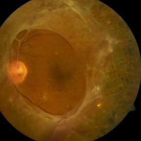
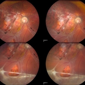
 Inferior Rhegmatogenous Retinal Detachment with Subretinal Fibrosis
Aug 23 2012 by Gabriela Lopezcarasa Hernandez, MD
Asymptomatic 25-year-old woman with high myopia.
Photographer: Gabriela Lopezcarasa Hernandez, Hospital Angeles Lomas
Imaging device: FF4
Condition/keywords: high myopia, subretinal fibrosis
-
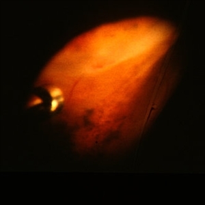
 PDR Fibrosis
Oct 8 2012 by David R. Chow, MD, FRCS(C)
PDR contracting diabetic hyaloid with evolving TRD
Condition/keywords: hyaloid, tractional retinal detachment
-
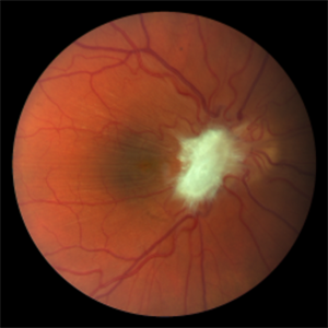
 Central Serous Chorioretinopathy (CSC)
Oct 16 2012 by S. Natarajan, MD, FASRS, FRCS (GLASGOW) , FICO, D.Sc, FELA
Middle-aged male came with small PED 4 months back; now this has progressed to a larger PED with SRF underneath the fovea.
Photographer: Prof. Dr. S. Natarajan
Condition/keywords: central serous chorioretinopathy (CSCR), central serous retinopathy (CSR), pigment epithelial detachment (PED), subretinal fibrosis
-
 NVD with fibrosis from PDR
Jan 1 2013 by John T. Thompson, MD
Fibrovascular proliferation from PDR, florid NVD.
Condition/keywords: fibrous proliferation, neovascularization of the disc (NVD)
-
 Diffuse Choroidal Hemangioma
Nov 7 2012 by Rajiv Anand, MD, FRCS, FASRS
Fundus photo shows classic 'tomato-ketchup' red appearance of diffuse hemangioma. Due to chronic SRF , there is subretinal fibrosis.
Condition/keywords: subretinal fibrosis
-
 Preretinal Hemorrhage due to Proliferative Diabetic Retinopathy
Oct 17 2012 by Sharon Fekrat, MD FACS FASRS
Fluorescein angiography of right eye with preretinal hemorrhage from neovascularization elsewhere associated with proliferative diabetic retinopathy. Note associated fibrosis.
Photographer: John Reaves, Ophthalmic Photographer, Durham VA Medical Center, Durham, NC
Condition/keywords: preretinal hemorrhage
-
 Angioid streaks - PXE
Jan 11 2013 by Alex P. Hunyor, MD
Pseudoxanthoma elasticum with angioid streaks, left eye - note subretinal fibrosis adjacent to disc.
Condition/keywords: angioid streaks, pseudoxanthoma elasticum (PXE)
-
 Angioid Streaks with Macular Fibrosis Secondary to Inactive CNV
Oct 11 2012 by Gabriela Lopezcarasa Hernandez, MD
50-year-old female with decrease in VA in right eye
Photographer: Ricardo Montoya, Mexico
Imaging device: Zeiss FF4
Condition/keywords: angioid streaks
-
 Anterior Capsule Opacification
Jun 26 2016 by Jared Watson
49-year-old male with anterior capsule fibrosis and wrinkling S/P PPV/PPL/C3f8. Patient will have secondary IOL after retinal issues resolve.
Photographer: Jared Watson COT/CRA University of Virginia
Condition/keywords: anterior capsule opacification
-
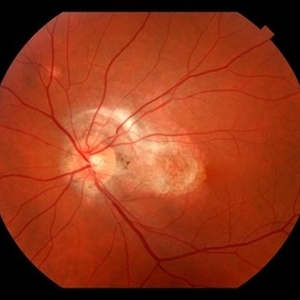
 NVD in Proliferative Diabetic Retinopathy
Sep 4 2014 by Ratimir Lazic, MD, PhD
A color fundus photography of a 59- year-old male. Diffuse neovascularizations of the disc can be seen. Epiretinal fibrosis in inferior temporal quadrant can be seen. The fundus correspond with proliferative diabetic retinopathy. Intravitreal bevacizumab was applied.
Photographer: Ivan Boras, MD Eye Clinic Svjetlost, Heinzelova street 39, Zagreb, Croatia
Imaging device: Zeis Visucam Lite 2
Condition/keywords: diabetic retinopathy, neovascularization of the disc (NVD)
-
 peripheral pre-retinal fibrosis and neovascularization with vitreous cutter
Feb 14 2013 by From the Collections of Thomas M. Aaberg, MD and Thomas M. Aaberg Jr., MD
intraoperative photo of peripheral pre-retinal fibrosis and neovascularization with vitreous cutter
Condition/keywords: pars planitis, peripheral retinal neovascularization
-
 Subretinal Fibrosis (PPCNVM and POHS) OS
Sep 18 2019 by John S. King, MD
57-year-old white male with history of PPCNVM OS and POHS OU here for a routine visit. History of avastin in 2014, and stable since then. Va OS 20/20. PP scar with macular subretinal fibrosis. No heme or exudates. CR spot supero-nasally.
Photographer: Shelly Blair
Imaging device: Topcon 50
Condition/keywords: choroidal neovascular membrane (CNVM), ocular histoplasmosis syndrome (OHS), peripapillary choroidal neovascularization (PPCNVM), presumed ocular histoplasmosis syndrome (POHS)
-
 multifocal choroiditis
Feb 14 2013 by From the Collections of Thomas M. Aaberg, MD and Thomas M. Aaberg Jr., MD
color fundus photos showing healed chorioretinal scars, pigment deposition, and subretinal fibrosis consistent with regressed multifocal choroiditis
Condition/keywords: multifocal choroiditis, posterior segment inflammation, subretinal fibrosis, white dot syndrome
-
 Lupus Retinopathy
Mar 27 2014 by Jason S. Calhoun
Female patient in for evaluation on lupus retinopathy. Has poor vision in the right eye. VA is hand motion in the right eye. Fundus photos show fibrosis along the temporal arcades and narrowing of the arteries. No macular edema found.
Photographer: Jason S. Calhoun, Mayo Clinic Jacksonville, Department of Ophthalmology
Imaging device: TOPCON TRC 50-EX
Condition/keywords: lupus, retinopathy
-
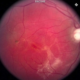
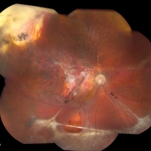
 Rhegmatogenous Retinal Detachment
Oct 11 2013 by Jason S. Calhoun
Patient in for a second opinion on RD, right eye. VA is NLP in the right eye. Fundus photography shows inferior retinal detachment with holes and subretinal fibrosis. No further surgery is suggested at this time.
Photographer: Jason S. Calhoun, Ophthalmic Photographer, Department of Ophthalmology, Mayo Clinic Jacksonville
Imaging device: TOPCON TRC 50-EX
-
 Angioid Streaks With CNV
Mar 11 2014 by Andrew M Hendrick, MD
Fundus photography of the right eye of a 50-year-old African American male with a remote history of minor trauma. Serial anti-VEGF injections failed to improve the subfoveal CNV and his condition is now being observed.
Photographer: Jannah Dobbs
Condition/keywords: angioid streaks, subretinal fibrosis
-
 Pre-Retinal Fibrosis Following Endogenous Fungal Endophthalmitis
Aug 23 2018 by Matthew Dombrow, MD
20-year-old male presents with posterior uveitis with a chain of white infiltrates stemming from the optic nerve. While treated, his infiltrates retracted and hemorrhage occured. Neovascularization of the disc developed and underwent Avastin treatment in addition to his oral anti-fungals and intravitreal anti-fungals. Patient was lost to follow up and presented with severe pre-retinal fibrosis 6 years later. Acuity is 20/40-1 with significant metamorphopsia.
Photographer: Patricia Candrea, COA, Connecticut Retina Consultants, LLC
Imaging device: Canon
Condition/keywords: pre-retinal membrane
-
 Aniridic Fibrosis Syndrome - #5 of 7
Jan 24 2013 by Christopher D. Riemann, MD
6-year-old pseudophakic girl with aniridic fibrosis syndrome. Inferior view with HD endoscope. Note: severe inferior fibrosis fibrosis clearly extending across the ciliary body and obliterating the inferior ciliary processes and migrating onto the anterior pars plana .
Photographer: Christopher Riemann MD, Cincinnati Eye Institute, University of Cincinnati
Imaging device: Endoscope
Condition/keywords: aniridia, epiciliary membrane
-
 Lupus Retinopathy
Mar 27 2014 by Jason S. Calhoun
Female patient in for evaluation on lupus retinopathy. Has poor vision in the right eye. VA is hand motion in the right eye. Fundus photos show fibrosis along the temporal arcades and narrowing of the arteries. No macular edema found.
Photographer: Jason S. Calhoun, Mayo Clinic Jacksonville, Department of Ophthalmology
Imaging device: TOPCON TRC 50-EX
Condition/keywords: lupus, retinopathy
-
---thumb.jpg/image-square;max$300,300.ImageHandler) peripheral pre-retinal fibrosis and neovascularization with vitreous cutter
peripheral pre-retinal fibrosis and neovascularization with vitreous cutter
Feb 14 2013 by From the Collections of Thomas M. Aaberg, MD and Thomas M. Aaberg Jr., MD
intraoperative photo of peripheral pre-retinal fibrosis and neovascularization with vitreous cutter
Condition/keywords: pars planitis, peripheral retinal neovascularization
-
 Aniridic Fibrosis Syndrome - #3 of 7
Jan 24 2013 by Christopher D. Riemann, MD
6-year-old pseudophakic girl with aniridic fibrosis syndrome. Nasal view with HD endoscope. Note: increasing fibrosis fibrosis clearly extending onto the ciliary body and enveloping ciliary processes.
Photographer: Christopher Riemann MD, Cincinnati Eye Institute, University of Cincinnati
Imaging device: Endoscope
Condition/keywords: aniridia, epiciliary membrane
-
 Rhegmatogenous Retinal Detachment
Oct 11 2013 by Jason S. Calhoun
Patient in for second opinion on RD, right eye. VA is NLP in the right eye. Fundus photography shows inferior retinal detachment with holes and subretinal fibrosis. No further surgery is suggested at this time.
Photographer: Jason S. Calhoun, Ophthalmic Photographer, Department of Ophthalmology, Mayo Clinic Jacksonville
Imaging device: TOPCON TRC 50-EX
-
---thumb.jpg/image-square;max$300,300.ImageHandler) peripheral pre-retinal fibrosis and neovascularization with vitreous cutter
peripheral pre-retinal fibrosis and neovascularization with vitreous cutter
Feb 14 2013 by From the Collections of Thomas M. Aaberg, MD and Thomas M. Aaberg Jr., MD
intraoperative photo of peripheral pre-retinal fibrosis and neovascularization with vitreous cutter
Condition/keywords: pars planitis, peripheral retinal neovascularization
-
 Aniridic Fibrosis Syndrome #6 of 7
Jan 24 2013 by Christopher D. Riemann, MD
6-year-old girl with Aniridic Fibrosis Syndrome. Note: endoscopic view of 20 gauge vitreous cutter engaging epiciliary membrane.
Photographer: Christopher Riemann MD, Cincinnati Eye Institute, University of Cincinnati
Imaging device: Endoscope
Condition/keywords: aniridia, epiciliary membrane
-
 Aniridic Fibrosis Syndrome - #1 of 7
Jan 24 2013 by Christopher D. Riemann, MD
6-year-old pseudophakic girl with aniridic fibrosis syndrome. Superior view with HD endoscope. Note: complete absence of fibrosis, a normal ciliary body, normal pars plana and normal anterior retina.
Photographer: Christopher Riemann MD, Cincinnati Eye Institute, University of Cincinnati
Imaging device: Endoscope
Condition/keywords: aniridia, epiciliary membrane
 Loading…