Search results (72 results)
-
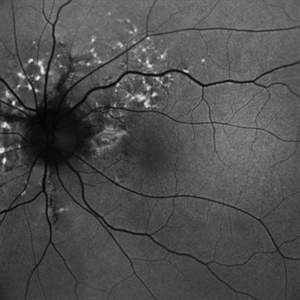
 Hypertensive Retinopathy
Feb 24 2020 by Brian K. Horsman, MD, FRCS(C) FASRS
Macular star, disc edema, intra vitreous bubble of Bevacizumab
Condition/keywords: bevacizumabhypertension
-
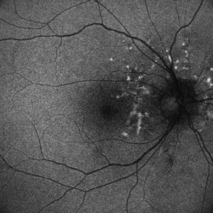
 Hypertensive Retinopathy
Feb 24 2020 by Brian K. Horsman, MD, FRCS(C) FASRS
Macular star, disc edema, intra vitreous bubble of Bevacizumab
Condition/keywords: bevacizumabhypertension
-
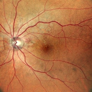
 Punctate Inner Choroidopathy with CNV Treated with Bevacizumab # 1 of 7
Feb 28 2013 by Gregory R. Blaha, MD, PhD
Fundus photograph in a 31-year-old female with vision loss from a choroidal neovascular membrane (CNV) from punctate inner choroidopathy. Note the CNV and hemorrhage superotemporal to the fovea.
Photographer: Gerard Gauthier, Spindel Eye Associates, Derry, NH
Imaging device: Zeiss FF 450 Plus
Condition/keywords: bevacizumabchoroidal neovascularization (CNV)punctate inner choroidopathy (PIC)
-
 Punctate Inner Choroidopathy with CNV Treated with Bevacizumab # 2 of 7
Feb 28 2013 by Gregory R. Blaha, MD, PhD
Red-free fundus photograph in a 31-year-old female with vision loss from a choroidal neovascular membrane (CNV) from punctate inner choroidopathy. Note the CNV and hemorrhage superotemporal to the fovea.
Photographer: Gerard Gauthier, Spindel Eye Assoc., Derry, NH
Imaging device: Zeiss FF 450 Plus
Condition/keywords: bevacizumabchoroidal neovascularization (CNV)punctate inner choroidopathy (PIC)
-
 Punctate Inner Choroidopathy with CNV Treated with Bevacizumab # 3 of 7
Feb 28 2013 by Gregory R. Blaha, MD, PhD
Early-phase fluorescein angiogram in a 31-year-old female with vision loss from a choroidal neovascular membrane (CNV) from punctate inner choroidopathy.
Photographer: Gerard Gauthier, Spindel Eye Assoc., Derry, NH
Imaging device: Zeiss FF 450 Plus
Condition/keywords: bevacizumabchoroidal neovascularization (CNV)punctate inner choroidopathy (PIC)
-
 Punctate Inner Choroidopathy with CNV Treated with Bevacizumab # 4 of 7
Feb 28 2013 by Gregory R. Blaha, MD, PhD
Mid-phase fluorescein angiogram in a 31-year-old female with vision loss from a choroidal neovascular membrane (CNV) from punctate inner choroidopathy.
Photographer: Gerard Gauthier, Spindel Eye Assoc., Derry, NH
Imaging device: Zeiss FF 450 Plus
Condition/keywords: bevacizumabchoroidal neovascularization (CNV)punctate inner choroidopathy (PIC)
-
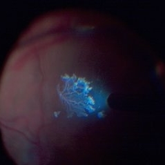
 Punctate Inner Choroidopathy with CNV Treated with Bevacizumab # 5 of 7
Feb 28 2013 by Gregory R. Blaha, MD, PhD
Late-phase fluorescein angiogram in a 31-year-old female with vision loss from a choroidal neovascular membrane (CNV) from punctate inner choroidopathy. Note the leakage from the CNV.
Photographer: Gerard Gauthier, Spindel Eye Assoc., Derry, NH
Imaging device: Zeiss FF 450 Plus
Condition/keywords: bevacizumabchoroidal neovascularization (CNV)punctate inner choroidopathy (PIC)
-
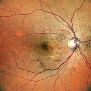
 Punctate Inner Choroidopathy with CNV Treated with Bevacizumab # 6 of 7
Feb 28 2013 by Gregory R. Blaha, MD, PhD
Fundus photo following treatment with bevacizumab in a 31-year-old female with vision loss from a choroidal neovascular membrane (CNV) from punctate inner choroidopathy. The vision improved and was stable following a single injection.
Photographer: Gerard Gauthier, Spindel Eye Assoc., Derry, NH
Imaging device: Zeiss FF 450 Plus
Condition/keywords: bevacizumabchoroidal neovascularization (CNV)punctate inner choroidopathy (PIC)
-
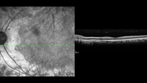
 Punctate Inner Choroidopathy with CNV Treated with Bevacizumab # 7 of 7
Feb 28 2013 by Gregory R. Blaha, MD, PhD
Improvement in OCT following treatment with bevacizumab in a 31-year-old female with vision loss from a choroidal neovascular membrane (CNV) from punctate inner choroidopathy. The vision improved and was stable following a single injection.
Photographer: Gerard Gauthier, Spindel Eye Assoc., Derry, NH
Imaging device: Zeiss Cirrus
Condition/keywords: bevacizumabchoroidal neovascularization (CNV)punctate inner choroidopathy (PIC)
-
 Satisfactory Long-Term Visual Outcome after Radiation Retinopathy Treatment
Jun 5 2020 by Sophia El Hamichi, MD
A 71-year-old female treated with plaque brachytherapy for choroidal melanoma in her left eye 10 years ago. She subsequently developed radiation retinopathy OS for which she was regularly receiving intravitreal injections of bevacizumab. The result is a stable visual acuity at 20/30. The patient continues to be regularly monitored.
Photographer: Belinda Rodriguez
Condition/keywords: bevacizumaboptical coherence tomography (OCT)radiation retinopathy
-
 Angioid Streaks with Regressing CNV s/p AntiVEGF Injections (LE)
Sep 20 2024 by Anand Temkar
A 45 year old male came to our OPD with chief complaints of DOV in BE since 2 months and wavy vision in periphery. Patient was diagnosed with (BE) CNVM in a case of Angioid Streaks and has already received (BE) bevacizumab x 2.
Photographer: Dr.Anand Temkar- Retina Foundation, Ahmedabad
Imaging device: Mirante
Condition/keywords: Angioid Streakschoroidal neovascularization (CNV)fundus autofluorescence (FAF)
-
 Angioid Streaks with Regressing CNV s/p AntiVEGF Injections (RE)
Sep 20 2024 by Anand Temkar
A 45 year old male came to our OPD with chief complaints of DOV in BE since 2 months and wavy vision in periphery. Patient was diagnosed with (BE) CNVM in a case of Angioid Streaks and has already received (BE) bevacizumab x 2.
Photographer: Dr.Anand Temkar- Retina Foundation, Ahmedabad
Imaging device: Mirante
Condition/keywords: Angioid Streakschoroidal neovascularization (CNV)fundus autofluorescence (FAF)
-
 Central Retinal Vein Occlusion with Macular Edema in Antiphospholipid Syndrome
Dec 24 2023 by Nikhil K Bommakanti, MD
A man in his thirties presented with a central retinal vein occlusion with macular edema in the right eye. Vision improved from 20/70 to 20/25 after 1 treatment with intravitreal bevacizumab. Laboratory testing revealed the presence of lupus anticoagulant.
Condition/keywords: antiphospholipid antibody syndromecentral retinal vein occlusion (CRVO)cystoid macular degenerationmacular edema
-
 Choroidal Osteoma
Mar 10 2020 by David L Kilpatrick, MD
Fundus photograph demonstrating a choroidal osteoma in the posterior pole. This patient had a drop in vision from 20/30 to 20/60 secondary to a new CNV that was treated with intravitreal bevacizumab.
Photographer: Retina Consultants of Alabama, P. C.
Condition/keywords: choroidal osteoma
-
 Choroidal Osteoma and Secondary Choroidal Neovascular Membrane
Sep 21 2012 by Allen Chiang, MD, FASRS
Fundus photograph of a 44-year old woman with a choroidal osteoma complicated by secondary choroidal neovascular membrane, regressed after serial intravitreal bevacizumab injections. The tumor exhibits areas of decalcification.
Imaging device: Topcon
Condition/keywords: choroidal neovascularization (CNV)choroidal osteomamacular choroidal osteoma
-
 CNV due to AMPPE
Oct 16 2012 by Ratimir Lazic, MD, PhD
OCT image of 58-year- old male. Total resolution of fluid one and a half month after treatment can be seen. The patient was treated with intravitreal bevacizumab.
Photographer: Marko Lukic, MD
Imaging device: OCT Copernicus
Condition/keywords: acute posterior multifocal placoid pigment epitheliopathy (APMPPE)anti-VEGFchoroidal neovascularization (CNV)
-
 Congenital Retinal Macrovessel
Oct 13 2023 by Jacob D. Grodsky, MD
41 y/o male who presented with acute onset of blurred vision OD. Visual acuity was 20/200 OD; 20/25 OS. Examination was consistent with congenital retinal macrovessel through the macula with intraretinal hemorrhage as seen in the fundus photo. Intravitreal bevacizumab was injected, and visual acuity improved to 20/40 at 4-week follow-up. MRA head and neck was ordered to rule out other vascular anomalies.
Condition/keywords: congenital retinal macrovesselRETINAL MACROVESSEL
-
---thumb.JPG/image-square;max$300,300.ImageHandler) CRVO in the Young
CRVO in the Young
Dec 13 2013 by Mallika Goyal, MD
Left eye CRVO in a 40-year-old male with no systemic disease or risk factors. Extreme macular edema resolved with intravitreal bevacizumab.
Photographer: Mallika Goyal, MD, Apollo Health City, Hyderabad, India
Condition/keywords: central retinal vein occlusion (CRVO)
-
 Detached NVE During PVD induction
Apr 27 2018 by Michael J. Koss, MD, PhD, MBA
A 73-year-old woman with macular pucker underwent a pars plana vitrectomy with membrane peeling. Additionally the patient suffers from diabetic retinopathy after being diagnosed with type 2 diabetes mellitus sixteen years ago. Prior to the procedure she was treated with a series of intravitreal Bevacizumab-injections due to diabetic macular edema. There was no history of a proliferative DRP. During the vitrectomy a branch of an obliterated NVE spontaneously detached and floated freely in the vitreous. The 3D shot was captured via Alcon’s NGENUITY® 3D Visualization System in form of photograph and video providing an outstandingly detailed image of the branched NVE.
Photographer: Michael Koss, Augenzentrum Nymphenburger Hoefe
Imaging device: Alcon’s NGENUITY® 3D Visualization System
Condition/keywords: diabetesdiabetic retinopathyneovascularization elsewhere (NVE)pars plana vitrectomy (PPV)PVD induction
-
 Early Phase PDR Post-Treatment
Aug 10 2014 by Thomas A. Ciulla, MD, MBA, FASRS
Intravitreal bevacizumab (1.25 mg) was administered intravitreally. When she returned 8 weeks later, her macular edema resolved and NV regressed.
Condition/keywords: intravitreal bevacizumabproliferative diabetic retinopathy (PDR)
-
 Early Phase PDR Pre-Treatment
Aug 10 2014 by Thomas A. Ciulla, MD, MBA, FASRS
Note the active NVD and NVE.
Condition/keywords: intravitreal bevacizumabproliferative diabetic retinopathy (PDR)
-
 Fundus Autofluorescence Showing Angioid Streaks with Regressing CNV s/p AntiVEGF Injections (LE)
Sep 20 2024 by Anand Temkar
A 45 year old male came to our OPD with chief complaints of DOV in BE since 2 months and wavy vision in periphery. Patient was diagnosed with (BE) CNVM in a case of Angioid Streaks and has already received (BE) bevacizumab x 2.
Photographer: Dr.Anand Temkar- Retina Foundation, Ahmedabad
Imaging device: Mirante
Condition/keywords: Angioid Streakschoroidal neovascularization (CNV)fundus autofluorescence (FAF)
-
 Fundus Autofluorescence Showing Angioid Streaks with Regressing CNV s/p AntiVEGF Injections (RE)
Sep 20 2024 by Anand Temkar
A 45 year old male came to our OPD with chief complaints of DOV in BE since 2 months and wavy vision in periphery. Patient was diagnosed with (BE) CNVM in a case of Angioid Streaks and has already received (BE) bevacizumab x 2.
Photographer: Dr.Anand Temkar- Retina Foundation, Ahmedabad
Imaging device: Mirante
Condition/keywords: Angioid Streakschoroidal neovascularization (CNV)fundus autofluorescence (FAF)
-
 Fundus Photo Macular Choroidal Hemangioma Treated with Laser
Nov 11 2019 by Sophia El Hamichi, MD
A 51-year-old female that presented with a macular choroidal hemagioma complicated by focal exudative retinal detachment OD. The patient was treated with vitrectomy and laser therapy of the choroidal hemagioma along with bevacizumab intravitreal injection during and after the surgery. The patient evolved well with resolution of the subretinal fluid OD. VA 20/200
Photographer: Sophia El Hamichi,MD, Murray Ocular Oncology and Retina, Miami
Condition/keywords: laser photocoagulationsubretinal fluid
-
 Hemorrhagic Age Related Macular Degeneration
Mar 25 2013 by Ratimir Lazic, MD, PhD
Color fundus photography of a 71- year-old female. Severe subretinal and sub RPE hemorrhage in macular area and inferior mid periphery. The patient was treated with 5 consecutive bevacizumab injections.
Photographer: Marko Lukic, MD
Imaging device: Zeis Visucam Lite 2
 Loading…