Search results (292 results)
-
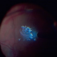
25 Gauge Vitrectomy Membrane Shaving
Jan 31 2015 by Thomas A. Ciulla, MD, MBA, FASRS
Membrane shaving of dense membranes in diabetic traction detachment using 25 gauge vitrectomy.
Condition/keywords: diabetes, pars plana vitrectomy (PPV), retina surgery, tractional retinal detachment, vitreoretinal surgery
-
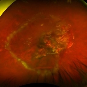
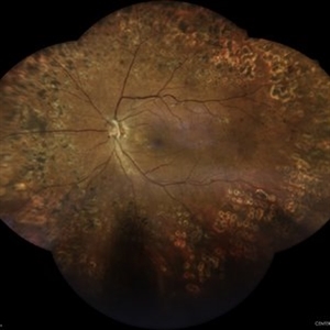 Active Laser Modified Proliferative Diabetic Retinopathy
Mar 17 2024 by Hector Gabriel Moreno Solano, MD, MHA
Wide-field composite fundus images of a 78-year-old male patient with a single functional eye due to poorly controlled diabetic retinopathy with activity data and multiple photocoagulation traces.
Photographer: Héctor Gabriel Moreno-Solano, MD, MHA
Imaging device: Centervue Eidon
Condition/keywords: Diabetes, proliferative diabetic retinopathy (PDR), retinopathy
-
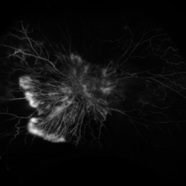
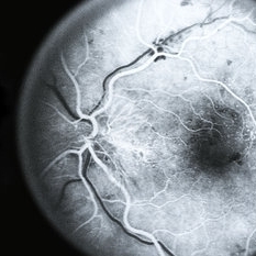
 Active neovascularization in Proliferative Diabetic Retinopathy
Jan 10 2018 by Peter H. Tang, MD, PhD
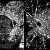
Fluorescein angiography image from a 46-year-old woman with uncontrolled proliferative diabetic retinopathy shows extensive dye leakage from active neovascularization.
Imaging device: Optos California
Condition/keywords: diabetes, diabetic retinopathy, fluorescein leakage, neovascularization elsewhere (NVE), neovascularization of the disc (NVD), pan-retinal photocoagulation (PRP), proliferative diabetic retinopathy (PDR)
-
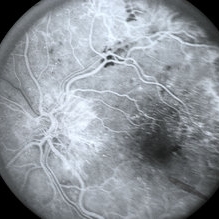
 Active Proliferative Diabetic Retinopathy
Aug 16 2022 by Donnie Willis
51 yo female. Uncontrolled Diabetes. Active Proliferative Diabetic Retinopathy.
Photographer: Donnie Willis, Tennessee Retina
Imaging device: Optos
Condition/keywords: capillary dropouts, Diabetes, fluorescein angiogram (FA), OPTOS, proliferative diabetic retinopathy (PDR), tractional retinal detachment
-
---thumb.jpg/image-square;max$300,300.ImageHandler) Acute optic nerve edema due to JODM
Acute optic nerve edema due to JODM
Apr 4 2014 by H. Michael Lambert, MD
23-year-old white female. Acute optic disc edema if JODM. VA 20/40 OU.
Photographer: Donald Lowd
Condition/keywords: diabetes, posterior hyaloid contraction
-
---thumb.jpg/image-square;max$300,300.ImageHandler) Acute optic nerve edema due to JODM
Acute optic nerve edema due to JODM
Apr 4 2014 by H. Michael Lambert, MD
23-year-old white female. Acute optic disc edema if JODM. VA 20/40 OU. Pregnant.
Photographer: Donald Lowd
Condition/keywords: diabetes, posterior hyaloid contraction
-
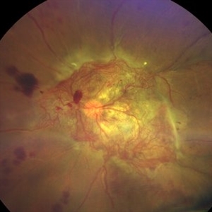
 Advanced Proliferative Diabetic Retinopathy
Nov 4 2017 by Hamid Ahmadieh, MD
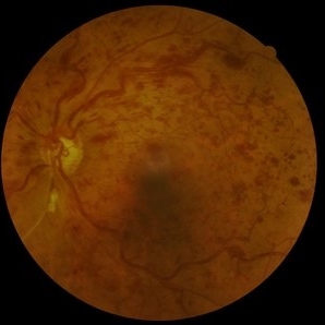
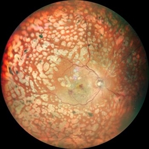
Merged color fundus photograph of the left eye of a 30-year-old woman with type1 diabetes since childhood. Note laser scars, severe fibrous proliferation, traction RD and macular dragging.
Photographer: Shabnam Poureh, Negah Eye Center, Tehran, Iran
Condition/keywords: color fundus photograph, diabetes, fibrous proliferation, proliferative diabetic retinopathy (PDR), severe traction
-
 Amalric triangular sign (posterior ciliary artery occlusion)
Feb 8 2023 by Bruno DECAY, MD
Fundus photograph of a 61-year-old female (routine examination)
Photographer: Amélie DULAC , Centre Ophtalmologique Vic-Montaner, Vic en Bigorre, France
Imaging device: Centervue Eidon
Condition/keywords: Diabetes, Hypertension
-
 An Intricate Web of Vasculature
Jan 5 2022 by SHISHIR VERGHESE, MS, FVRS, FAICO (Retina)
Fundus photograph of a 55-year-old gentleman with decreased vision in the left eye for 6 months. History of uncontrolled diabetes and hypertension for 15 years. Best corrected visual acuity in the left eye was 5/60.
Photographer: SHISHIR VERGHESE
Imaging device: ZEISS CLARUS
Condition/keywords: diabetes, proliferative diabetic retinopathy (PDR)
-
 Asteroid Hyalosis
Apr 9 2024 by Hector Gabriel Moreno Solano, MD, MHA
Slit lamp photograph of a 48-year-old female patient with long-standing diabetes attending consultation due to the sensation of moving spots in her vision.
Photographer: Héctor Gabriel Moreno-Solano
Condition/keywords: asteroid hyalosis, diabetes, slit lamp photo
-
 Before and After Vitrectomy
Nov 17 2023 by Bradley T. Smith, MD, FASRS
Middle age male diabetic retinopathy and resolving exudate following repair of tractional detachment with membrane peeling.
Condition/keywords: coats-like response, Diabetes, fibrotic neovascularization, fibrovascular proliferation, pars plana vitrectomy (PPV), proliferative diabetic retinopathy (PDR), tractional retinal detachment
-
 Bilateral Proliferative Diabetic Retinopathy OU
Feb 21 2025 by Drew Mitchell
OCT-Angiography 8x8 Montage OU. PDR with active NVE OD. 37 year old male with no visual complaints. Vision is 20/20 in both eyes.
Photographer: Drew Mitchell OCT-C
Imaging device: Zeiss Cirrus 5000
Condition/keywords: CIRRUS 5000 ANGIOPLEX, Diabetes, NVE, OCT Angiography, proliferative diabetic retinopathy (PDR)
-
 BRVO, FA, Hemorrhage, Diabetic
Mar 13 2014 by James B. Soque, CRA, OCT-C, COA, FOPS
51-year-old white male, diabetes, and with BRVO left eye, early phase 36 seconds. Flame heme from ON, showing microaneurysims, and fine capillary detail of this FA.
Photographer: James B Soque, CRA COA
Imaging device: Topcon TRC 50DX with MERGE software
Condition/keywords: branch retinal vein occlusion (BRVO), diabetes, FA early phase, microaneurysms
-
 Candy Stripe Sign
Mar 30 2023 by pedro fernandes souza neto
Candy Stripe Sign, patient with proliferative diabetic retinopathy progressing to vitreous hemorrhage and subsequently to ghost cell glaucoma.
Photographer: Marlos Henrique Oliveira Junior, Federal University of Bahia.
Condition/keywords: dehemoglobinized hemorrhage, diabetes, diabetic glaucoma
-
 Central Retinal Artery Occlusion
Apr 20 2018 by Kim Barrett
64-year-old female woke with no vision in her right eye. This image was taken at 6:11 minutes and the vessels have not filled. Patient has been treated with PRP laser and anti-VEGF therapy. Current vision is CF @ 2 ft.
Photographer: Kim Barrett C.O.A.
Imaging device: Heidelberg
Condition/keywords: central retinal artery occlusion (CRAO), diabetes, hypertension, smoker, uncontrolled
-
 Central retinal vein obstruction (CRVO)
May 29 2022 by Eduardo Javier Pinuer Alvarado
Fundus photograph of a 63 year-old woman with central retinal vein obstruction (CRVO). The patient didn´t notice the decrease of VA, we found this in a routine exam for diabetic patients.
Photographer: Eduardo Pinuer A, Universidad Austral de Chile.
Imaging device: CR-2 AF Digital Non-Mydriatic Retinal Camera, Canon.
Condition/keywords: central retinal vascular obstruction, diabetes, fundus photograph
-
 CNVM in Pan-retinal Photocoagulated Patient
Dec 30 2020 by ASRS Staff
Wide fundus photograph of 65-year-old, female, diabetic patient.
Imaging device: Nidek Mirante
Condition/keywords: age-related macular degeneration (AMD), diabetes, pan-retinal photocoagulation (PRP)
-
 Combined Tractional and Rhegmatogenous Retinal Detachment
Jan 30 2023 by Olivia Rainey
Ultra-widefield fluorescein angiography of a combined tractional and rhegmatogenous retinal detachment repair affecting the left eye. The retina is attached following silicone oil placement during most recent surgery. The patient was seeing CF at the time the image was taken.
Photographer: Olivia Rainey, OCT-C, COA
Imaging device: Optos California
Condition/keywords: diabetes, diabetic macular edema, diabetic retinopathy, fluorescein angiogram (FA), hyperfluorescence, right eye, scleral buckle, silicone oil, tractional retinal detachment, ultra-wide field imaging, ultra-widefield image
-
 Detached NVE During PVD induction
Apr 27 2018 by Michael J. Koss, MD, PhD, MBA
A 73-year-old woman with macular pucker underwent a pars plana vitrectomy with membrane peeling. Additionally the patient suffers from diabetic retinopathy after being diagnosed with type 2 diabetes mellitus sixteen years ago. Prior to the procedure she was treated with a series of intravitreal Bevacizumab-injections due to diabetic macular edema. There was no history of a proliferative DRP. During the vitrectomy a branch of an obliterated NVE spontaneously detached and floated freely in the vitreous. The 3D shot was captured via Alcon’s NGENUITY® 3D Visualization System in form of photograph and video providing an outstandingly detailed image of the branched NVE.
Photographer: Michael Koss, Augenzentrum Nymphenburger Hoefe
Imaging device: Alcon’s NGENUITY® 3D Visualization System
Condition/keywords: diabetes, diabetic retinopathy, neovascularization elsewhere (NVE), pars plana vitrectomy (PPV), PVD induction
-
 Diabetes
Jul 11 2013 by Jerald A. Bovino, MD
No history, fluorescein angiogram, early phase, active with PDR.
Condition/keywords: diabetic mellitus
-
 Diabetes
Jul 11 2013 by Jerald A. Bovino, MD
No history, fluorescein angiogram, early phase, active with PDR, same eye 354.
Condition/keywords: diabetic mellitus
-
---thumb.jpg/image-square;max$300,300.ImageHandler) Diabetes with Cortical Cataract
Diabetes with Cortical Cataract
Apr 4 2014 by H. Michael Lambert, MD
Diabetes. Cortical Spoking
Photographer: Donald Lowd
Condition/keywords: cataract, diabetes
-
---thumb.jpg/image-square;max$300,300.ImageHandler) Diabetes with Flame Hemorrhage FA
Diabetes with Flame Hemorrhage FA
Apr 4 2014 by H. Michael Lambert, MD
Diabetes.
Photographer: Donald Lowd
Condition/keywords: diabetes, flame shaped retinal hemorrhage
-
 Diabetic Papillopathy
Apr 6 2024 by Hector Gabriel Moreno Solano, MD, MHA
Optic nerve photo of a 53-year-old woman with proliferative diabetic retinopathy.
Photographer: Héctor Gabriel Moreno-Solano, MD, MHA
Imaging device: Smartphone (IPhone 11 pro max)
Condition/keywords: diabetes, proliferative diabetic retinopathy (PDR)
-
 Diabetic Retinopathy
Dec 11 2019 by Lauren Whaley
44-year-old male diabetic patient had an acute change in A1C over 9 months and ended up with a tractional retinal detachmen in right eye. This photo is 2 weeks post operative with current vision level at hand motion. He had extensive laser, retinectomy, and silicone oil fill.
Photographer: Lauren R. Whaley, COA
Imaging device: Optos Wide Field
Condition/keywords: diabetes, diabetic retinopathy, fibrosis, laser scarring, proliferative vitreoretinopathy (PVR), retinectomy, silicone oil, tractional retinal detachment
 Loading…