Search results (139 results)
-
 Retinal Detachment Following Scleral Buckling, Retinectomy, Laser, and Oil
Jan 31 2022 by Ahmad B. Tarabishy, MD
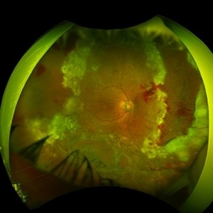
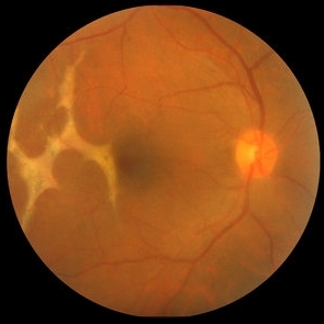
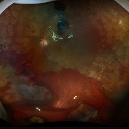
Ultra wide-field fundus photograph of a 55-year-old gentleman who is 4 days after surgery with scleral buckling, pars plana vitrectomy, perfluoron tamponade, membrane peeling, direct fluid-PFO-oil exchange, nasal and temporal retinectomies, and endolaser photocoagulation. Visual acuity was 20/150 under oil.
Photographer: Megan McLandsborough, Lakeland Eye Clinic
Imaging device: Optos California UWF Camera
Condition/keywords: endolaser, Membrane Peel, PPV, proliferative retinopathy, proliferative vitreoretinopathy (PVR), Retinal Detachment, retinal detachment with retinal defect, scleral buckle, submacular perfluorocarbon liquid (PFO)
-
 PPV retained cataract
Apr 19 2023 by Denica Rodriguez
A 46-year-old male with hypermature dense cataract. Patient got a piece of metal in his eye when he was 5 years old and was not able to see since. Patient was having cataract surgery and phacodonesis was present. The lens dropped to the back of the eye. Patient had to have another surgery to do vitrectomy. The lens removal was done with a fragmatome handpiece.
Photographer: Denica Rodriguez COA, ST
Imaging device: Zeiss Microscope with resight
Condition/keywords: cataract, dropped nucleus, fragmatome, lens capsule, ocular trauma, pars plana vitrectomy (PPV), retained lens fragments, Retina, retina surgery, traumatic cataract
-
 10 Days Post Subretinal TPA
Jan 9 2019 by John S. King, MD
76-year-old white male with history of treat/extend with Eylea OD for a PPCNVM; also monocular due to large scar in fellow eye. Two months since last injection, had acute decrease in vision OD and was seen that day. Vision CF; moderate SRH involving the fovea. Discussed monotherapy with anti-VEGF vs displacement, and elected for PPV, srTPA, AFx, SF6. Total of 0.2 cc of 25 microgram/0.1 ml of srTPA administered from two different areas in the temporal macula. 10 days post-op vision is improving; 20/200 J7; displacement of heme (photo)
Photographer: Kay Dalby
Imaging device: Topcon 50
Condition/keywords: choroidal neovascular membrane (CNVM), peripapillary hemorrhage, presumed ocular histoplasmosis syndrome (POHS)
-
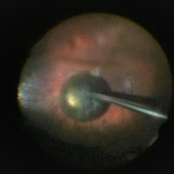
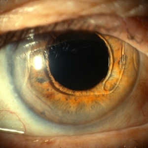
 24 Hours Post Scleral Wound Closure+ Scleral Buckle+25 g Vitrectomy+Silicon Oil
Jan 23 2015 by Carlos Quezada-Ruiz, MD, FASRS
24 hours post op fundus photograph of a 43-year-old man who had perforating injury to the right eye with a small piece of plastic while he was hammering. OD LP, subconjunctival hemorrhage, clear cornea, hyphema, irido and ciclodyalisis as well as a luxated lens with traumatic cataract and a dense vitreous hemorrhage. B-US showed rhegmatogenous retinal detachment with a tear and a big inferior hemorrhagic choroidal detachment. 360 peritomy revealed 2-entry scleral wounds were found in zone II (M V and M VI) and closure was performed. 25 G PPV was performed with the infusion canal placed in the AC through the limbus. Lensectomy and removal of a dense recent vitreous hemorrhage revealed a white detached retina with an exit wound through the temporal inferior segment of the optic nerve with a nasal GRT and sub retinal hemorrhage as well as temporal inferior choroidal, PVD was induced and PFOs helped stabilizing the retina while vitrectomy and sub-retinal hemorrhage was removed through the GRT. Fluid air exchange was made and 360 endolaser over the buckle indentation was done and silicon oil was used as endotamponade. This picture was taken 24 hrs after the surgery.
Photographer: Lilibeth Rodriguez, Instituto de la Visión. Torreon, Mexico.
Condition/keywords: central retinal artery occlusion (CRAO), giant retinal tear, trauma
-
25 Gauge Vitrectomy Membrane Shaving
Jan 31 2015 by Thomas A. Ciulla, MD, MBA, FASRS
Membrane shaving of dense membranes in diabetic traction detachment using 25 gauge vitrectomy.
Condition/keywords: diabetes, pars plana vitrectomy (PPV), retina surgery, tractional retinal detachment, vitreoretinal surgery
-
 25G PPV Without Scleral Buckling for RRD, PVR, Giant Breaks
Dec 10 2012 by Yale L. Fisher, MD
Dr. Steve Charles shares his approach to 25G PPV without scleral buckling for RRD, PVR and giant breaks. NOTE: A narration by Dr. Steve Charles will soon be available for this movie- please check back periodically.
Condition/keywords: video
-
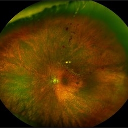
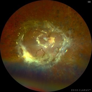 360 Retinotomy in a closed Funnel combined Tractional and rhegmatogenous retinal detachment
Jan 1 2023 by Malek Yassine, MD
This is the results at 6 months of a Bimanual 23 G-PPV with a very extensive and posterior 360 retinotomy for the management of a combined longstanding closed funnel RD, with submacular membranes, intraretinal PVR. Preop VA was a doubtful light perception. Borders of the retinotomy are stable at 6 months under 1300 Cs Silicon oil with some pigmented PVR developping the edges. Macula appears spared. Silicon oil emulsification droplets are well visualized beneath the superior temporal arcade.
Imaging device: Zeiss Clarus 700
Condition/keywords: combined retinal detachment, retinotomy, silicone oil
-
 4 Point Scleral Fixation Akreos AO60 With Gore Tex Suture
May 20 2021 by Jesus Lozano, MD
Optos Silverstone fundus image of a 54-year-old man after 4 point scleral fixation Akreos AO60 with Gore Tex suture plus PPV who had a severe traumatic iris defect and was aphakic after ocular trauma.
Photographer: Yair Bet Yosef, Hadassah Medical Center. Israel
Imaging device: Optos Silverstone
Condition/keywords: aphakia, globe perforation, lens, pars plana vitrectomy (PPV), penetrating trauma, vitreous hemorrhage
-
 4 Point Scleral Fixation Akreos AO60 With Gore Tex Suture
May 21 2021 by Jesus Lozano, MD
Anterior segment photo of a 54-year-old man after 4 point scleral fixation Akreos AO60 with Gore Tex suture plus PPV who had a severe traumatic iris defect and was aphakic after ocular trauma.
Photographer: Luigi Zinn, Hadassah Medical Center, Jerusalem.
Condition/keywords: aphakia, cornea rupture, lens, penetrating trauma
-
 Anterior Capsule Opacification
Jun 26 2016 by Jared Watson
49-year-old male with anterior capsule fibrosis and wrinkling S/P PPV/PPL/C3f8. Patient will have secondary IOL after retinal issues resolve.
Photographer: Jared Watson COT/CRA University of Virginia
Condition/keywords: anterior capsule opacification
-
 Anterior Capsule Opacification in Eye
Oct 11 2012 by Jeffrey G. Gross, MD, FASRS
Anterior capsule opacification in eye, s/p PPV lensectomy, without IOL.
Condition/keywords: anterior capsule opacification, lensectomy, without intraocular lens
-
 Anterior Chamber Intraocular Lens
Sep 20 2012 by Jeffrey G. Gross, MD, FASRS
AC-IOL, s/p PPV, lensectomy for dislocated crystalline lens, 20/20
Condition/keywords: anterior chamber, dislocated crystalline lens, intraocular lens (IOL), lensectomy
-
 Before and After Vitrectomy
Nov 17 2023 by Bradley T. Smith, MD, FASRS
Middle age male diabetic retinopathy and resolving exudate following repair of tractional detachment with membrane peeling.
Condition/keywords: coats-like response, Diabetes, fibrotic neovascularization, fibrovascular proliferation, pars plana vitrectomy (PPV), proliferative diabetic retinopathy (PDR), tractional retinal detachment
-
 Branch Retinal Vein Occlusion
Sep 11 2018 by Olivia Rainey
Ultra-wide field pseudocolor montage of an 84-year-old female with a branch retinal vein occlusion affecting her left eye. Patient recently had a PPV for a epiretinal membrane in her left eye and shortly after developed an occlusion.
Photographer: Olivia Rainey
Imaging device: Optos
Condition/keywords: branch retinal artery occlusion (BRAO), hemorrhage, left eye, montage, Optos, pseudocolor, ultra-wide field imaging
-
 Branch Retinal Vein Occlusion With Peripheral Pigmentary Change
Jan 15 2019 by Olivia Rainey
Ultra-wide field fluorescein angiogram of an 85-year-old female with a branch retinal vein occlusion with peripheral pigmentary changes. Patient developed a BRVO after a PPV for an epiretinal membrane.
Photographer: Olivia Rainey
Imaging device: Optos
Condition/keywords: branch retinal vein occlusion (BRVO), epiretinal membrane (ERM), fluorescein angiogram (FA), left eye, Optos, pigmentary retinal dystrophy
-
 Broken macroaneurysm
Nov 27 2022 by Nassim Alejandro Abreu Arbaje, MD
Fundus video frame of a 58 year old male who had a PPV on his left eye because a retinal macroaneurysm that broke a bled on all 3 retinal planes.
Photographer: Nassim Abreu, Hospital Dr. Elías Santana
Imaging device: NGenuity 3D system
Condition/keywords: broken macroanerysm, intraretinal hemorrhage, macroaneurysm, subretinal hemorrhage, vitreous hemorrhage
-
 Carotid Artery Plaque
Sep 9 2020 by John S. King, MD
66-year-old white male, former smoker, with a history of femoral artery stent a plaque removal in 2017, triple bypass 2019 (at that time there was no high grade carotid stenosis), diabetes significant for SNPDR OD and PDR OS (NVD). He underwent PRP OS and two months later developed a vitreous hemorrhage and had a PPV OS. Early in the post-operative period vision dropped to LP due to acute CRAO with retinal embolus present. He was found to have progressed to high grade carotid stenosis (versus imaging 6 months ago) and a left carotid endarterectomy was performed (see picture of the large plaque) .
Condition/keywords: carotid artery occlusion
-
 Choroid Detachment
Jul 7 2021 by Patrik Rajs
This eye was a tough one. The patient underwent PPV twice, the second one with silicone oil (SO) for retinal re-detachment. Due to the development of secondary glaucoma, silicone oil evacuation and lavage of the anterior chamber were performed. Because of the high IOP even after the evacuation, the XEN was implanted. The surgery was followed by choroidal detachment presented in the picture on the left side along with the residual silicone bubble superiorly. The retinal tear is captured inferiorly surrounded by laser spots. The second image (on the right) was taken only 7 days later and it shows that choroidal detachment in the eye resolved completely.
Photographer: Patrik Rajs, EYE CLINIC of Jan Evangelista Purkyne University and Masaryk Hospital, Czech Republic, Ústí nad Labem
Condition/keywords: choroid, detachment, glaucoma, retina, silicone oil, tear
-
 Choroidal Rupture
Oct 16 2012 by Jeffrey G. Gross, MD, FASRS
Choroidal rupture, s/p PPV, with TPA, and gas pneumatic displacement.
Condition/keywords: 10L technique, choroidal rupture, gas pneumatic displacement
-
 Combined RD in PDR patient
Sep 8 2017 by Eitae Kim, MD
52-year-old woman with diabetes visited visual disturbance. Fundus exam shows superior tractional-rhegmatogenous retinal detachment with proliferative vitreoretinopathy. I performed PPV, membranectomy, endolaser and silicone oil injection.
Photographer: Eitae Kim, BOIM retina center, Pureun eye hospital
Condition/keywords: ultra-wide field imaging
-
 Cutter Segmentation in a case of Diabetic Combined Retinal Detachment | Intra-Operative Still
Apr 25 2023 by Veer Singh, MS, FVRS, FMRF, FICO (Retina)
Cutter Segmentation in a case of Diabetic Combined Retinal Detachment | Intra-Operative Still Patient underwent Vitrectomy with Silicone Oil
Photographer: Dr. Veer Singh
Condition/keywords: combined retinal detachment, cutter, diabetic retinopathy, intraoperative, pars plana vitrectomy (PPV)
-
 Detached NVE During PVD induction
Apr 27 2018 by Michael J. Koss, MD, PhD, MBA
A 73-year-old woman with macular pucker underwent a pars plana vitrectomy with membrane peeling. Additionally the patient suffers from diabetic retinopathy after being diagnosed with type 2 diabetes mellitus sixteen years ago. Prior to the procedure she was treated with a series of intravitreal Bevacizumab-injections due to diabetic macular edema. There was no history of a proliferative DRP. During the vitrectomy a branch of an obliterated NVE spontaneously detached and floated freely in the vitreous. The 3D shot was captured via Alcon’s NGENUITY® 3D Visualization System in form of photograph and video providing an outstandingly detailed image of the branched NVE.
Photographer: Michael Koss, Augenzentrum Nymphenburger Hoefe
Imaging device: Alcon’s NGENUITY® 3D Visualization System
Condition/keywords: diabetes, diabetic retinopathy, neovascularization elsewhere (NVE), pars plana vitrectomy (PPV), PVD induction
-
 Diabetic Macular Edema
May 28 2016 by Olivia Rainey
Optical coherence tomography of an 54-year-old female with diabetic macular edema affecting both eyes. Patient has a history of proliferative diabetic retinopathy s/p PRP/PPV/MP/EL, and glaucoma s/p tube shunt in both eyes. There has been a persistence of her macular edema and limited response to antiVEGF therapy, which puts into question whether there is another cause for her edema. Leading the possible causes is her renal insufficiency and fluid retention. Patient was seeing 20/50 in the right eye and 20/80 in the left eye.
Photographer: Olivia Rainey
Imaging device: Heidelberg Spectralis
Condition/keywords: anti-VEGF, diabetic macular edema, edema, glaucoma, optical coherence tomography (OCT), pan-retinal photocoagulation (PRP), proliferative diabetic retinopathy (PDR)
-
 Diabetic Tractional Retinal Detachment 1 week s/p SO fill
Aug 14 2024 by Virginia Gebhart
21 year old male 1 week s/p PPV/laser/STR/SO. Eye is stable, PRHs inferior and superior, possible traction from PRH/membrane. Will observe and let clot liquify, will consider scleral buckle if no improvement
Photographer: Virginia Gebhart
Imaging device: Optos California
Condition/keywords: Diabetic Tractional Detachment, retinal detachment of the macula, silicone oil
-
 Diamond Dust from Retinal Scraper
Oct 15 2012 by Jeffrey G. Gross, MD, FASRS
Diamond dust from retinal scraper, s/p PPV.
Condition/keywords: diamond dust, pars plana vitrectomy (PPV), retinal scraper
 Loading…