Search results (139 results)
-
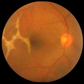
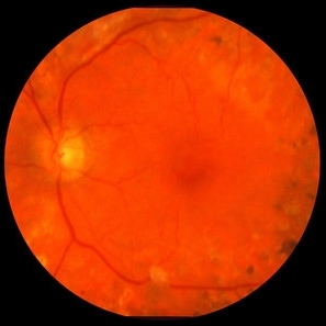
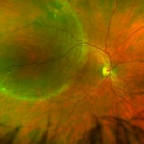 Retinal Detachment Right Eye Optomap
Mar 31 2014 by James B. Soque, CRA, OCT-C, COA, FOPS
36-year-old white male presented with non traumatic retinal detachment OD, with six very distinct demarcation lines and isolated tear, and detachment parameters. Patient underwent PPV OD on 12/3/13 with 20% SF6 gas placement and face down in his first 1 month post op period.
Photographer: James Soque, CRA, COA
Imaging device: Optos Daytona
Condition/keywords: Cryopexy, demarcation line, gas pneumatic displacement, Optomap, Optos, pars plana vitrectomy (PPV), retinal tear, scanning laser ophthalmoscope
-
 Anterior Chamber Intraocular Lens
Sep 20 2012 by Jeffrey G. Gross, MD, FASRS
AC-IOL, s/p PPV, lensectomy for dislocated crystalline lens, 20/20
Condition/keywords: anterior chamber, dislocated crystalline lens, intraocular lens (IOL), lensectomy
-
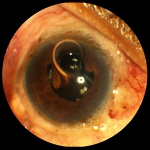 Gas Bubble Extending into Anterior Chamber
Oct 12 2012 by Jeffrey G. Gross, MD, FASRS
Gas bubble extending into anterior chamber in aphakic eye, after PPV.
Condition/keywords: anterior chamber, aphakic eye, gas bubble, pars plana vitrectomy (PPV)
-
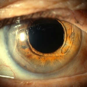
 Anterior Capsule Opacification in Eye
Oct 11 2012 by Jeffrey G. Gross, MD, FASRS
Anterior capsule opacification in eye, s/p PPV lensectomy, without IOL.
Condition/keywords: anterior capsule opacification, lensectomy, without intraocular lens
-
 Optos Picture With Speculum: Dislocated Natural Lens
Oct 9 2018 by John S. King, MD
55-year-old white female with history of pathologic myopia+, lattice (laser), SB OU (1990s), and dislocated natural lenses OU that had been watched for years. In the fellow eye she developed phacolytic glaucoma and a PPV, PPL was performed. Plan for both eyes are monitoring. I wanted to get a good picture of her lens today with the optos machine, as the other pics had artifact from the lower lid. It worked out well to use a speculum in the left eye. Vision cc is 20/400 J1+ OD and 20/40 J2 OS; aphakic OU; vitreous clear OD; dislocated lens OS (see pic); retinas attached.
Photographer: Maisee Yang
Imaging device: Optos California
Condition/keywords: dislocated crystalline lens, pathologic myopia, scleral buckle, staphyloma
-
 Retinoschisis/Retinal Detachment
Oct 16 2012 by Jeffrey G. Gross, MD, FASRS
Retinoschisis/RD s/p PPV and laser.
Condition/keywords: laser, pars plana vitrectomy (PPV), retinoschisis
-
 Choroidal Rupture
Oct 16 2012 by Jeffrey G. Gross, MD, FASRS
Choroidal rupture, s/p PPV, with TPA, and gas pneumatic displacement.
Condition/keywords: 10L technique, choroidal rupture, gas pneumatic displacement
-
 Intravitreal Gas Bubble after PPV
Oct 12 2012 by Jeffrey G. Gross, MD, FASRS
Intravitreal gas bubble after PPV, with mirroring reflection of ON.
Condition/keywords: intravitreal gas bubble, mirroring reflection of ON, pars plana vitrectomy (PPV)
-
 Diamond Dust from Retinal Scraper
Oct 15 2012 by Jeffrey G. Gross, MD, FASRS
Diamond dust from retinal scraper, s/p PPV.
Condition/keywords: diamond dust, pars plana vitrectomy (PPV), retinal scraper
-
 Retinoschisis/Retinal Detachment
Oct 16 2012 by Jeffrey G. Gross, MD, FASRS
Retinoschisis/RD, s/p PPV and laser.
Condition/keywords: retinoschisis
-
 Proliferative Diabetic Retinopathy with Severe Subhyaloid Hemorrhage
Oct 15 2012 by Jeffrey G. Gross, MD, FASRS
PDR with severe subhyaloid hemorrhage post-op, PPV.
Condition/keywords: pars plana vitrectomy (PPV), post-op, subhyaloid hemorrhage
-
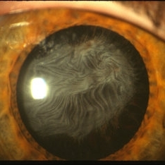
 Anterior Capsule Opacification
Jun 26 2016 by Jared Watson
49-year-old male with anterior capsule fibrosis and wrinkling S/P PPV/PPL/C3f8. Patient will have secondary IOL after retinal issues resolve.
Photographer: Jared Watson COT/CRA University of Virginia
Condition/keywords: anterior capsule opacification
-
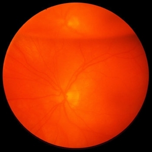
 24 Hours Post Scleral Wound Closure+ Scleral Buckle+25 g Vitrectomy+Silicon Oil
Jan 23 2015 by Carlos Quezada-Ruiz, MD, FASRS
24 hours post op fundus photograph of a 43-year-old man who had perforating injury to the right eye with a small piece of plastic while he was hammering. OD LP, subconjunctival hemorrhage, clear cornea, hyphema, irido and ciclodyalisis as well as a luxated lens with traumatic cataract and a dense vitreous hemorrhage. B-US showed rhegmatogenous retinal detachment with a tear and a big inferior hemorrhagic choroidal detachment. 360 peritomy revealed 2-entry scleral wounds were found in zone II (M V and M VI) and closure was performed. 25 G PPV was performed with the infusion canal placed in the AC through the limbus. Lensectomy and removal of a dense recent vitreous hemorrhage revealed a white detached retina with an exit wound through the temporal inferior segment of the optic nerve with a nasal GRT and sub retinal hemorrhage as well as temporal inferior choroidal, PVD was induced and PFOs helped stabilizing the retina while vitrectomy and sub-retinal hemorrhage was removed through the GRT. Fluid air exchange was made and 360 endolaser over the buckle indentation was done and silicon oil was used as endotamponade. This picture was taken 24 hrs after the surgery.
Photographer: Lilibeth Rodriguez, Instituto de la Visión. Torreon, Mexico.
Condition/keywords: central retinal artery occlusion (CRAO), giant retinal tear, trauma
-
 Proliferative Diabetic Retinopathy with Macular Traction
Oct 15 2012 by Jeffrey G. Gross, MD, FASRS
PDR with macular traction, post-op, PPV and PRP.
Condition/keywords: macular traction, pan-retinal photocoagulation (PRP), pars plana vitrectomy (PPV), post-op
-
 25G PPV Without Scleral Buckling for RRD, PVR, Giant Breaks
Dec 10 2012 by Yale L. Fisher, MD
Dr. Steve Charles shares his approach to 25G PPV without scleral buckling for RRD, PVR and giant breaks. NOTE: A narration by Dr. Steve Charles will soon be available for this movie- please check back periodically.
Condition/keywords: video
-
 Tools & Techniques for 23, 25, and 27G PPV
Dec 10 2012 by Yale L. Fisher, MD
Dr. Steve Charles reviews the tools and techniques he uses for 23G, 25G, and 27G PPV. NOTE: A narration by Dr. Steve Charles will soon be available for this movie- please check back periodically.
Condition/keywords: pars plana vitrectomy (PPV), video
-
 Silicone Oil Bubble in Anterior Chamber - 30 Degree Angle
Apr 11 2016 by Zach Dupureur
30 % silicone oil bubble involving central visual axis. Occurred after a PPV with silicone oil. Oil from vitreous moved into the anterior chamber.
Photographer: Zachary Dupureur, OCT-C
Imaging device: Heidelberg Spectralis
Condition/keywords: anterior chamber, detachment, infrared image, pars plana vitrectomy (PPV), scleral buckle, silicone oil
-
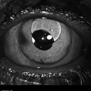
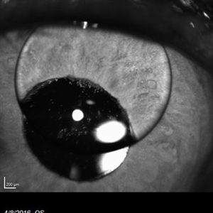
 Subluxated Sulcus IOL With Small AC Bubble
Aug 21 2018 by Russell Pokroy, MD
Anterior segment photograph of 71-year-old woman with 1-piece soft IOL decentered nasally with the haptics in the ciliary sulcus. A small area of the capsule remnant is evident suprotemporally. Small gas bubble in the AC is evident two weeks after PPV. The vitreous gas bubble decentered this IOL, with some spontaneous improvement of the IOL centering after gas dissipation.
Photographer: Russell Pokroy, Assaf Harofe Medical Center, Israel
Condition/keywords: intraocular lens (IOL)
-
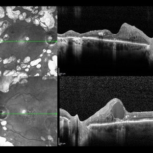 Diabetic Macular Edema
May 28 2016 by Olivia Rainey
Optical coherence tomography of an 54-year-old female with diabetic macular edema affecting both eyes. Patient has a history of proliferative diabetic retinopathy s/p PRP/PPV/MP/EL, and glaucoma s/p tube shunt in both eyes. There has been a persistence of her macular edema and limited response to antiVEGF therapy, which puts into question whether there is another cause for her edema. Leading the possible causes is her renal insufficiency and fluid retention. Patient was seeing 20/50 in the right eye and 20/80 in the left eye.
Photographer: Olivia Rainey
Imaging device: Heidelberg Spectralis
Condition/keywords: anti-VEGF, diabetic macular edema, edema, glaucoma, optical coherence tomography (OCT), pan-retinal photocoagulation (PRP), proliferative diabetic retinopathy (PDR)
-
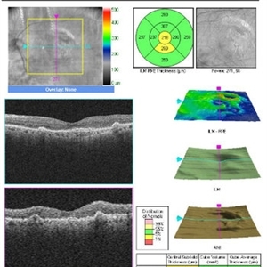 Pars Plana Vitrectomy TPA 2
Oct 29 2012 by John S. King, MD
About 2 months after PPV, sub-retinal tPA, for the second time a large sub-retinal hemorrhage appeared. Visual acuity 20/40-20/50 after second ppv c subretinal tpa (vs cf prior to PPV), but less than result following first surgery (20/30).
Photographer: Kristin Konecki, OcuSight Eye Care Center, Rochester, NY
Imaging device: Cirrus
Condition/keywords: subretinal hemorrhage
-
25 Gauge Vitrectomy Membrane Shaving
Jan 31 2015 by Thomas A. Ciulla, MD, MBA, FASRS
Membrane shaving of dense membranes in diabetic traction detachment using 25 gauge vitrectomy.
Condition/keywords: diabetes, pars plana vitrectomy (PPV), retina surgery, tractional retinal detachment, vitreoretinal surgery
-
 Silicone Oil Bubble in Anterior Chamber - 20 Degree Angle
Apr 11 2016 by Zach Dupureur
30 % silicone oil bubble involving central visual axis. Occurred after a PPV with silicone oil. Oil from vitreous moved into the anterior chamber.
Photographer: Zachary Dupureur, OCT-C
Imaging device: Heidelberg Spectralis
Condition/keywords: anterior chamber, detachment, infrared image, pars plana vitrectomy (PPV), scleral buckle, silicone oil
-
 Macular Hole
Oct 8 2012 by Jeffrey G. Gross, MD, FASRS
Macular hole, s/p PPV, not closed, with residual subretinal gass bubble, in cuff.
Condition/keywords: cuff, macular hole
-
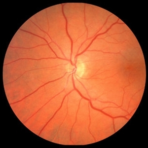
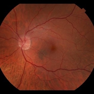
 Encircling Buckle Effect
Jul 7 2015 by Hamid Ahmadieh, MD
Late FA image of the right eye of a 30-year-old man who underwent pars plana vitrectomy , endolaser photocoagulation and an encircling band placement a couple of years before following a penetrating trauma at the vitreous base area at the 7 o'clock meridian.
Photographer: Nayereh Hadipour, Negah Eye Center,Tehran, Iran
Imaging device: Specteralis
Condition/keywords: pars plana vitrectomy (PPV)
-
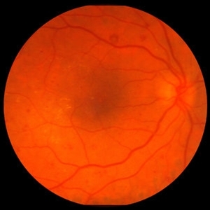
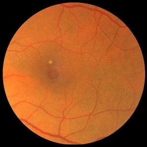
 NAION, ERM
Sep 7 2018 by John S. King, MD
70-year-old white male with background history of fovea involving RD repaired with PPV 5 months ago, and history of HTN. Some recent photopsias, mild scotoma. Focal ERM superiorly and swollen superior part of the optic disc.
Photographer: Macey
Imaging device: Topcan
Condition/keywords: drusen, epiretinal membrane (ERM), ischemic optic neuropathy
 Loading…