Search results (139 results)
-
 Double Trouble
Jun 28 2025 by Tejaswita Verma
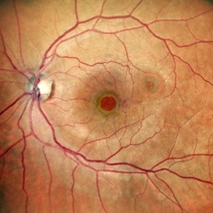
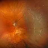
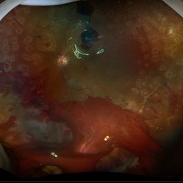
Fundus image of a 60 year old diabetic female with double macular holes with 6/60 vision status post LE PPV+gas 4 months ago. Other eye also had an unoperated large macular hole. Known case of glaucoma
Photographer: Dr. Tejaswita Verma
Imaging device: MIRANTE
Condition/keywords: macular hole
-
 Double Trouble
Jun 28 2025 by Tejaswita Verma
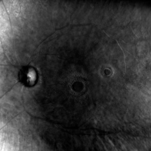
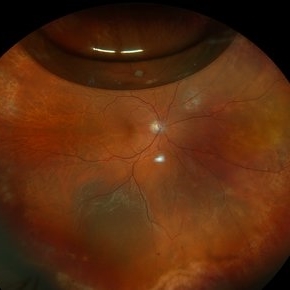
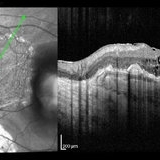
Retro image of aa double macular hole in a 60 yr old diabetic female status post PPV + gas 4 months ago. Vision was 6/60 in LE.
Photographer: Dr. Tejaswita Verma
Imaging device: MIRANTE
Condition/keywords: macular hole, retro mode
-
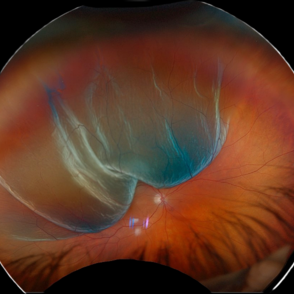
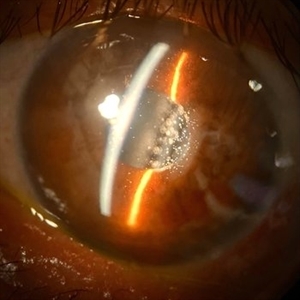
 Dislocation of the Crystalline Lens with a Retinal Detachment
Apr 21 2025 by Hrishikesh Naik, MS
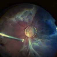
An intraoperative screen grab shows a dislocation of the crystalline lens along with an associated rhegmatogenous retinal detachment in a case of Marfan’s syndrome. The case was managed by a combined PPV-SB procedure. A vitrectomy cutter is seen at the left.
Photographer: Hrishikesh Naik
Condition/keywords: intraoperative, lens dislocation, Marfan's syndrome, Retinal Detachment, vitrectomy
-
 Repaired Retinal Detachment with Scleral Buckle
Mar 25 2025 by Kimberly Wakester
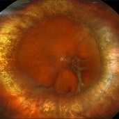
Optomap RGB montage of an 64-year-old woman with a repaired retinal detachment with scleral buckle in the right eye. There is nasal and inferior pre-retinal membranes with traction. PPV was recommended but patient defers to proceed with sx at this time. Will continue to follow patient closely for worsening traction. Patient was educated on how to monitor their peripheral vision and was advised to report any changes immediately.
Photographer: Kimberly Wakester, COA, OCT-C
Imaging device: Optos California
Condition/keywords: pre-retinal membrane with traction, repaired RD, scleral buckle
-
 Retinal Detachment (Mac-Off)
Feb 20 2025 by Virginia Gebhart
63 year old male with a mac-off retinal detachment from 4:00 to 1:30 with a single break at 10:00. Pt schedule for PPV/GFE. Guarded prognosis for visual recovery.
Photographer: Virginia Gebhart, Retina Consultants of Carolina
Imaging device: Optos California
Condition/keywords: horseshoe tear, retinal detachment, retinal detachment of the macula
-
 Mac-on Retinal Detachment (Barely!)
Feb 6 2025 by Virginia Gebhart
FAF of 46 year old male with a mac-on retinal detachment from 1:00 to 6:00 with a single break at 3:00. Pt scheduled for emergent PPV/Laser/GFE
Photographer: Virginia Gebhart, Retina Consultants of Carolina
Imaging device: Optos California
Condition/keywords: autofluorescence imaging, retinal detachment
-
 Retinal Detachment with Single Break
Feb 5 2025 by Virginia Gebhart
61 year old male with mac-off retinal detachment with single horseshoe tear. Macula has been off for several days and has developed associated cystic edema. Visual prognosis guarded. Pt schedule for PPV/Laser/GFE
Photographer: Virginia Gebhart, Retina Consultants of Carolina
Imaging device: Optos California
Condition/keywords: horseshoe tear, PVD, retinal detachment
-
 Retinal Detachment with Multiple Breaks
Feb 3 2025 by Kimberly Wakester
Fundus photograph of a 67-year-old man with a retinal detachment with multiple breaks in the right eye. Patient is doing well s/p PPV and will continued to be observed during PO period.
Photographer: Kimberly Wakester, COA
Imaging device: Optos California
Condition/keywords: horseshoe tear, multiple retinal tears, retinal detachment
-
 Recurrent Retinal Detachment with Single Break
Nov 2 2024 by Virginia Gebhart
84 year old male with recurrent detachment s/p PPV/RD repair 2 weeks ago. Retinotomy is opened and appears to be the source of the fluid. Pt scheduled for emergency repair with scleral buckle.
Photographer: Virginia Gebhart
Imaging device: Optos California
-
 Persistent Fetal Vasculature
Sep 23 2024 by Carlos Augusto Moreira, MD, PhD
Persistent Fetal Vasculature - an intraocular cotton swab appearance.
Photographer: Carlos Augusto Moreira-Neto, Hospital de Olhos do Paraná
Imaging device: NGENUITY Visualization System
Condition/keywords: pars plana vitrectomy (PPV), persistent fetal vasculature (PFV)
-
 New RD with Multiple Breaks
Aug 26 2024 by Virginia Gebhart
59 year old male with superior, bullous, mac off RD with multiple breaks. Pt scheduled for PPV, laser, GFE.
Photographer: Virginia Gebhart
Imaging device: Optos California
Condition/keywords: retinal detachment of the macula, retinal tear, retinal tear with detachment
-
 Diabetic Tractional Retinal Detachment 1 week s/p SO fill
Aug 14 2024 by Virginia Gebhart
21 year old male 1 week s/p PPV/laser/STR/SO. Eye is stable, PRHs inferior and superior, possible traction from PRH/membrane. Will observe and let clot liquify, will consider scleral buckle if no improvement
Photographer: Virginia Gebhart
Imaging device: Optos California
Condition/keywords: Diabetic Tractional Detachment, retinal detachment of the macula, silicone oil
-
 RPE-Transplantation
Jul 25 2024 by Gabriel Costa Andrade, PhD
Postoperative period of RPE-transplantation in a patient with neovascular AMD after RPE tear.
Photographer: Gabriel Andrade
Condition/keywords: neovascular age-related macular degeneration (AMD), pars plana vitrectomy (PPV), wet age-related macular degeneration (wet AMD)
-
 Dislocated IOL
Jun 27 2024 by Vishal Agrawal, MD, FRCS,FACS,FASRS
Fundus picture of a 65-year-old male patient presenting with posteriorly dislocated IOL & Soemmerring ring in the right eye. PPV + IOL removal + secondary IOL (Yamane technique) was performed.
Photographer: Dr Ayushi
Imaging device: Clarus 700
Condition/keywords: IOL, Soemmering's ring
-
 Rhegmatogenous Macula Off Retinal Detachment with Multiple Breaks
May 29 2024 by Alexis Singstock
Ultra widefield fundus photograph of a 66 year old male with rhegmatogenous macula off retinal detachment with multiple breaks. Patient presented emergently for a curtain/veil in inferonasal visual field. Patient reports the curtain/veil in left eye started about 1 week prior, yet denied seeing flashes and floaters. Patient's vision was hand motion. Dr. Edward Korot examined the patient and scheduled him for a scleral buckle along with pars plana vitrectomy surgery.
Photographer: Alexis Singstock, Retina Specialists of Michigan
Imaging device: Optos California
Condition/keywords: fundus photography, left eye, macula off retinal detachment, OPTOS CALIFORNIA, pars plana vitrectomy (PPV), rhegmatogenous retinal detachment, scleral buckle, ULTRA WIDE FIELD
-
 Emulsificated Silicon Oil in Vitreous Cavity
Apr 18 2024 by JULIAN VILLARREAL, MD
62 -year-old male that underwent a PPV with silicon oil for a mac-off retinal detachment in March 2023.
Photographer: Julián Villarreal Olivares, Instituto Mexicano de Oftalmología IAP
Condition/keywords: silicon oil emulsification in vitreous cavity
-
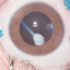
 Silicone Oil in the Anterior Chamber
Mar 27 2024 by Emma Paulina Carrillo Haro
Anterior chamber photograph of a 67-year-old male with silicone oil in the anterior chamber after pars plana vitrectomy (PPV) and silicone oil injection into the vitreous cavity.
Photographer: Emma Paulina Carrillo Haro
Imaging device: Iphone
Condition/keywords: anterior chamber, silicone oil
-
 Dislocated Crystalline Lens
Mar 19 2024 by Annaka Gooding
Ultra Wide field fundus photography of a 70 year old male who presented to clinic with a sudden increase of vision due to dropped crystalline lens secondary to severely dense cataract. Patient reported seeing a full black circle in his inferior visual field. Patient's visual acuity at time of visit was 20/100 with a +5.00 diopter lens. The physician recommended surgical intervention, and discussed surgery for PPV/PPL/IOL implantation with an ACIOL.
Photographer: Annaka Gooding, CPO
Imaging device: Optos California RGB
Condition/keywords: dislocated crystalline lens, fundus photography, inferior retina, OPTOS CALIFORNIA RGB, Right Eye, Ultra-wide field retinal imaging
-
 Serous Retinal Detachment in Advanced Proliferative Diabetic Retinopathy
Feb 15 2024 by Annaka Gooding
Ultra-Wide fundus photograph of a 29 year old female with a Serous Retinal Detachment in Advanced PDR. Patient present to clinic with LP vision following PPV and fill in PRP. Physician recommended oral prednisone treatment and to reassess at their following visit.
Photographer: Annaka Gooding, CPO
Imaging device: Optos California RGB
Condition/keywords: Diabetes, diabetic macular edema, fundus photography, OPTOS CALIFORNIA, pan-retinal photocoagulation (PRP), pars plana vitrectomy (PPV), proliferative diabetic retinopathy (PDR), serous retinal detachment, ultra-wide field imaging
-
 Dislocated Lens, Posterior OD
Jan 26 2024 by Corey Grant
OPTOS California photo presents a 71 year old male patient with a dislocated lens, posterior in the right eye. Presented on 1/26/24 with posteriorly dislocated SN60WF with a Soemmerring ring. Associated retinal hemorrhage within retinoschisis as well. This will result in a PPV/IOL exchange/SFIOL/STK for the right eye.
Photographer: Corey Grant, Ophthalmic Imager, Retina Specialist of Michigan
Imaging device: OPTOS California
Condition/keywords: color photo, IOL, OD, Optos, OPTOS CALIFORNIA, pars plana vitrectomy (PPV), retina
-
 Macula off Retinal Detachment
Jan 23 2024 by Annaka Gooding
Ultra-widefield fundus photograph of an 81-year-old male with a Macula Off Retinal Detachment affecting his right eye. Patient presented at office with complaints of flashes of light for about 2 weeks accompanied by a curtain veil covering inferior visual field. Patient had total vision loss 24 hours prior to visit. His vision was scHM. The physician recommended Retinal Detachment Repair with PPV.
Photographer: Annaka Gooding, CPO
Imaging device: Optos California
Condition/keywords: detachment, fundus photography, macula off retinal detachment, Optos, retinal detachment of the macula, right eye, ultra-wide field imaging
-
 Mac off Retinal Detachment with Horseshoe Tear
Dec 5 2023 by Virginia Gebhart
68 year old male presented with HM vision in OD. Near total detachment with multiple breaks. Scheduled PPV with GFE. Visual prognosis guarded
Photographer: Virginia Gebhart
Imaging device: Topcon
Condition/keywords: Retinal Detachment, retinal detachment of the macula, Retinal Detachment with multiple breaks
-
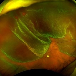
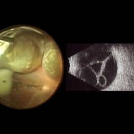 Macrocysts in Kickboxer
Nov 17 2023 by Bradley T. Smith, MD, FASRS
Intraoperative photo and preoperative b scan of chronic retinal detachment with macrocysts in a kickboxer
Condition/keywords: B scan ultrasound, chronic retinal detachment, ocular trauma, pars plana vitrectomy (PPV), retinal macrocyst
-
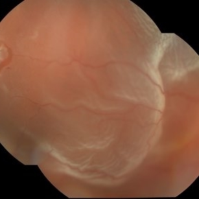
 Before and After Vitrectomy
Nov 17 2023 by Bradley T. Smith, MD, FASRS
Middle age male diabetic retinopathy and resolving exudate following repair of tractional detachment with membrane peeling.
Condition/keywords: coats-like response, Diabetes, fibrotic neovascularization, fibrovascular proliferation, pars plana vitrectomy (PPV), proliferative diabetic retinopathy (PDR), tractional retinal detachment
-
 New Retinal Detachment 6w s/p RD repair
Nov 16 2023 by Virginia Gebhart
13 year old male presented with new blind spot 6 weeks s/p RD repair with cryo/scleral buckle/prophylaxis laser with gas bubble. New RD involving the macula, posterior to scleral buckle, secondary to PVD. Small gas bubble remaining. Pt was brought back to OR for repeat PPV and silicone oil repair
Photographer: Virginia Gebhart
Imaging device: Optos
Condition/keywords: gas bubble, Retinal Detachment, retinal detachment of the macula, scleral buckle
 Loading…