Search results (191 results)
-
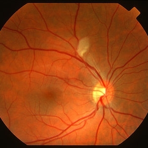
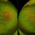
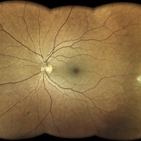 Amalric triangular sign (posterior ciliary artery occlusion)
Feb 8 2023 by Bruno DECAY, MD
Fundus photograph of a 61-year-old female (routine examination)
Photographer: Amélie DULAC , Centre Ophtalmologique Vic-Montaner, Vic en Bigorre, France
Imaging device: Centervue Eidon
Condition/keywords: Diabetes, Hypertension
-
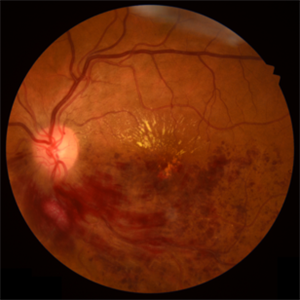
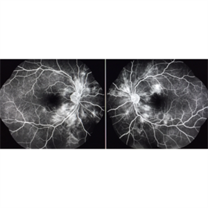
 Arterial Occlusion With Preservation of Cilioretinal Network
Aug 29 2016 by JEFFERSON R SOUSA, Tecg.º (Biomedical Systems Technology)
Patient male, 52-years-old, denies hypertension, blood systemic, and diabetes. Refers low subtle of vision, field loss and stain central.
Photographer: JEFFERSON R SOUSA - Institute Dr. Suel Abujamra / São Paulo - Brazil
Imaging device: Acquisition of the image in the Camera background Topcon TRC-50 Dx - IA, Keystone field photo of 50 Degrees. Composition automatic of Imaginet with manual adjustment.
Condition/keywords: arterial occlusion, hypertension
-
 Branch Retinal Vein Occlusion
Apr 15 2023 by Yousef A Fouad, MBBCh, MSc
BRVO in a young female with uncontrolled hypertension
Photographer: Yousef Fouad, Ain Shams University, Egypt
Condition/keywords: branch retinal vein occlusion (BRVO), fundus photograph, Hypertension, venous occlusion
-
 Central Retinal Artery Occlusion
Apr 20 2018 by Kim Barrett
64-year-old female woke with no vision in her right eye. This image was taken at 6:11 minutes and the vessels have not filled. Patient has been treated with PRP laser and anti-VEGF therapy. Current vision is CF @ 2 ft.
Photographer: Kim Barrett C.O.A.
Imaging device: Heidelberg
Condition/keywords: central retinal artery occlusion (CRAO), diabetes, hypertension, smoker, uncontrolled
-
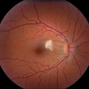 Cotton Wool Spot
Jul 10 2013 by Jason S. Calhoun
Fundus photograph shows a young male with a single cotton wool spot just nasal to the macula in the right eye.
Photographer: Jason S. Calhoun, Department of Ophthalmology, Mayo Clinic Jacksonville, Florida
Condition/keywords: cotton wool spots, hypertension
-
 HTN / Macular Star / CWS
Feb 17 2015 by David Callanan, MD
HTN / Macular star / CWS.
Condition/keywords: cotton wool spots, hypertension, macular star
-
 HTN / Macular Star / CWS
Feb 17 2015 by David Callanan, MD
HTN / Macular star / CWS.
Condition/keywords: cotton wool spots, hypertension, macular star
-
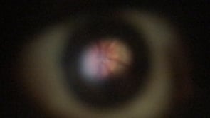 Hypertension
May 20 2021 by MICHAEL HAMBRICK
Video obtained from right eye of a 28-year-old male (utilizing the D-Eye lens attached to an iPhone 6) with a blood pressure of 180/120 who presented with a HA and intermittent blurry vision for 3-4 days. VA: OD 20/20 OS 20/20 OD 20/20 at 6 feet with Snellen Card Visual fields intact to confrontation. PERRLA. EOMI No RAPD. Decreased red perception on the left.
Condition/keywords: hypertension
-
 Hypertension With Arterio-Venous Nicking and a Superior Macular BRVO with CME
Aug 7 2018 by Jennifer I. Lim, MD, FARVO, FASRS
Fundus photograph of a 50-year-old man with hypertensive retinal changes: arterio-venous nicking and a superior macular BRVO with CME.
Photographer: Norb Jednock, University of Illinois at Chicago (1992)
Condition/keywords: arteriovenous nipping, hypertension, macular branch retinal vein occlusion (BRVO)
-
 Hypertensive Changes
Aug 26 2019 by Narciso F. Atienza, MD, MBA, FASRS, FPCS, FPAO.
Fundus photo of the left eye showing hypertensive changes in the vessels.
Photographer: Narciso F Atienza, Jr. MD, MBA
Imaging device: Topcon TRC
Condition/keywords: fundus photograph, hypertension
-
 Hypertensive Retinopathy
Mar 29 2013 by Henry J. Kaplan, MD
Cotton wool spot in grade III hypertensive retinopathy.
Condition/keywords: hypertension, hypertensive retinopathy
-
 Hypertensive Retinopathy
Feb 24 2020 by Brian K. Horsman, MD, FRCS(C) FASRS
Macular star, disc edema, intra vitreous bubble of Bevacizumab
Condition/keywords: bevacizumab, hypertension
-
 Hypertensive Retinopathy
Feb 24 2020 by Brian K. Horsman, MD, FRCS(C) FASRS
Macular star, disc edema, intra vitreous bubble of Bevacizumab
Condition/keywords: bevacizumab, hypertension
-
 Hypertensive Retinopathy
Feb 25 2013 by Suber S. Huang, MD, MBA, FASRS
32-year-old African American male with Grade IV hypertensive retinopathy and acute renal failure. Vision OD 20/70, OS 20/25. Creatine 7.1. BP: 250/150.
Photographer: Geoffrey Pankhurst, University Hospitals, Eye Institute/Dept. Ophthalmology and Visual Sciences Case Western Reserve University Cleveland, OH
Imaging device: Topcon TRC 50x
Condition/keywords: acute renal failure, disc edema, exudate, hypertension, hypertensive retinopathy, ischemia, macular edema, macular ischemia, optic disc edema
-
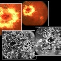
 Hypertensive Retinopathy
Nov 14 2019 by Jennifer Schiefer, CRA, COA
46-year-old male who suffered CVA from hypertensive crisis prior to these images. Patient presented in office reporting episode of dim Va OS that happened several days prior. Va was 20/25 OU upon examination.
Photographer: Jennifer Schiefer
Condition/keywords: exudates, fluorescein angiogram (FA), hypertension, hypertensive retinopathy, ischemia, late phase
-
 Hypertensive Retinopathy
Oct 20 2012 by Hyung-Woo Kwak, MD
The sudden appearance of cotton wool spots with hypertension retinopathy is known as accelerated hypertension. This patient has acute hypertension of rapid onset and measured systolic blood pressure was more than 200mmhg at this time.
Imaging device: Zeiss F450 plus
Condition/keywords: cotton wool spots, hypertension
-
 Hypertensive Retinopathy
May 6 2017 by Mitzy E Torres Soriano, MD
Hypertensive retinopathy associated with malignant hypertension in a female patient with scleroderma.
Photographer: Mitzy Torres Soriano
Condition/keywords: hypertension, hypertensive retinopathy
-
 Large Sub-ILM Macular Hemorrhage
Jan 15 2020 by Deepak Bhojwani, MS
Montage image of a young hypertensive gentlemen with huge macular hemorrhage. OCT is pointing the plane of heme to be sub-ILM. High reflective taut membrane (block arrow) The line arrow shows the posterior hyaloid (low reflective band).
Photographer: Deepak Bhojwani, Raghudeep Eye Hospital, Ahmedabad
Imaging device: ZEISS VISUCAM 500
Condition/keywords: hypertension, macula lesion, ruptured macroaneurysm
-
 Left Eye Hypertension
May 20 2021 by MICHAEL HAMBRICK
Video obtained from right eye of a 28-year-old male (utilizing the D-Eye lens attached to an iPhone 6) with a blood pressure of 180/120 who presented with a HA and intermittent blurry vision for 3-4 days. VA: OD 20/20 OS 20/20 OD 20/20 at 6 feet with Snellen Card Visual fields intact to confrontation. PERRLA. EOMI No RAPD. Decreased red perception on the left.
Condition/keywords: hypertension
-
 Macroaneurism
Aug 29 2016 by JEFFERSON R SOUSA, Tecg.º (Biomedical Systems Technology)
Patient female, 55-years-old, the bearer of hypertension, blood systemic and diabetes. Visual acuity 20/60 c/c. ja with the treatment of laser photocoagulation.
Photographer: JEFFERSON R SOUSA - Institute Dr. Suel Abujamra / São Paulo - Brazil
Imaging device: Topcon TRC-50 Ex - Angulation of field photo of 35 Degrees. Digital system OphthaVision
Condition/keywords: hypertension, macroaneurysm
-
 Malignant Hypertension
Oct 23 2012 by Larry Halperin, MD
Malignant hypertension
Condition/keywords: hypertension, malignant hypertension
-
 Serous Retinal Detachment Due to Systemic Hypertension (Pre-Eclampsia)
Mar 13 2013 by Jose Dalma-Weiszhausz, MD
Fundus photo of 22-year-old female, 8 months pregnant with severe poorly controlled hypertension due to pre-eclampsia.
Photographer: José Dalma, MD, Dalma & Asoc.. Mexico City, Mexico
Condition/keywords: hypertension, preeclampsia, serous retinal detachment
-
 Small subILM Hemorrhage
Oct 26 2019 by Navneet Mehrotra, DNB
44-year-old hypertensive male with sudden decrease in vision showing small sub ILM hemorrhage at macula.
Photographer: Navneet Mehrotra
Imaging device: NidekRS330
Condition/keywords: hypertension, subILM hemorrhage
-
 Advanced RP
Nov 5 2024 by rahul saradge
A man, 58, arrived complaining of BOV for both near and distance vision in both eyes, with a 6/9 BCVA in each eye. For a year, the patient had been taking medication for both diabetes and hypertension. In both eyes, the dilated ophthalmoscopic retina revealed waxy disc pallor paired with bony spicules in the mid-periphery. The patient was prescribed spectacles and given counseling regarding the nature of the illness.
Photographer: Lokesh Dukare ,Isha Netralaya Thane
Imaging device: optos
Condition/keywords: bone spicule, optic disc pallor, Optos, Retinitis Pigmentosa
 Loading…