Search results (191 results)
-
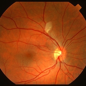
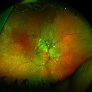
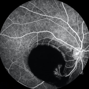 Sub-ILM Hemorrhage with Neovessels
Apr 30 2020 by Saurabh Deshmukh, MBBS, DNB, FVRS, MNAMS
Late arteriovenous phase FA showing a large sub-internal limiting membrane hemorrhage with overlying neovessels. This hypertensive patient presented with a visual acuity of counting fingers at 2 meters. The patient was advised intravitreal anti-VEGF injection, Nd: YAG Membranotomy, and systemic control of hypertension.
Photographer: Saurabh Deshmukh, Sri Sankaradeva Nethralaya, Guwahati, India
Imaging device: Topcon TRC-50 DX
Condition/keywords: hypertensive retinopathy, neovascularization elsewhere (NVE), subILM hemorrhage
-
 Siegrist Streak
Nov 6 2012 by F. Ryan Prall, MD
32-year-male with history of hypertension, recent admission for malignant hypertension.
Photographer: Tom Egnatz, Indiana University
Condition/keywords: hypertensive retinopathy, malignant hypertension
-
 Malignant Hypertension
Oct 23 2012 by Larry Halperin, MD
Malignant hypertension
Condition/keywords: hypertension, malignant hypertension
-
 Cotton Wool Spot
Jul 10 2013 by Jason S. Calhoun
Fundus photograph shows a young male with a single cotton wool spot just nasal to the macula in the right eye.
Photographer: Jason S. Calhoun, Department of Ophthalmology, Mayo Clinic Jacksonville, Florida
Condition/keywords: cotton wool spots, hypertension
-
 Oliferative Diabetic Retinopathy, Neovascularization of Disc
Mar 28 2018 by awaneesh m upadhyay, MBBS, DNB
Fundus photograph of right eye of a 56-year-old gentleman having diabetes, hypertension and chronic kidney disease.
Photographer: Dr Awaneesh Upadhyay
Imaging device: Zeiss
Condition/keywords: proliferative diabetic retinopathy (PDR)
-
 Hypertensive Retinopathy
Feb 25 2013 by Suber S. Huang, MD, MBA, FASRS
32-year-old African American male with Grade IV hypertensive retinopathy and acute renal failure. Vision OD 20/70, OS 20/25. Creatine 7.1. BP: 250/150.
Photographer: Geoffrey Pankhurst, University Hospitals, Eye Institute/Dept. Ophthalmology and Visual Sciences Case Western Reserve University Cleveland, OH
Imaging device: Topcon TRC 50x
Condition/keywords: acute renal failure, disc edema, exudate, hypertension, hypertensive retinopathy, ischemia, macular edema, macular ischemia, optic disc edema
-
 Serous Retinal Detachment Due to Systemic Hypertension (Pre-Eclampsia)
Mar 13 2013 by Jose Dalma-Weiszhausz, MD
Fundus photo of 22-year-old female, 8 months pregnant with severe poorly controlled hypertension due to pre-eclampsia.
Photographer: José Dalma, MD, Dalma & Asoc.. Mexico City, Mexico
Condition/keywords: hypertension, preeclampsia, serous retinal detachment
-
 OCT in Patient With IIH Showing Thickened RNFL
Jan 16 2019 by John S. King, MD
18-year-old African American female with increased BMI with a history of headaches, nausea, transient diplopia and vision loss that she notices when getting up from her bed (and goes away after standing upright) for the last two weeks. Went to PCP and was treated for the flu, and after no improvement and visual symptoms known, was sent to ED. MRI did not show any masses and showed empty sella turcia. Vision 20/30 OD and 20/20 OS; no RAPD; IOP 15OU; no anterior segment or vitreous inflammation; discs are elevated with obscuration of the disc margins and some of the smaller vessels; there are no SVPs; there are mild Patton's lines temporally (see Initial Photos). The optic disc cube shows 360 degrees of RNFL thickening (see OCT). Was referred to near-ophthalmologist, Dr. Doyle. She obtained additional work-up, and LP opening pressure was high, and MRV showed bilateral transverse sinus stenosis. Patient showed steady improvement with medical therapy, that included weight loss and oral diamox. On her last visit with Dr. Doyle, vision has remained stable at 20/20-20/25 without an enlarged blindspot; there are SVPs and optic disc edema has resolved (see Post Treatment Photos); she is currently on 1000 mg of diamox and has lost 15 pounds, and no stinting procedure needed.
Imaging device: Cirrus
Condition/keywords: benign idiopatic intracranial hypertension, optic disc edema, papilledema
-
 Hypertensive Retinopathy
Oct 20 2012 by Hyung-Woo Kwak, MD
The sudden appearance of cotton wool spots with hypertension retinopathy is known as accelerated hypertension. This patient has acute hypertension of rapid onset and measured systolic blood pressure was more than 200mmhg at this time.
Imaging device: Zeiss F450 plus
Condition/keywords: cotton wool spots, hypertension
-
 Hypertensive Retinopathy
Feb 25 2013 by Suber S. Huang, MD, MBA, FASRS
32-year-old African American male with Grade IV hypertensive retinopathy and acute renal failure. Vision OD 20/70, OS 20/25. Creatine 7.1. BP: 250/150.
Photographer: Geoffrey Pankhurst, University Hospitals, Eye Institute/Dept. Ophthalmology and Visual Sciences Case Western Reserve University Cleveland, OH
Imaging device: Topcon TRC 50x
Condition/keywords: acute renal failure, disc edema, exudate, hypertension, hypertensive retinopathy, ischemia, macular edema, macular ischemia, optic disc edema
-
 Papilledema
Sep 21 2012 by Suber S. Huang, MD, MBA, FASRS
Fundus photograph of a 24-year-old obese woman with severe papilledema secondary to idiopathic intracranial hypertension.
Condition/keywords: dilated tortuous vessels, exudate, idiopathic intracranial hypertension, Paton's lines, peripapillary hemorrhage, pseudotumor cerebri
-
 Hypertensive Choroidopathy
Dec 7 2012 by F. Ryan Prall, MD
32-year-old male with decreased vision, admitted for malignant hypertension.
Photographer: Tom Egnatz, Indiana University
Condition/keywords: hypertensive choroidopathy, malignant hypertension
-
 Hypertensive Retinopathy
Mar 29 2013 by Henry J. Kaplan, MD
Cotton wool spot in grade III hypertensive retinopathy.
Condition/keywords: hypertension, hypertensive retinopathy
-
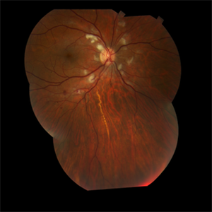
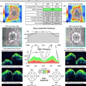
 Mild Patton's Lines in IIH - Initial Photos
Jan 16 2019 by John S. King, MD
18-year-old African American female with increased BMI with a history of headaches, nausea, transient diplopia and vision loss that she notices when getting up from her bed (and goes away after standing upright) for the last two weeks. Went to PCP and was treated for the flu, and after no improvement and visual symptoms known, was sent to ED. MRI did not show any masses and showed empty sella turcia. Vision 20/30 OD and 20/20 OS; no RAPD; IOP 15OU; no anterior segment or vitreous inflammation; discs are elevated with obscuration of the disc margins and some of the smaller vessels; there are no SVPs; there are mild Patton's lines temporally (see Initial Photos). The optic disc cube shows 360 degrees of RNFL thickening (see OCT). Was referred to near-ophthalmologist, Dr. Doyle. She obtained additional work-up, and LP opening pressure was high, and MRV showed bilateral transverse sinus stenosis. Patient showed steady improvement with medical therapy, that included weight loss and oral diamox. On her last visit with Dr. Doyle, vision has remained stable at 20/20-20/25 without an enlarged blindspot; there are SVPs and optic disc edema has resolved (see Post Treatment Photos); she is currently on 1000 mg of diamox and has lost 15 pounds, and no stinting procedure needed.
Photographer: Gretchen Harper
Imaging device: Topcon 50
Condition/keywords: idiopathic intracranial hypertension, optic disc edema, papilledema, Patton's Lines
-
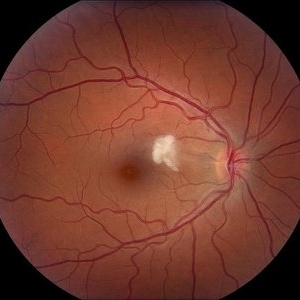
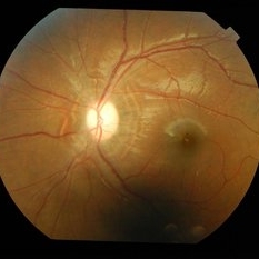 Post Papilledema High- Water Marks
Sep 22 2014 by Mallika Goyal, MD
Left fundus of a 21-year-old lady with benign intracranial hypertension shows circumpapillary high-water marks 2 months after initiating oral steroidsfor elevated intracranial pressure with severe papilledema. These marks identify extent of prior retinal elevation due to disc edema.
Photographer: Mallika Goyal, MD, Apollo Health City, Jubilee Hills, Hyderabad-500033
Condition/keywords: papilledema
-
 Incomplete Central Retinal Vein Occlusion V Papillophlebitis OD
Feb 20 2013 by From the Collections of Thomas M. Aaberg, MD and Thomas M. Aaberg Jr., MD
46-year-old; 20/20 OU; hypertension.
Condition/keywords: central retinal vein occlusion (CRVO), papillophlebitis
-
 Paracentral Acute Middle Maculopathy (PAMM)
Mar 21 2019 by Jonathan C. Tsui, MD
26-year-old female with hypertension presenting with chief complaint of "darkening" in her nasal visual field in the right eye. No flashes, floaters, or vision loss. Va 20/60 and nasal VF defect OD. SD-OCT demonstrated hyperreflectivity in the INL consistent with paracentral acute middle maculopathy. She was referred to her PCP for blood pressure optimization and a cardiovascular work-up. She returned for follow-up two months later with 20/80 OD, 20/20 OS. Repeat SD-OCT demonstrated inner retinal atrophy.
Photographer: Zellers, Diane
Condition/keywords: paracentral acute middle maculopathy
-
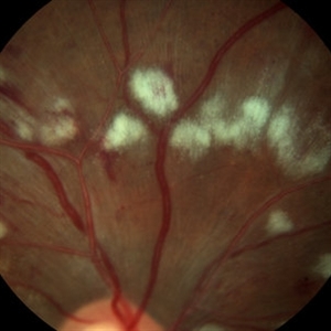 Cotton Wool Spots
Mar 1 2014 by Homayoun Tabandeh, MD, FASRS
Cotton wool spots in a patient with hypertension and diabetes
Condition/keywords: cotton wool spots, diabetic retinopathy, hypertensive retinopathy
-
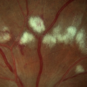 Cotton Wool Spots
Mar 1 2014 by Homayoun Tabandeh, MD, FASRS
Cotton wool spots in a patient with hypertension and diabetes.
Condition/keywords: cotton wool spots, diabetic retinopathy, hypertensive retinopathy
-
 HTN / Macular Star / CWS
Feb 17 2015 by David Callanan, MD
HTN / Macular star / CWS.
Condition/keywords: cotton wool spots, hypertension, macular star
-
 Branch Retinal Vein Occlusion
Oct 17 2012 by Sharon Fekrat, MD FACS FASRS
Fluorescein angiography of an inferior perfused branch retinal vein occlusion in the left eye of a middle aged male with hypertension. The foveal avascular zone is irregular. Subretinal hemorrhage is present.
Photographer: John Reaves, Ophthalmic Photographer, Durham VA Medical Center Eye Clinic Imaging Suite, Durham, NC
Imaging device: Fluorescein Angiography
Condition/keywords: branch retinal vein occlusion (BRVO), subretinal hemorrhage
-
 Hypertensive Retinopathy
Jun 28 2013 by Jason S. Calhoun
Patient came in complaining of spots in vision in both eyes. VA was 20/25 - right eye and 20/20- left eye. Fundus exam reveals little hemorrhages with cotton wool spots due to hypertension and anemia.
Photographer: Jason S. Calhoun, Mayo Clinic Jacksonville, Florida
Imaging device: TOPCON TRC 50-EX
Condition/keywords: hypertensive retinopathy
-
 Malignant Hypertension
Jan 29 2015 by H. Michael Lambert, MD
Malignant hypertension with cotton wool spots and flame hemorrhages.
Condition/keywords: cotton wool spots, hemorrhage, malignant hypertension
-
 HTN / Macular Star / CWS
Feb 17 2015 by David Callanan, MD
HTN / Macular star / CWS.
Condition/keywords: cotton wool spots, hypertension, macular star
-
 Hypertensive Retinopathy, Right
Feb 23 2017 by Alla Goldberg, MD
Fundus photograph of 35-year-old man with severe hypertension (182/128).
Photographer: Sofia Rutiaga, UT Health McGovern Medical School, Cizik Eye Clinic
Condition/keywords: cotton wool spots, Elschnig's spots, hypertensive choroidopathy, hypertensive retinopathy, serous retinal detachment
 Loading…