Search results (191 results)
-
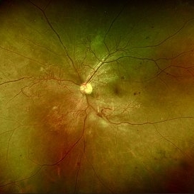
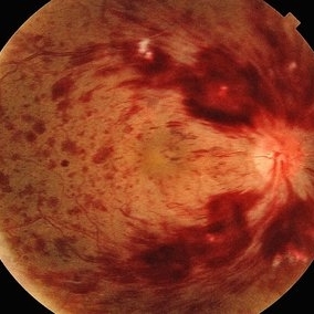
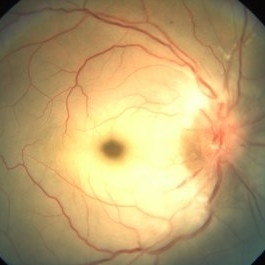
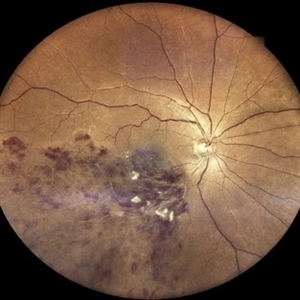 Branch Retinal Vein Occlusion
Mar 10 2025 by T. P . VIGNESH, MBBS,MS
Fundus photograph of a 52-year-old man with history of hypertension for the past 10 years revealing Inferotemporal branch retinal vein occlusion (ITBRVO) with macular edema.
Photographer: Sivanath
Imaging device: EIDON
Condition/keywords: branch retinal vein occlusion (BRVO)
-
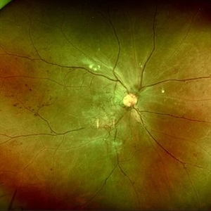
 Central Retinal Vein Occlusion
Feb 25 2025 by Prithvi Chandrakanth
A 61-year-old woman with a history of hypertension noticed a sudden painless blurring of vision in her left eye. Over the next few days, the blurriness persisted, and she experienced a mild central scotoma. On examination, Fundoscopic evaluation revealed dilated, tortuous retinal veins, retinal hemorrhages, and macular oedema.
Photographer: DR.PRITHVI CHANDRAKANTH, DR.CHANDRAKANTH NETHRALAYA, KOZHIKODE
Imaging device: EIDON
Condition/keywords: CRVO, CRVO WITH MACULA EDEMA, flame shaped retinal hemorrhage
-
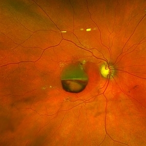
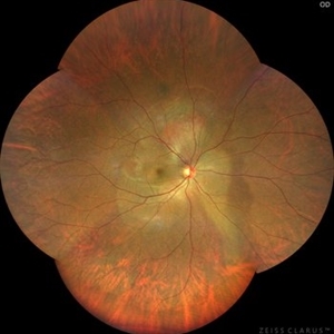 Vortex-pattern Exudative Retinal Detachment
Feb 22 2025 by CUI YUELING
Patient: Male, 40 years old. Chief Complaint: Blurred vision and metamorphopsia in the left eye for more than 10 days. Past Medical History Hypertension for 4 years, with a highest recorded blood pressure of 160/80 mmHg. Currently controlled with oral "Nifedipine Sustained-Release Tablets, 2 tablets daily." Long-term history of heavy alcohol consumption and smoking. Ophthalmic Examination: Visual Acuity: Right eye (OD): 0.4 (uncorrected, no improvement with correction). Left eye (OS): 0.5 (-1.5DS = 1.0). Intraocular Pressure (IOP): OD: 15 mmHg. OS: 17 mmHg. Anterior Segment:Unremarkable. Fundus Examination: Right eye: Optic disc margins are clear, with a slightly reddish hue. Cup-to-disc ratio (C/D) = 0.2. A scalloped, orange-red elevated lesion is observed superior to the optic disc, with anterior displacement of the focal point. This is accompanied by a secondary, turbine-like exudative retinal detachment centered around the optic disc, involving the macula. The macular region shows scattered punctate yellow-white exudates. Diagnosis: Choroidal hemangioma with secondary exudative retinal detachment(OD).
Photographer: Cui yueling The First People's Hospital of Zunyi, Guizhou, Zunyi, China
Imaging device: Zeiss Clarus 500
Condition/keywords: choroidal hemangioma, exudative retinal detachment
-
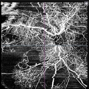
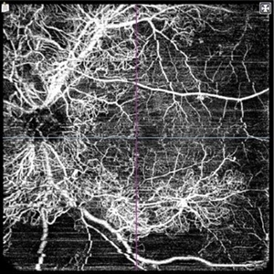 Vascular maze- Proliferative Diabetic Retinopathy
Feb 7 2025 by Hemanth Murthy, MBBS, MD, FASRS
OCTA image left eye. A 32 year male with history of blurring of vision in right eye since 4 months. History of Diabetes and Hypertension since 2 years. Vision 6/36 in right eye and 6/9 in left eye
Photographer: Veda Vyas
Condition/keywords: proliferative diabetic retinopathy (PDR)
-
 Vascular Maze-Proliferative Diabetic Retinopathy
Feb 7 2025 by Hemanth Murthy, MBBS, MD, FASRS
OCTA image right eye-A 32 year male with history of blurring of vision in right eye since 4 months. History of Diabetes and Hypertension since 2 years. Vision 6/36 in right eye and 6/9 in left eye
Photographer: Veda Vyas
Condition/keywords: OCT Angiography, proliferative diabetic retinopathy (PDR)
-
 Vascular Maze-Proliferative Diabetic Retinopathy
Feb 7 2025 by Hemanth Murthy, MBBS, MD, FASRS
Fundus photo left eye. A 32 year male with history of blurring of vision in right eye since 4 months. History of Diabetes and Hypertension since 2 years. Vision 6/36 in right eye and 6/9 in left eye
Photographer: Veda Vyas
Condition/keywords: proliferative diabetic retinopathy (PDR)
-
 Vascular Maze-Proliferative Diabetic Retinopathy
Feb 7 2025 by Hemanth Murthy, MBBS, MD, FASRS
Fundus photo of right eye. A 32 year male with history of blurring of vision in right eye since 4 months. History of Diabetes and Hypertension since 2 years. Vision 6/36 in right eye and 6/9 in left eye
Photographer: Veda Vyas
Condition/keywords: proliferative diabetic retinopathy (PDR)
-
 Treatment of Ocular Ischemic Syndrome with Hyperbaric Oxygen Therapy
Dec 2 2024 by Catherine S Kang
A 66-year-old female with past medical history significant for hypertension and ocular ischemic syndrome. She presented in emergency department (ED) reporting eye pain and blurred vision in both eyes since earlier that morning. On examination, best corrected visual acuity in each eye was counting fingers (20cm). Further investigation was performed and fluorescein angiography revealed a delay in choroidal filling. The patient has been followed for ocular ischemic syndrome since the onset of the condition and hyperbaric oxygen therapy was promptly initiated. Final best corrected visual acuity was 20/150 and macula developed atrophy.
Photographer: Catherine Kang
Condition/keywords: hyperbaric oxygen therapy, ocular ischemic syndrome
-
 Advanced RP
Nov 5 2024 by rahul saradge
A man, 58, arrived complaining of BOV for both near and distance vision in both eyes, with a 6/9 BCVA in each eye. For a year, the patient had been taking medication for both diabetes and hypertension. In both eyes, the dilated ophthalmoscopic retina revealed waxy disc pallor paired with bony spicules in the mid-periphery. The patient was prescribed spectacles and given counseling regarding the nature of the illness.
Photographer: Lokesh Dukare ,Isha Netralaya Thane
Imaging device: optos
Condition/keywords: bone spicule, optic disc pallor, Optos, Retinitis Pigmentosa
-
 Hypertensive Retinopathy
Oct 27 2024 by César Adrián Gómez Valdivia, MD
Fundus photograph of a 62 year-old woman with history of untreated hypertension and chronic kidney disease. Findings were bilateral.
Photographer: @eyemissu2
Imaging device: TOPCON TRC-50DX
Condition/keywords: hypertensive retinopathy
-
 Ruptured Retinal Arterial Macro-Aneurysm
Oct 27 2024 by César Adrián Gómez Valdivia, MD
Ruptured retinal arterial macro-aneurysm found in a 56 YO female patient with history of untreated hypertension. Round or fusiform dilation of a retinal arteriole is usually seen within a third degree branch of one of the four main arcade arteries. Most common location for a symptomatic macroaneurysm is from a branch of the superotemporal arcade.
Photographer: @eyemissu2
Imaging device: TOPCON TRC-50DX
Condition/keywords: ruptured macroaneurysm
-
 Branch Retinal Artery Occlusion
Oct 1 2024 by Angel Enrique Flores Pineda
Fundus photograph of a 78-year-old woman with poorly controlled systemic arterial hypertension and dyslipidemia. Hollenhorst plaque can be observed.
Photographer: Angel Enrique Flores Pineda, Hospital General de Zona #20
Imaging device: Smartphone (IPhone 15 plus)
Condition/keywords: branch retinal artery occlusion (BRAO)
-
 Subhyaloid Hemorrhage
Jul 31 2024 by Arthi Mohankumar , MS,MRCS ED, FICO,FAICO
A 35 year old male presented with complaints of seeing a black spot in left eye for past one day after working out in the gym the previous day. He has history of uncontrolled diabetes and hypertension. Fundus exam of the left eye revealed a sub hyaloid hemorrhage nasal to the disc with minimal background Diabetic and hypertensive changes. His baseline CBG was 200 mg/dl and BP was 170/100 He was suggested observation initially considering the nasal location. But patient found the scotoma very disturbing and eventually underwent yag hyaloidotomy
Photographer: Arthi Mohankumar
Condition/keywords: Sub hyaloid haemorrhage, valsalva retinopathy
-
 Central Retinal Vein Occlusion
Jul 21 2024 by César Adrián Gómez Valdivia, MD
Central Retinal Vein Occlusion found in a 72 year old patient with history of uncontrolled Hypertension. Non-Ischemic Variant.
Photographer: Erika Paulina Ornelas Cazares
Imaging device: TOPCON TRC-50DX
Condition/keywords: central retinal vein occlusion (CRVO)
-
 Macular OCT Image of a Patient With Central Retinal Artery Occlusion
Jul 7 2024 by Thiago Mazzeo
This is a macular OCT image of a patient that presented sudden visual loss in the right eye (Light perception) after leaving the hospital due to uncontrolled systemic arterial hypertension.
Photographer: Thiago Mazzeo
Imaging device: Zeiss Cirrus 5000
Condition/keywords: Central Retinal Artery Occlusion, macular changes, OCT
-
 Hypertensive Retinopathy
May 1 2024 by Marco Antonio Sauza
36 year old male with uncontrolled systemic hypertension.
Photographer: MARCO SAUZA CASTILLEJOS
Imaging device: VISUCAM ZEISS
Condition/keywords: choroidal infarction, hypertensive retinopathy, macular star
-
 Hypertensive Retinopathy Grade IV
May 1 2024 by Marco Antonio Sauza
Fundus photograph of an 36-year-old male with uncontrolled systemic hypertension, >200/100mmhg, presenting decreased vision in the left eye.
Photographer: MARCO SAUZA CASTILLEJOS
Imaging device: VISUCAM ZEISS
Condition/keywords: choroidal infarction, hypertensive retinopathy, macular star
-
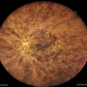
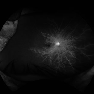
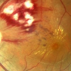
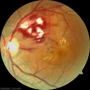
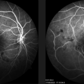 Buds on Tree Appearance on FFA: Old BRVO
Mar 12 2024 by MEENAL SONI
A middle-aged man with idiopathic hypertension presented with old IT BRVO, sclerosed vein on with hemorrhages on fundus examination. FFA reveals delayed filling of vein with pruning of venules resembling buds on tree.
Photographer: Dr. Meenal Soni, Fellow VR, ASG eye Hospital Jodhpur
Imaging device: ZEISS Visucam 400
Condition/keywords: non-perfused branch retinal vein occlusion (BRVO)
-
 Central Retinal Artery Occlusion
Mar 11 2024 by Dr.Pavithra Subramanian
A 51 year old male with defective vision in right eye for past 4days.On examination RE RAPD present and Fundus examination found to be Right eye Central retinal artery occlusion with Grade 4 Hypertensive Retinopathy.
Photographer: Dr Pavithra Subramanian
Condition/keywords: central retinal artery occlusion (CRAO), malignant hypertension
-
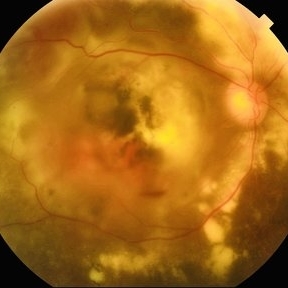
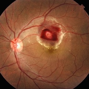
 Ruptured Macroaneurysm
Mar 6 2024 by Mari Ann Z. Keithahn, MD, FASRS
Fundus Photograph of 73 year-old female with hypertension.
Photographer: JaTori Maxwell, Missouri Retina Consultants, PC
Imaging device: OPTOS Silverstone
Condition/keywords: Rupture Macroaneurysm
-
 Blistered Retina
Jan 27 2024 by prathibha hande, MS DNB
Fundus photo of a 32 year old male presenting with blurred vision. Undiagnosed renal hypertension. Blood pressure at the time of presentation 210/120 mmhg.
Photographer: Mr Prathap K
Imaging device: Mirante SLO fundus camera
Condition/keywords: hypertensive choroidopathy
-
 CRAO
CRAO
Jan 8 2024 by ANKIT JAIN
RIGHT EYE FUNDUS IMAGE OF A 68 YEARS OLD MALE WITH SUDDEN LOSS OF VISION, WHO IS A KNOWN CASE OF HYPERTENSION FOR 15 YEARS
Photographer: Dr Ankit Jain
Condition/keywords: central retinal artery occlusion (CRAO), cherry red spot
-
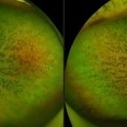
 Malignant Hypertension
Sep 28 2023 by ANKIT JAIN
Fundus photograph of RE of a 13 year old male with malignant hypertension
Photographer: Dr. Ankit Jain
Imaging device: Mirante
Condition/keywords: hypertensive retinopathy, malignant hypertension
-
 Malignant Hypertension
Sep 28 2023 by ANKIT JAIN
Fundus photograph of LE of a 13 year old male with malignant hypertension
Photographer: Dr. Ankit Jain
Imaging device: Mirante
Condition/keywords: hypertensive retinopathy, malignant hypertension
-
 Malignant Hypertension
Sep 28 2023 by ANKIT JAIN
Widefield photograph of LE of a 13 year old male with malignant hypertension
Photographer: Dr. Ankit Jain
Imaging device: Mirante
Condition/keywords: hypertensive retinopathy, malignant hypertension
 Loading…