Search results (5 results)
-
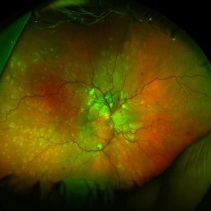
 Blistered Retina
Jan 27 2024 by prathibha hande, MS DNB
Fundus photo of a 32 year old male presenting with blurred vision. Undiagnosed renal hypertension. Blood pressure at the time of presentation 210/120 mmhg.
Photographer: Mr Prathap K
Imaging device: Mirante SLO fundus camera
Condition/keywords: hypertensive choroidopathy
-
 Vein Occlusion Zoom in a BRVO
Apr 29 2020 by Gabriel Castilho S Barbosa, MD
A zoomed vein occlusion in a young patient with systemic arterial hypertension.
Photographer: Gabriel Castilho, Suel Abujamra Institute, São Paulo.
Condition/keywords: branch retinal vein occlusion (BRVO), macular branch retinal vein occlusion (BRVO), non-perfused branch retinal vein occlusion (BRVO)
-
 Small subILM Hemorrhage
Oct 26 2019 by Navneet Mehrotra, DNB
44-year-old hypertensive male with sudden decrease in vision showing small sub ILM hemorrhage at macula.
Photographer: Navneet Mehrotra
Imaging device: NidekRS330
Condition/keywords: hypertension, subILM hemorrhage
-
 Hypertensive Retinopathy, Right
Feb 23 2017 by Alla Goldberg, MD
Fundus photograph of 35-year-old man with severe hypertension (182/128).
Photographer: Sofia Rutiaga, UT Health McGovern Medical School, Cizik Eye Clinic
Condition/keywords: cotton wool spots, Elschnig's spots, hypertensive choroidopathy, hypertensive retinopathy, serous retinal detachment
-
 Chronical Submacular Hemorrhage in the Setting of Neovascular AMD
Mar 23 2015 by Rita Couceiro, MD, MS
An 80-year-old male, with a history of hypertension and high cholesterol, complained of acute and painless vision loss in his left eye (OS) in the previous 5 months. On observation best corrected visual acuity in OS was hand motion. A dense vitreous opacity in OS precluded fundus examination. Ocular ultrasound revealed vitreous hemorrhage and thickening of the macular area. The patient was submitted to pars plana vitrectomy, which disclosed a large submacular hemorrhage with chronical features and disciform scarring in the setting of neovascular AMD.
Imaging device: Intraoperative fundus photograph
Condition/keywords: neovascular age-related macular degeneration (AMD), submacular hemorrhage, wet age-related macular degeneration (wet AMD)
 Loading…