Search results (421 results)
-
 Busacca nodules
May 2 2013 by Henry J. Kaplan, MD
Typical Busacca iris stromal nodules in sarcoid uveitis; notice the ps formation.
Condition/keywords: busacca nodulaes, granulomatous uveitis, iris nodules, sarcoid bussaca iris nodules
-
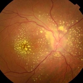
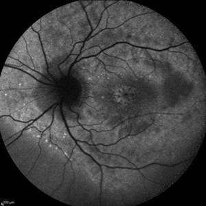
 Siegrist Streak
Nov 6 2012 by F. Ryan Prall, MD
32-year-male with history of hypertension, recent admission for malignant hypertension.
Photographer: Tom Egnatz, Indiana University
Condition/keywords: hypertensive retinopathy, malignant hypertension
-
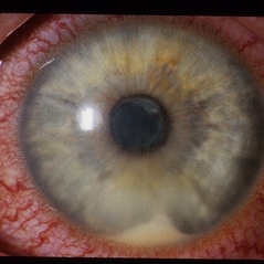
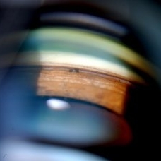
 Ozurdex implant
Aug 23 2012 by Daniel A. Adelberg, MD, FASRS
Anterior Segment photograph of a 50 year old with Uveitis and Cystoid Macular Edema status post Intravitreal injection of an Ozurdex dexamethasone implant
Photographer: Robert Ramsey, Southwestern Eye Center, Mesa Arizona
Condition/keywords: Ozurdex implant
-
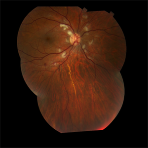
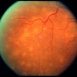
 Uveitis Posterior
Jul 19 2019 by JEFFERSON R SOUSA, Tecg.º (Biomedical Systems Technology)
A 23-year-old male patient attended the clinic with low vision of the right eye. In the evaluation it presented important fundoscopical alterations like retinal exudations in the posterior pole and nasal retina, aspects of macular star. It was proven that it was a posterior uveitis.
Photographer: JEFFERSON R SOUSA - Study Center and Ophthalmological Research Dr. Andre M V Gomes, Institute Dr. Suel Abujamra São Paulo-Brazil
Imaging device: Topcon TRC-50 DX, Imaginet 4.0, angle de 50 graus. Flash 50w-s
Condition/keywords: uveitis
-
 Toxoplasmosis Uveitis with Mutton Fat KP and Broken Posterior Synechia
Oct 10 2012 by Jeffrey G. Gross, MD, FASRS
Toxooplasmosis uveitis with mutton fat KP and broken posterior synechia.
Condition/keywords: mutton-fat keratic precipitates (KP), posterior synechiae, toxoplasmosis uveitis
-
---thumb.jpg/image-square;max$300,300.ImageHandler) Multifocal Choroiditis and Panuveitis Syndrome
Multifocal Choroiditis and Panuveitis Syndrome
Feb 26 2013 by Henry J. Kaplan, MD
Multifocal choroiditis, left eye: multiple punched out scar formations in the posterior pole.
Condition/keywords: multifocal choroiditis, panuveitis
-
 Epiretinal Membrane
Oct 11 2012 by Michael P. Kelly, FOPS
This is a patient with idiopathic panuveitis who developed a visually significant epiretinal membrane. Pars plana vitrectomy with membrane peeling was performed to remove the epiretinal proliferation. I recommend magnifying the image to see the exquisite detail centrally.
Photographer: Michael P. Kelly, FOPS Director, Duke Eye Center Labs, Duke Universtiy Hospital
Imaging device: Zeiss 450Plus
Condition/keywords: epiretinal membrane (ERM), panuveitis
-
---thumb.jpg/image-square;max$300,300.ImageHandler) Multifocal Choroiditis & Panuveitis Syndrome
Multifocal Choroiditis & Panuveitis Syndrome
Feb 26 2013 by Henry J. Kaplan, MD
Multifocal choroiditis (MFC) and panuveitis.
Condition/keywords: multifocal choroiditis, panuveitis
-
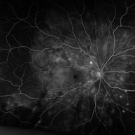 "Starry Sky" Fundus in Vogt-Koyanaki-Harada Syndrome
Jan 10 2018 by Peter H. Tang, MD, PhD
Fluorescein angiography imaging of a 27-year-old male with acute inflammation as part of Vogt-Koyanagi-Harada Syndrome.
Imaging device: Optos California
Condition/keywords: chorioretinal inflammations, retina, uveitis, Vogt-Koyanagi-Harada
-
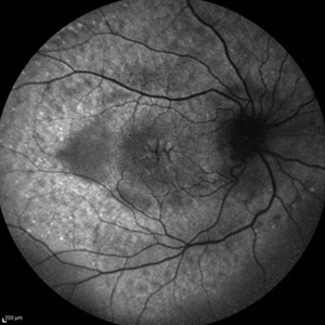
 Cystoid Macular Edema (CME)
Sep 11 2012 by Hamid Ahmadieh, MD
Autofluorescence imaging of the left eye of a 17-year-old boy with chronic intermediate uveitis showing CME.
Photographer: Hamid Ahmadieh, MD, Ophthalmic Research Center, Labbafinejad Medical Center, Shahid Beheshti University of Medical Sciences
Imaging device: Heidelberg Spectralis
Condition/keywords: autofluorescence imaging, cystoid macular edema (CME), intermediate uveitis
-
 BSC CME OS
Nov 10 2012 by Pauline T Merrill, MD, FASRS
Fundus photograph left eye of a 42-year-old Caucasian male with birdshot retinochoroidopathy (HLA-A29+) and cystoid macular edema (CME)
Condition/keywords: birdshot retinochoroidopathy, cystoid macular edema (CME), posterior uveitis, uveitis
-
---thumb.jpg/image-square;max$300,300.ImageHandler) Multifocal Choroiditis and Panuveitis Syndrome
Multifocal Choroiditis and Panuveitis Syndrome
Feb 26 2013 by Henry J. Kaplan, MD
Multifocal choroiditis and panuveitis: left eye. Acute stage: haziness of the media due to vitritis and multiple active yellow and also inactive choroidal lesions are present.
Condition/keywords: multifocal choroiditis
-
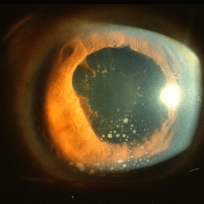
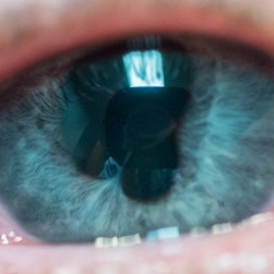
 HLA-B27 Associated Uveitis
Jun 4 2014 by Henry J. Kaplan, MD
Severe anterior uveitis with fibrinous reaction and hypopyon formation related to HLA-B27. Notice the membrane on the lens surface.
Condition/keywords: acute anterior uveitis, HLA-B27, hypopyon
-
 Cystoid Macular Edema (CME)
Sep 11 2012 by Hamid Ahmadieh, MD
Fundus autofluorescence (FAF) of the right eye a 17-year-old boy with chronic intermediate uveitis showing CME.
Photographer: Hamid Ahmadieh, MD, Ophthalmic Research Center, Labbafinejad Medical Center, Shahid Beheshti University of Medical Sciences
Imaging device: Heidelberg Spectralis
Condition/keywords: cystoid macular edema (CME), fundus autofluorescence (FAF), intermediate uveitis
-
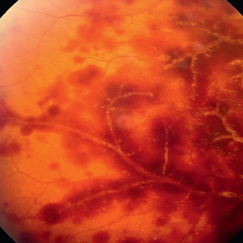 Acute Retinal Periphlebitis and Panuveitis OD
Jul 16 2014 by Deepayan Kar
Hemorrhagic retinopathy + frosted retinal angitis OD. Exudative sheathing of the major retinal blood vessels Px 25-year-old woman (VA-OD 20/40/ OS 20/25): recovering from URTI. AC ++ Vit Cells +.
Photographer: Deepayan Kar
Condition/keywords: exudative sheathing, frosted branch angiitis, hemorrhage, retinopathy
-
---thumb.jpg/image-square;max$300,300.ImageHandler) Multifocal Choroiditis & Panuveitis Syndrome
Multifocal Choroiditis & Panuveitis Syndrome
Feb 26 2013 by Henry J. Kaplan, MD
Multifocal choroiditis, MFC. Lesions look similar to POHS but the patient has vitritis in contrast to the former.
Condition/keywords: multifocal choroiditis, panuveitis
-
 Iritis With Breaking Posterior Synechiae
Jul 4 2013 by Nathan E. Podoll
Iritis with breaking posterior synechiae.
Photographer: Nathan E. Podoll, M.D., University of Louisville
Condition/keywords: acute anterior uveitis, posterior synechiae
-
 Serpiginous
Aug 29 2012 by F. Ryan Prall, MD
70-year-old female with reactivation of serpiginous choroiditis.
Photographer: Tom Egnatz, Indiana University
Condition/keywords: serpiginous choroiditis
-
 Tuberculosis Panuveitis
Feb 25 2013 by Henry J. Kaplan, MD
Peripheral vascular sheathing and multiple choroidiris foci in a patient with tuberculosis panuveitis.
Condition/keywords: panuveitis, tuberculosis
-
 Gonioscopy; Scattered Peripheral Anterior Synechiae
Jul 8 2013 by Jason S. Calhoun
Patient came in for evaluation for glaucoma. Patient also has a history of uveitis. Last flare up was back in 1990. Patient's VA was 20/30, Right eye and 20/40-1, Left eye. Slit Lamp Gonioscopy reveals iris bow with scattered PAS around the angles of the anterior chamber. You can also see pigmentation in the trabecular meshwork. Patient will follow up in 3 months.
Photographer: Jason S. Calhoun, Department of Ophthalmology, Mayo Clinic Jacksonville, Florida
Condition/keywords: gonioscopy, goniosynechiae
-
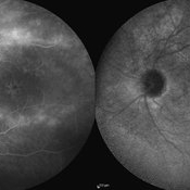 Cystoid Macular Edema (CME)
Sep 11 2012 by Hamid Ahmadieh, MD
Late phase FA & ICG angiography imagings of the left eye a 17-year-old boy with CME & retinal periphlebitis secondary to chronic intermediate uveitis.
Photographer: Hamid Ahmadieh, MD, Ophthalmic Research Center, Labbafinejad Medical Center, Shahid Beheshti University of Medical Sciences
Imaging device: Heidelberg Spectralis
Condition/keywords: cystoid macular edema (CME), indocyanine green (ICG) angiography, intermediate uveitis
-
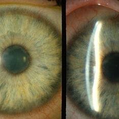 pupillary block; periph uveitis
Feb 14 2013 by From the Collections of Thomas M. Aaberg, MD and Thomas M. Aaberg Jr., MD
diffuse and slit-beam anterior segment photographs showing pupillary-block angle closure associated with uveitis. pupillary block; angle closure; uveitis; posterior synechiae
Condition/keywords: angle closure, posterior synechiae, pupillary block, uveitis
-
 Pars Planitis - Peripheral Uveitis
Nov 9 2012 by Norman Byer
This 25-year-old man had pars planitis, peripheral uveitis bilaterally. In this eye it produced a small tractional oval tear of the retina and an inferior retinal detachment. The typical creamy yellow exudates of pars planitis can be seen in the lower right very close to the ora serrata.
Condition/keywords: creamy yellow exudates, inferior retinal detachment, pars planitis, peripheral uveitis, tractional retinal tear
-
---thumb.jpg/image-square;max$300,300.ImageHandler) Multifocal Choroiditis & Panuveitis Syndrome
Multifocal Choroiditis & Panuveitis Syndrome
Feb 26 2013 by Henry J. Kaplan, MD
Multifocal choroiditis, MFC, old scars.
Condition/keywords: multifocal choroiditis, panuveitis
-
 Multifocal Choroiditis
Sep 16 2012 by Ivan R. Batlle, MD
71-year-old male with bilateral decreased vision and uveitis
Condition/keywords: disseminated chorioretinitis, generalized chorioretinitis, uveitis
 Loading…