Search results (13 results)
-
 Retinitis Pigmentosa Bullseye Appearing Autofluorescence
Feb 4 2025 by Isaac Agranoff
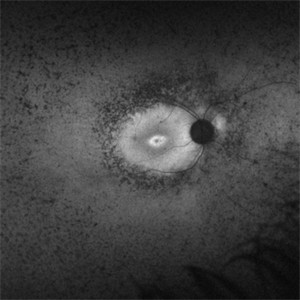
Fundus Autofluorescence of a 14-year-old boy with suspected RP. ERG performed afterwards was almost flat. VA measured at 20/30 but with extensive constriction of confrontational visual fields. Currently awaiting genetic testing.
Photographer: Isaac Agranoff
Imaging device: Optos California
Condition/keywords: fundus autofluorescence (FAF), retinitis pigmentosa, RP
-
 Neovascular vessels
Sep 22 2022 by Filip Kecer
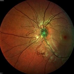
Multicolor widefield scan of a 16-year-old girl with a neovascularization from disc to vitreous space
Photographer: Filip Kecer, National Institute of Childrens Diseases
Imaging device: Spectralis, Heidelberg Engineering
Condition/keywords: neovascularization (NV), neovascularization at the disc, uveitis, vitreous
-
 Idiopathic retinal vasculitis, aneurysms and neuroretinitis
Apr 24 2022 by Aniruddha K Agarwal, MD
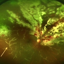
Ultra-wide field fundus fluorescein angiography (FFA) of the left eye from an asymptomatic, healthy 33-year-old woman who was referred to the retina clinic from a refractive surgery unit due to the presence of vascular anomalies and hard exudates in both eyes. FFA revealed the characteristic sacular aneurysms at the bifurcation of retinal arterioles in the posterior pole, together with microvascular anomalies and capillary closure peripherally.
Photographer: Julio J GONZALEZ-LOPEZ, MD, PhD, FEBO and Teresa GONZALEZ-LOMAS, RN
Imaging device: Optos California
Condition/keywords: IRVAN Syndrome, IUSG, neuroretinitis, retinal vasculitis, uveitis
-
 Stage 3 Vogt-Koyanagi-Harada (VKH) Disease
Oct 20 2021 by Bryon R McKay, MD, PhD, FRCSC, DRCPSC - Retina
27yF presented with sub-acute findings of VKH, she has an interesting pattern of perivascular changes. She was successfully treated with immunosuppressive agents and maintains 20/20 vision.
Photographer: Dr. Vaezi, University of British Columbia
Imaging device: Optos Imaging System
Condition/keywords: uveitis
-
 Multifocal Choroiditis and Panuveitis- Schlaegel lines
Nov 16 2021 by Manuel Ángel Alcántara Delgado, MD
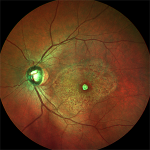
Optomap ultra-widefield retinal imaging of an 52-year-old woman showed multiple punched-out chorioretinal lesions and 2 rows of peripheral curvilinear pigmented chorioretinal streaks (Schlaegel lines).
Photographer: Manuel Ángel Alcántara Delgado. Conde de Valenciana.
Condition/keywords: multifocal choroiditis, myopia, retina, uveitis
-
 Serous Retinal Detachment in Vogt Koyanagi Harada Patient
Apr 26 2021 by Pablo Baquero Ospina, MD
24-year-old woman with bilateral panuveitis and serous retinal detachment, headache and tinnitus.
Photographer: Pablo Baquero-Ospina, Asociación Para Evitar la Ceguera en México
Imaging device: Heidelberg Spectralis
Condition/keywords: serous retinal detachment, Vogt-Koyanagi-Harada
-
 CMV Retinitis with Frosted Branch Angiitis
Sep 23 2020 by Nimesh A. Patel, MD, FASRS
Fundus photo showing peri-vascular inflammation of both arteries and veins with translucent exudation (yellow arrow). Superior nasally, there is classic retinal whitening with retinal hemorrhages superior. This patient was found to have a low CD4 count and a diagnosis of AIDS was made.
Condition/keywords: cytomegalovirus (CMV), HIV, uveitis
-
 Uveitis Posterior
Jul 19 2019 by JEFFERSON R SOUSA, Tecg.º (Biomedical Systems Technology)
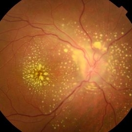
A 23-year-old male patient attended the clinic with low vision of the right eye. In the evaluation it presented important fundoscopical alterations like retinal exudations in the posterior pole and nasal retina, aspects of macular star. It was proven that it was a posterior uveitis.
Photographer: JEFFERSON R SOUSA - Study Center and Ophthalmological Research Dr. Andre M V Gomes, Institute Dr. Suel Abujamra São Paulo-Brazil
Imaging device: Topcon TRC-50 DX, Imaginet 4.0, angle de 50 graus. Flash 50w-s
Condition/keywords: uveitis
-
 Macular Scar Due to Cysticercosis Fundus Photo
Aug 8 2017 by Manuel A Paez-Escamilla, MD, FICO
Fundus photograph of a 69-year-old patient with a long history of eye inflammation and progressive decrease in vision. Multiple trips to Asia and eating undercooked pork.
Photographer: Mark Erickson CRA, COT. The Macula Center. Clearwater, Florida
Condition/keywords: cysticercosis, uveitis
-
 Suspected Multiple Evanescent White Dot Syndrome
Mar 3 2015 by Stuart Alfred, CRA, OCT-C
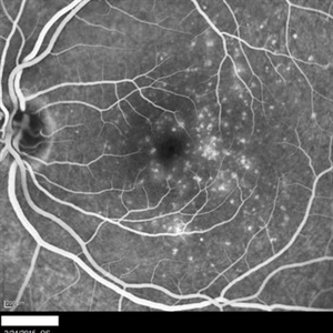
30 degree, late phase angiogram image of left fundus of a 28-year-old Caucasian female.
Photographer: Stuart Alfred, CRA, OCT-C, Midwest Eye Institute, Greenwood, Indiana
Imaging device: cSLO by Heidelberg engineering (Spectralis)
Condition/keywords: dry age-related macular degeneration (dry AMD), multiple evanescent white dot syndrome (MEWDS), punctate inner choroidopathy (PIC), uveitis
-
 Acute Retinal Periphlebitis and Panuveitis OD
Jul 16 2014 by Deepayan Kar
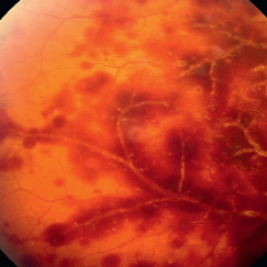
Hemorrhagic retinopathy + frosted retinal angitis OD. Exudative sheathing of the major retinal blood vessels Px 25-year-old woman (VA-OD 20/40/ OS 20/25): recovering from URTI. AC ++ Vit Cells +.
Photographer: Deepayan Kar
Condition/keywords: exudative sheathing, frosted branch angiitis, hemorrhage, retinopathy
-
---thumb.jpg/image-square;max$300,300.ImageHandler) Multifocal Choroiditis and Panuveitis (MCP) (OS)
Multifocal Choroiditis and Panuveitis (MCP) (OS)
Apr 1 2014 by Min Kim, MD, PhD, MBA, FASRS
Wide field fundus photograph of a 61-year-old female with decreased vision in both eyes due to multifocal choroiditis and panuveitis.
Condition/keywords: multifocal choroiditis, panuveitis
-
---thumb.jpg/image-square;max$300,300.ImageHandler) Central Retinal Vascular Obstruction
Central Retinal Vascular Obstruction
Oct 9 2013 by Maurice F. Rabb
Thirty five year old black male with low-grade uveitis. An unusual cause of central retinal vascular obstruction.
Condition/keywords: central retinal vascular obstruction
 Loading…