Search results (421 results)
-
 Ozurdex
Nov 22 2025 by Gabriel Costa Andrade, PhD
Anterior segment photograph of a Ozurdex implant in a 53-year-old man with macular edema due to intermediate uveitis.
Photographer: Gabriel Andrade
Condition/keywords: Ozurdex implant
-
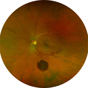
 Retinal Detachment with Macular Hole
Nov 10 2025 by Korey Starkey
A 58 year-old female presented with re-detached retina through macular hole. Planned for surgical intervention.
Photographer: Korey Starkey
Imaging device: Optos
Condition/keywords: gas bubble, intermediate uveitis, macular hole, pars plana vitrectomy (PPV), retinal detachment, scleral buckle, traction detachment
-
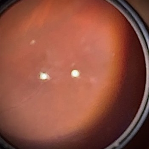
 Pearl in the Eye
Nov 7 2025 by Hemanth Murthy, MBBS, MD, FASRS
42 year male patient presented with sudden blurring in vision in left eye. His vision was 6/18 (OD) and 1/60 (OS). His IOP was normal with no signs of inflammation. His Fundus revealed healed posterior uveitis in both eyes. This is the image through operating microscope.
Photographer: Mr Vyas
Imaging device: Operating microscope
Condition/keywords: spontaneous lens dislocation
-
 Pearl in the Eye
Nov 7 2025 by Hemanth Murthy, MBBS, MD, FASRS
42 year male patient presented with sudden blurring in vision in left eye. His vision was 6/18 (OD) and 1/60 (OS). His IOP was normal with no signs of inflammation. His Fundus revealed healed posterior uveitis in both eyes. This is the image through operating microscope.
Photographer: Mr Vyas
Imaging device: Optos Daytona
Condition/keywords: spontaneous lens dislocation
-
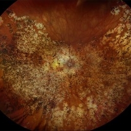 Multifocal Choroiditis with Panuveitis
Oct 16 2025 by Virginia Gebhart
39 year old female diagnosed with MCP in 2009. Extensive RPE changes and hypertrophy, arterial attenuation and pale nerve. Currently no active inflammation.
Photographer: Virginia Gebhart, Retina Consultants of Carolina
Imaging device: Optos California
Condition/keywords: corticosteroid-induced glaucoma, hypertrophy, multifocal chorioretinitis (MCP), PALE DISC
-
 Kyrieleis Plaques
Aug 18 2025 by Helder Vasconcelos
Kyrieleis Plaques and vitritis in a patient with posterior uveitis secondary to presumed ocular toxoplasmosis.
Photographer: Helder Vasconcelos
Imaging device: Smartphone Fundoscopy
Condition/keywords: kyrieleis plaques, toxoplasmosis chorioretinitis
-
 Synechiae
-
 Synechiae
-
 Synechiae
Jul 26 2025 by oren reuven
Uveitis causing Synechiae leading to Glaucoma in a 74 year-old male.
Photographer: oren reuven
Condition/keywords: synechiae
-
 Synechiae
Jul 26 2025 by oren reuven
Uveitis causing Synechiae leading to Glaucoma in a 74 year-old male.
Photographer: oren reuven
Condition/keywords: synechiae
-
 Synechiae
Jul 26 2025 by oren reuven
Uveitis causing Synechiae leading to Glaucoma in a 74 year-old male.
Photographer: oren reuven
Condition/keywords: synechiae
-
 Synechiae
Jul 26 2025 by oren reuven
Uveitis causing Synechiae leading to Glaucoma in a 74 year-old male.
Photographer: oren reuven
Condition/keywords: synechiae
-
 Synechiae
Jul 26 2025 by oren reuven
74 year-old male, 4 years of uveitis casing Synechiae that closed the chamber angle and caused glaucoma. Suffering minor field loss
Photographer: oren reuven
Imaging device: EIDON
Condition/keywords: glaucoma and uveitis
-
 Synechiae
Jul 26 2025 by oren reuven
74 year-old male, 4 years of uveitis casing Synechiae that closed the chamber angle and caused glaucoma. Suffering minor field loss.
Photographer: oren reuven
Imaging device: EIDON
Condition/keywords: synechiae, uveitis
-
 Large Subhyaloid Hemorrhage
Jul 11 2025 by Jessilla Phou
This is a fundus photograph depicting a large subhyaloid hemorrhage in the mid periphery of the left eye. The patient, a 53-year-old female, presented with a sudden onset of floaters, headache, and blurred vision. The image also demonstrates associated optic disc hemorrhage, vitreous hemorrhage, retinal hemorrhage, and venous tortuosity. Despite the extensive workup performed and the severity of the hemorrhage, no underlying cause was determined.
Photographer: Jessilla Phou
Imaging device: Optos California
Condition/keywords: fundus photograph, optic disc hemorrhage, retinal hemorrhage, venous tortuosity, vitreous hemorrhage
-
 Sialidosis
Jul 10 2025 by Jessilla Phou
These are fundus photographs capturing an 18 year old male with Type 1 Sialidosis, a rare inherited lysosomal storage disorder caused by a deficiency in the neuraminidase 1 (Neu1) enzyme. Currently, there are fewer than 1,000 people in the USA who have this disorder. It is characterized by a cherry red spot in the macula which occurs when lipids accumulate in the retinal ganglion cells. This causes the macula to appear red as seen in these fundus images. The patient presented at our office with ataxia, depth perception issues, and slow reaction time. His visual acuity was 20/40, suggestive of early stage Sialidosis.
Photographer: Jessilla Phou
Imaging device: Optos California
Condition/keywords: cherry red spot, fundus photograph, Sialidosis
-
 Acute Retinal Necrosis (ARN)
Jul 3 2025 by Heitor Nogueira
Fundus photograph of an 63-year-old woman who reported unilateral visual acuity loss for 10 days associated with ocular pain. He presented conjunctival hyperemia with temporal and nasal nodular scleritis, anterior chamber reaction 2+/4+, Koeppe nodules, granulomatous PKs, vitreitis 2+/4+, multiple areas of vasculitis in the arcades and periphery, associated with hemorrhages and necrotizing retinitis in the temporal, inferior and nasal periphery. Positive serology for Herpes Virus
Photographer: Heitor Nogueira, Penido Burnier Institute, Campinas, São Paulo, Brazil
Imaging device: Optos Daytona
Condition/keywords: ARN complications, Herpes, progressive outer retinal necrosis (PORN), Uveitis
-
 Idiopathic Uveal Effusion Syndrome with Diabetic TRD
Jul 2 2025 by Virginia Gebhart
53 year old male with prominent choroidal effusions and significant diabetic tractional pathology/detachment. No visible breaks in presence pf Descemet's folds, unclear whether choroidals may be secondary to rhegmatogenous/tractional RD and low IOP, or separate process of exudative detachment/uveitis with some pre-existing diabetic traction and PDR. All labs WNL, pt was started on oral prednisone.
Photographer: Virginia Gebhart, Retina Consultants of Carolina
Imaging device: Optos California
Condition/keywords: Diabetic Tractional Detachment, idiopathic uveal effusion syndrome, TRD, uveal effusion syndrome
-
 Serpiginous Choroidopathy
Jun 23 2025 by César Adrián Gómez Valdivia, MD
Fundus photograph of a 29 year-old female patient diagnosed with Serpiginous Choroidopathy. Finings were bilateral. The most common complication of SC is choroidal neovascularization affecting up to 35% of patients. Other reported complications are subretinal fibrosis, cystoid macular edema, branch vein occlusion, serous retinal detachment, optic disc neovascularization ,and anterior uveitis.
Photographer: @eyemissu2
Imaging device: TOPCON TRC-50DX
Condition/keywords: serpiginous choroiditis
-
 Serpiginous Choroidopathy
Jun 23 2025 by César Adrián Gómez Valdivia, MD
Fundus photograph of a 29 year-old female patient diagnosed with Serpiginous Choroidopathy. Finings were bilateral. The most common complication of SC is choroidal neovascularization affecting up to 35% of patients. Other reported complications are subretinal fibrosis, cystoid macular edema, branch vein occlusion, serous retinal detachment, optic disc neovascularization, and anterior uveitis.
Photographer: @eyemissu2
Imaging device: California ICG OPTOS
Condition/keywords: serpiginous choroiditis
-
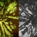 Fractal Pattern of Chronic Serpiginous Choroiditis
Jun 17 2025 by Guilherme Sturzeneker, MD, MSc
Ultra-widefield fundus photograph and autofluorescence of a 33-year-old woman with longstanding serpiginous choroiditis in the right eye. The image reveals centrifugal chorioretinal atrophy forming a dramatic fractal-like pattern, sparing the fovea. The patient is several years post-onset, with repeated negative workups, including for tuberculosis. Despite extensive lesions, the patient retains 20/20 vision in both eyes. Management included azathioprine monotherapy, as systemic steroids were contraindicated due to bipolar disorder.
Photographer: Andrea Almeida, IPEPO - Instituto da Visão
Imaging device: Optos Silverstone
Condition/keywords: autoimmune uveitis, azathioprine, chorioretinal atrophy, serpiginous choroiditis, ultra-wide field imaging
-
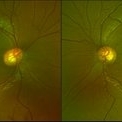
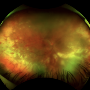
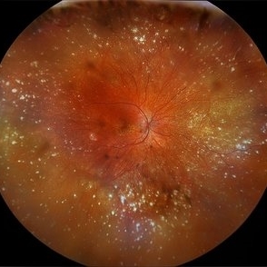 VKH Syndrome
Jun 12 2025 by Virginia Gebhart
22 year old male with VKH Syndrome. Pt has been experiencing severe headaches, distorted vision, hearing loss, weakness, and a large white patch of hair. Significant cell in AC and vitreous, multiple punched-out CR scars in periphery. Referred to rheumatology for possible immunomodulatory treatment
Photographer: Virginia Gebhart, Retina Consultants of Carolina
Imaging device: Optos California
Condition/keywords: montage, multifocal choroiditis, panuveitis, Vogt-Koyanagi-Harada
-
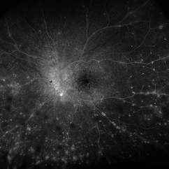 VKH Syndrome
Jun 12 2025 by Virginia Gebhart
Fluorescein angiogram of 22 year old male with VKH syndrome. Significant cell in AC and vitreous, multiple punched-out CR scars in periphery, mild vascular leakage. Pt referred to rheumatology for immunomodulatory treatment.
Photographer: Virginia Gebhart, Retina Consultants of Carolina
Imaging device: Optos California
Condition/keywords: FA, fluorescein angiogram (FA), multifocal choroiditis, panuveitis, VKH, Vogt-Koyanagi-Harada
-
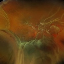
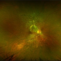 VKH Pseudotumor – Chronic Subretinal Fibrosis
May 11 2025 by Felipe Murati
Ultra-widefield fundus image from a 36-year-old woman with chronic VKH syndrome showing a pseudotumor-like subretinal fibrotic lesion in the right eye. The lesion developed after multiple relapses and remained stable over a 1-year follow-up with immunosuppressive treatment including prednisone, mycophenolate mofetil, and adalimumab. No active choroiditis or exudative detachment was observed. Multimodal imaging was essential for disease monitoring.
Photographer: Felipe A. Murati, MD, University of Arizona
Imaging device: Optos California ultra-widefield retinal imaging system, single-capture, color fundus modality.
Condition/keywords: adalimumab, chronic inflammation, granulomatous uveitis, OCT, Optos ultra-widefield imaging, pseudotumor, subretinal fibrosis, VKH, Vogt-Koyanagi-Harada
-
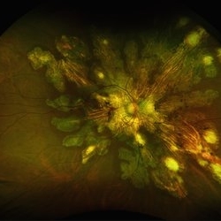
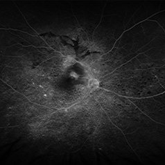 VKH Pseudotumor – Fluorescein Angiography
May 11 2025 by Felipe Murati
Fluorescein angiography image from a 36-year-old woman with chronic Vogt-Koyanagi-Harada (VKH) syndrome showing a pseudotumor-like lesion with late-phase staining and no active leakage. The image highlights subretinal fibrosis in the right eye, stable under long-term immunosuppressive therapy with mycophenolate mofetil and adalimumab. No signs of active choroiditis are present, confirming a quiescent phase.
Photographer: Felipe A. Murati, MD, University of Arizona
Imaging device: Optos California, fluorescein angiography modality
Condition/keywords: choroiditis, Fluorescein angiography, granulomatous uveitis, Optos FA, pseudotumor, subretinal fibrosis, VKH, Vogt-Koyanagi-Harada
 Loading…