Search results (363 results)
-
 Bilateral Retinoschisis Retinal Detachment
Sep 15 2012 by Barbara Parolini, MD
Fundus photograph of a case of bilateral retinoschisis and retinal detachment. The border of the external layer breaks and the border of the schisis have been treated with argon laser. An epiretinal membrane formed after the formation of retinal detachment.
Photographer: Dr Rino Frisina, Istituto Clinico S.Anna, Brescia, Italy
Imaging device: optos
Condition/keywords: epiretinal membrane formation, retinoschisis
-
 Retinoschisi and Retinal Detachment
Sep 15 2012 by Barbara Parolini, MD
Fundus photograph of an eye with retinoschisis and retinal detachment. The other eye has a retinoschisis and retinal detachment with epiretinal membrane.
Photographer: Dr Rino Frisina, Istituto Clinico S.Anna, Brescia, Italy
Imaging device: Optos ultra wide-field retinographer
Condition/keywords: epiretinal membrane formation, retinoschisis
-
 Retinoschisis
Oct 12 2012 by Jeffrey G. Gross, MD, FASRS
Retinoschisis with large outer layer break.
Condition/keywords: large outer layer break, retinoschisis
-
 Retinoschisis + Retinal Detachment
Mar 9 2017 by Uriel Rubin, MD
Retinal detachment at the posterior edge of a retinoschisis in a 45-year-old male patient.
Condition/keywords: bullous retinoschisis, retinoschisis
-
 Senile Retinoschisis
Nov 9 2012 by Norman Byer
This is the same case as seen in the previous photograph but is a different view with the scleral indentation moved more anterior. The retinoschisis is seen to be very peripheral coming at least grossly right up to the ora serrata. Please notice how clear a view one can get of the ora serrata and pars plana using indirect ophthalmoscopy with scleral indentation.
Condition/keywords: ora serrata, retinoschisis, scleral indentation
-
 Retinoschisis/Retinal Detachment with Outer Layer Holes
Oct 15 2012 by Jeffrey G. Gross, MD, FASRS
Retinoschisis/RD with outer layer holes.
Condition/keywords: outer layer hole, retinoschisis
-
 Retinoschisis/Retinal Detachment
Oct 16 2012 by Jeffrey G. Gross, MD, FASRS
Retinoschisis/RD s/p PPV and laser.
Condition/keywords: laser, pars plana vitrectomy (PPV), retinoschisis
-
 Juvenile X-linked retinoschisis
Jan 11 2013 by Alex P. Hunyor, MD
Juvenile X-linked retinoschisis - right macula.
Condition/keywords: juvenile retinoschisis, x-linked retinoschisis (XLRS)
-
 Retinoschisis
Nov 9 2012 by Norman Byer
This 51-year-old woman has retinoschisis with a large outer layer hole which has a white posterior rolled border. The left side of the posterior border of this hole can be seen to lie quite close to the inner layer showing that the outer layer is detached. This, therefore, is actually a combined schisis detachment which may safely be observed without treatment. This is an asymptomatic process, and the detachment of the outer layer is almost always localized and self limited.
Condition/keywords: intact inner layer, localized detachment of outer layer, outer layer hole, retinoschisis, rolled edges of retina, schisis detachment, white posterior
-
 Senile Retinoschisis
Nov 9 2012 by Norman Byer
Senile retinoschisis in a 40-year-old woman showing a very large outer layer hole and a smaller one just above it. The inner layer is intact as shown by the blood vessels running through this layer. Note the prominent, sharp, yellow border at the upper and left edges of the large hole. This is caused by an inward rolling of the border of the outer layer and tends to be much more common along the posterior edges of holes in the outer layer in retinoschisis. This is an example of a schisis detachment with onset before age 40. It extends posteriorly to a .2½ disc diameters from the macula and has remained essentially "arrested" at this location for more than 17 years. A similar symmetrical "schisis-detachment" is present in the fellow eye.
Condition/keywords: intact inner layer, outer layer hole, schisis detachment, senile retinoschisis
-
 Retinal Schisis Detachment
Nov 9 2012 by Norman Byer
This 57-year-old man has a combined retinal schisis detachment caused by an outer layer hole in the upper right. On the right half of this photograph, the outer layer is detached and represented by the prominent yellow line which is lying against the inner layer. On the left half the inner layer appears very thin and the schisis cavity lies just behind it as it was originally. This, therefore, represents a localized detachment of the outer layer and thus a true secondary retinal detachment. The reason these cases remain localized and nonprogressive is that the only fluid available to the subretinal space is that which is contained within the schisis cavity. Furthermore, this fluid tends to be quite viscous and is not readily passed through the retinal breaks. A clinical symptomatic progressive retinal detachment cannot occur unless the retinal schisis cavity is very large or a break occurs in the inner layer also.
Condition/keywords: intact inner layer, localized detachment of outer layer, outer layer hole, retinal schisis detachment, retinoschisis, secondary retinal detachment
-
 X-linked Retinoschisis (XLRS)
Oct 9 2012 by Audina M. Berrocal, MD FASRS
6-week-old baby with XLRS
Photographer: Ditte Hess CRA, BPEI
Imaging device: RETCAM/OIS
Condition/keywords: retinoschisis, x-linked retinoschisis (XLRS)
-
---thumb.jpg/image-square;max$300,300.ImageHandler) Retinoschisis
Retinoschisis
-
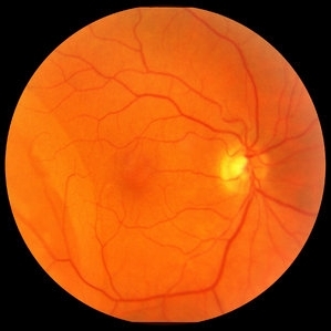
 Retinoschisis Right Eye
Oct 27 2014 by AnneMarie Smykowski
65-year-old white male, with hypertensive retinopathy. He has a stable retinoschisis for approximately 10 years.
Photographer: AnneMarie Smykowski C.O.A., Island Retina Shirley, NY
Imaging device: Optos Daytona
Condition/keywords: color photo, Daytona, Optos, retinoschisis
-
 Juvenile Retinoschisis
Nov 13 2012 by Mallika Goyal, MD
Fundus photograph of right eye of a 20-year-old gentleman showing inferotemporal retinoschisis. There was associated bilateral foveal schisis.
Photographer: Mallika Goyal, MD
Condition/keywords: foveal schisis, juvenile retinoschisis
-
 Retinoschisis Detachment
Nov 9 2012 by Norman Byer
Combined retinoschisis detachment, so-called schisis detachment, in a 47-year-old woman. The large outer layer hole in the center has a posterior yellow border which represents the position of the outer layer. Please observe superior to the hole the dark convexity of the scleral indentation. Just below the hole at the middle of the slide and going to the left the yellow zone comes to lie right against the inner layer and a fluid filled cavity lies deep to the outer layer. At this point, therefore, there is a true neurosensory detachment of the retina. On the right side of the hole, the yellow line slants up and to the right and lies close to the pigment epithelium. On the right side of the photograph, the original schisis cavity can be seen separating the yellow line of the outer layer above from the inner retinal layer below. The mechanism of this detachment is that some of the fluid from the schisis cavity passes through the outer layer hole and detaches the outer layer. This lesion has not been treated and has remained exactly the same for 13 years. A similar symmetrical "schisis-detachment" is present in the fellow eye.
Condition/keywords: neurosensory detachment of retina, outer layer hole, pigment epithelium, retinoschisis, schisis detachment, scleral indentation
-
---thumb.JPG/image-square;max$300,300.ImageHandler) Retinoschisis
Retinoschisis
Oct 26 2012 by Mallika Goyal, MD
Fundus photograph of left eye of a 9-year-old boy with juvenile retinoschisis with large inner retinal break .
Condition/keywords: juvenile retinoschisis, retinal break
-
---thumb.JPG/image-square;max$300,300.ImageHandler) Juvenile Retinoschisis
Juvenile Retinoschisis
Nov 13 2012 by Mallika Goyal, MD
Fundus photograph of right eye of a 20-year-old gentleman with bilateral inferotemporal retinoschisis with foveal schisis.
Photographer: Mallika Goyal, MD, Apollo Health City, Hyderabad, India
Condition/keywords: foveal schisis, inferotemporal retinoschisis
-
 Retinoschisis
Nov 9 2012 by Norman Byer
This 53-year-old man has retinoschisis involving the upper temporal quadrant but with no visible yellow dots or white lines to make it obvious. However, with scleral indentation you can see a large convex area showing the so-called white with pressure phenomenon. This area corresponds exactly to the area being indented and therefore must arise either from the outer layer of the retina or from some structure deep to it. White with pressure is an interesting optical phenomenon of uncertain origin but of no definite diagnostic or prognostic significance.
Condition/keywords: retinoschisis, scleral indentation, white with pressure
-
 Juvenile X-linked Retinoschisis
Jan 11 2014 by Caesar K. Luo, MD, FASRS
RetCam fluorescein angiography of child with JXLRS.
Photographer: Caesar Luo, Progressive Vision Institute, PA
Condition/keywords: juvenile retinoschisis
-
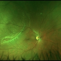
 Retinoschisis Left Eye
Oct 27 2014 by AnneMarie Smykowski
65-year-old white male, with hypertensive retinopathy. He has a stable retinoshisis for approximately 10 years.
Photographer: AnneMarie Smykowski C.O.A., Island Retina Shirley, NY
Imaging device: Optos Daytona
Condition/keywords: color photo, Daytona, Optos, retinoschisis
-
---thumb.JPG/image-square;max$300,300.ImageHandler) Retinoschisis
Retinoschisis
Oct 26 2012 by Mallika Goyal, MD
Fundus photograph of left eye of a 9-year-old boy with juvenile retinoschisis with large inner retinal break.
Condition/keywords: juvenile retinoschisis, retinal break
-
---thumb.JPG/image-square;max$300,300.ImageHandler) Juvenile Retinoschisis
Juvenile Retinoschisis
Nov 13 2012 by Mallika Goyal, MD
Fundus photograph of left eye of a 20-year-old gentleman with bilateral inferotemporal retinoschisis with foveal schisis.
Photographer: Mallika Goyal, MD
Condition/keywords: foveal schisis, inferotemporal retinoschisis
-
 Retinoschisis/Retinal Detachment
Oct 16 2012 by Jeffrey G. Gross, MD, FASRS
Retinoschisis/RD, s/p PPV and laser.
Condition/keywords: retinoschisis
-
 X-Linked Juvenile Retinoschisis
Dec 17 2016 by Young Hee Yoon, MD, PhD
Fundus photograph of a 27-year-old male with x-linked juvenile retinoschisis shows foveal retinoshisis with spokewheel pattern in his left eye.
Photographer: Ji Soo Kim, University of Ulsan, Asan Medical Center, Seoul, Korea
Condition/keywords: foveal schisis, spokewheel pattern
 Loading…