Search results (363 results)
-
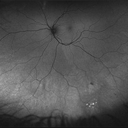 Autofluorescence of Peripheral Retinoschisis
Jul 26 2018 by Olivia Rainey
Ultra-wide field autofluorescence image of a 49-year-old male with non-progressive peripheral retinoschisis of his left eye. Patient was asymptomatic and had no prior trauma or surgery to his eye. Recommended observation at this time.
Photographer: Olivia Rainey
Imaging device: Optos
Condition/keywords: autofluorescence imaging, left eye, Optos, retinoschisis, ultra-wide field imaging
-
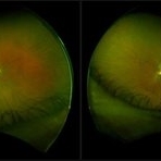
 Bilateral Retinoschisis Retinal Detachment
Sep 15 2012 by Barbara Parolini, MD
Fundus photograph of a case of bilateral retinoschisis and retinal detachment. The border of the external layer breaks and the border of the schisis have been treated with argon laser. An epiretinal membrane formed after the formation of retinal detachment.
Photographer: Dr Rino Frisina, Istituto Clinico S.Anna, Brescia, Italy
Imaging device: optos
Condition/keywords: epiretinal membrane formation, retinoschisis
-
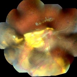
 Bilateral Retinoschisis with Retinal Detachment
Jun 21 2016 by Vishak J. John, MD
Fundus photograph of a 67-year-old female with retinoschisis in both eyes and retinal detachment in the right eye.
Photographer: Mark Clark, Wake Forest Baptist Eye Center
Imaging device: Optos California
Condition/keywords: retinoschisis
-
 Branch Retinal Vein Occlusion
Aug 13 2024 by Shakhzod Muratov
Fundus photograph of a 66 year old woman with a BRVO and Bullous retinoschisis.
Photographer: Shakhzod Muratov, S. Fyodorov Eye Microsurgery Federal State Institution
Imaging device: Zeiss Clarus 500
Condition/keywords: BRVO, retinoschisis
-
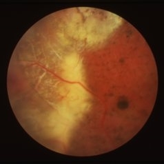
 Coats' Disease
Mar 28 2021 by JEFFERSON R SOUSA, Tecg.º (Biomedical Systems Technology)
A 14-year-old male patient was admitted for visual evaluation. Visual acuity s/c in the right eye and 20/80 in the left eye. According to family members, he reported low vision since childhood. He had already undergone treatment with photocoagulation in another service to which he had a diagnostic hypothesis of Coatas disease. Laboratory tests were requested (HIV, TOXO, TOXOCARIASIS, ECA, VDRL, PPD). In the evaluation it was observed important exudation in the posterior pole, some vascular irregularities in the right eye. In the left eye, there is retinoschisis affecting the entire posterior pole and the region nasal to the optic disc, macula with a characteristic aspect of a cartwheel. Well exemplified by OCT-A (Structrure Deep: IPL - 25, OPL - 25).
Photographer: JEFFERSON R SOUSA - Study Center and Ophthalmological Research Dr. Andre M V Gomes, Institute Dr. Suel Abujamra São Paulo-Brazil
Imaging device: Topcon TRC-50 DX, Imaginet 4.0, angle de 50 graus. Flash 50w-s
Condition/keywords: Coats' disease, retinoschisis
-
 Complex Retinal Detachment with PVR and Starfold
Jun 6 2025 by Jenn Geelan
57 year old male with a Complex Retinoschisis related retinal detachment with PVR and a Posterior Star Fold
Photographer: Jenn Geelan, Retina-Vitreous Surgeons of CNY
Imaging device: Optos California
Condition/keywords: proliferative vitreoretinopathy (PVR), rare, Retinal Detachment, retinoschisis, Starfolds, subretinal fluid
-
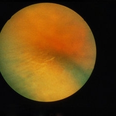 Degenerative Retinoschisis
Feb 19 2015 by H. Michael Lambert, MD
Color photo of degenerative retinoschisis.
Condition/keywords: color photo, retinoschisis
-
 Degenerative Retinoschisis
Feb 19 2015 by H. Michael Lambert, MD
Color photo of degenerative retinoschisis
Condition/keywords: color photo, retinoschisis
-
 Degenerative Retinoschisis
Feb 19 2015 by H. Michael Lambert, MD
Color photo of degenerative retinoschisis.
Condition/keywords: color photo, retinoschisis
-
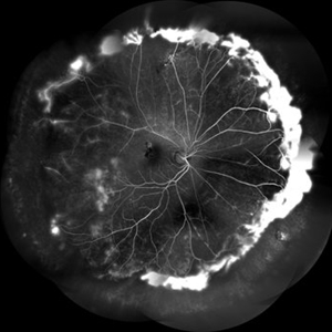 FFA in a Case of Retinoschisis With Fluid
Jul 28 2024 by Prashant K Bawankule, M.S.
Young male of 22 years presented with DOV. Examination showed retinoschisis with fluid in periphery. FFA showed massive leakage in the periphery
Photographer: Prashant Bawankule, Sarakshi Netralaya, Nagpur, Maharashtra , India
Imaging device: Mirante ( by Nidek)
Condition/keywords: retinoschisis
-
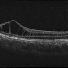
 Foveoschisis August 2016 OD
May 9 2018 by Aaron P. Appiah, MD
21-year-old male with congenital retinoschisis with bilateral macular involvement and large inner retinal hole OD.
Imaging device: Zeiss Cirrus 5000
Condition/keywords: retinal hole, retinoschisis
-
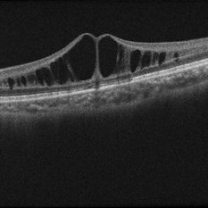
 Foveoschisis August 2016 OS
May 9 2018 by Aaron P. Appiah, MD
21-year-old male with congenital retinoschisis with bilateral macular involvement and large inner retinal hole OD . Foveoschisis reformed again OS, after it collapsed, compared to last visit in March 2016
Imaging device: Zeiss Cirrus 5000
Condition/keywords: retinoschisis
-
 Foveoschisis Collapsed March 2016 OS
May 9 2018 by Aaron P. Appiah, MD
21-year-old male with congenital retinoschisis with bilateral macular involvement and large inner retinal hole OD. Foveoschisis collapsed OS, since last exam in 07/2015.
Imaging device: Zeiss Cirrus 5000
Condition/keywords: retinal hole, retinoschisis
-
 Foveoschisis July 2015 OD
May 9 2018 by Aaron P. Appiah, MD
21-year-old male with congenital retinoschisis with bilateral macular involvement and large inner retinal hole OD.
Imaging device: Zeiss Cirrus 5000
Condition/keywords: retinal hole, retinoschisis
-
 Foveoschisis July 2015 OS
May 9 2018 by Aaron P. Appiah, MD
21-year-old male with congenital retinoschisis with bilateral macular involvement and large inner retinal hole OD
Imaging device: Zeiss Cirrus 5000
Condition/keywords: retinal hole, retinoschisis
-
 Foveoschisis March 2016 OD
May 9 2018 by Aaron P. Appiah, MD
21-year-old male with congenital retinoschisis with bilateral macular involvement and large inner retinal hole OD
Imaging device: Zeiss Cirrus 5000
Condition/keywords: retinal hole, retinoschisis
-
 Foveoschisis May 2018 OD
May 9 2018 by Aaron P. Appiah, MD
21-year-old male with congenital retinoschisis with bilateral macular involvement and large inner retinal hole OD.
Imaging device: Zeiss Cirrus 5000
Condition/keywords: retinoschisis
-
 Foveoschisis May 2018 OS
May 9 2018 by Aaron P. Appiah, MD
21-year-old male with congenital retinoschisis with bilateral macular involvement and large inner retinal hole OD.
Imaging device: Zeiss Cirrus 5000
Condition/keywords: retinal hole, retinoschisis
-
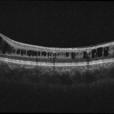
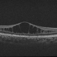
 Foveoschisis secondary to high myopia
Mar 13 2015 by Niloofar Piri, MD
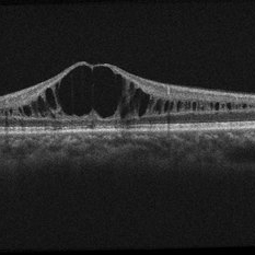
Infrared and HD-OCT of the right eye in a 55-year-old African American female with high myopia (more than -6.00 D), BCVA: 20/25 OU Cartwheel appearance of the fovea in the infrared imaging is visible. HD- OCT demonstartes schisis in different layers of the retina (both NFL and OPL; notice stretching of the Muller cells); VMT is also present . Outer retinal layers are preserved which explains the good vision . She had the same findings in OS.
Photographer: Niloofar Piri, MD
Imaging device: Heidelberg Spectralis
Condition/keywords: high myopia, retinoschisis
-
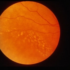
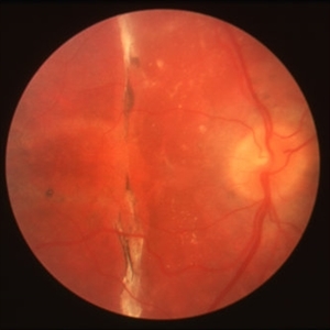
 Juvenile Retinoschisis
Apr 2 2019 by Gary R. Cook, MD, FACS
Temporal periphery of a 15-year-old white male with juvenile retinoschisis OD.
Condition/keywords: juvenile retinoschisis, retinoschisis
-
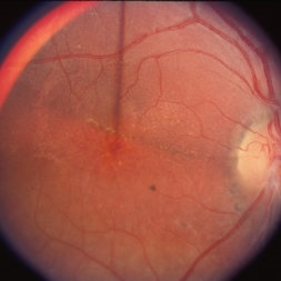
 Juvenile Retinoschisis
Apr 2 2019 by Gary R. Cook, MD, FACS
Posterior pole of the same 15-year-old white male with juvenile retinoschisis OD.
Condition/keywords: juvenile retinoschisis, retinoschisis
-
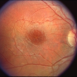
 Juvenile Retinoschisis
Apr 2 2019 by Gary R. Cook, MD, FACS
11-year-old male with juvenile retinoschisis with stellate maculopathy OD; V.A. = 20/40
Condition/keywords: juvenile retinoschisis, retinoschisis, stellate maculopathy
-
 Juvenile Retinoschisis
Apr 2 2019 by Gary R. Cook, MD, FACS
10-year old white male with stellate macular lesion OD secondary to juvenile retinoschisis; V.A. = 20/100
Condition/keywords: juvenile retinoschisis, retinoschisis, stellate maculopathy
-
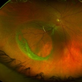
 Juvenile Retinoschisis with Localized Inferior Retinal Detachment
May 4 2020 by Giridhar Anantharaman, MS
Optos ultra-widefield retinal image of the left eye of a 11-year-old male child showing peripheral retinoschisis with localized inferior retinal detachment.
Photographer: Rakesh PR , Giridhar Eye Institute, Kerala, India
Imaging device: Optos UWF Daytona Plus
Condition/keywords: retinoschisis
 Loading…