Search results (359 results)
-
 Coats' Disease With Exudative Retinal Detachment and Retinal Macrocyst
Dec 9 2019 by Sophia El Hamichi, MD
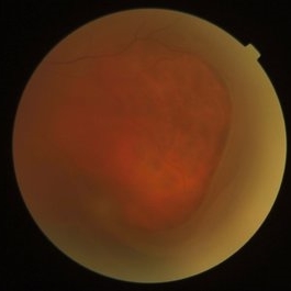
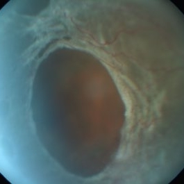
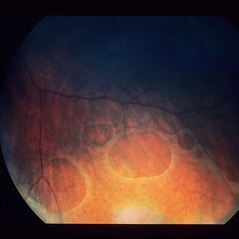
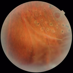
A 3-year-old male with a presentation of a complex Coats' disease in the left eye with exudative retinal detachment, abnormal telangiectatic vasculature, and inferotemporal retinal macrocyst/retinoschisis.
Photographer: Abby Orcutt-Hayes, Murray Ocular Oncology and Retina
Imaging device: RetCam
Condition/keywords: Coats' disease, exudative detachment, montage, retinal macrocyst
-
 Foveoschisis secondary to high myopia
Mar 13 2015 by Niloofar Piri, MD
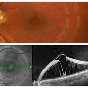
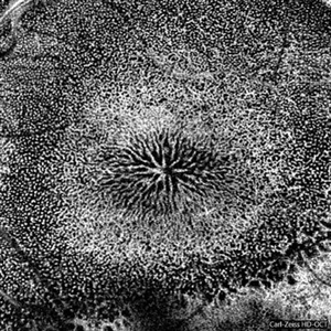
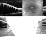
Infrared and HD-OCT of the right eye in a 55-year-old African American female with high myopia (more than -6.00 D), BCVA: 20/25 OU Cartwheel appearance of the fovea in the infrared imaging is visible. HD- OCT demonstartes schisis in different layers of the retina (both NFL and OPL; notice stretching of the Muller cells); VMT is also present . Outer retinal layers are preserved which explains the good vision . She had the same findings in OS.
Photographer: Niloofar Piri, MD
Imaging device: Heidelberg Spectralis
Condition/keywords: high myopia, retinoschisis
-
 Juvenile Retinoschisis OS
Jan 25 2017 by Manish Nagpal, MD, FRCS (UK), FASRS
Juvenile retinoschisis.
Photographer: Ashish Jain
Condition/keywords: juvenile retinoschisis
-
 Optic Disc Pit with Maculopathy
Feb 25 2021 by Niloofar Piri, MD
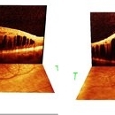
Color fundus photograph and SD OCT of a 6-year-old patient with optic disc pit associated with large retinoschisis involving the entire macula. SD OCT demonstrating large retinoschisis with ILM draping centrally giving it the appearance of pseudohole on the corresponding central area of color photo. Vision 20/80
Photographer: Lisa Breeding, St Louis University
Condition/keywords: maculopathy, optic disc
-
 X-Linked Retinoschisis
Nov 15 2019 by Nelson Chamma Capelanes, MD
SD-OCT of an 28-year-old man with X - linked retinoschisis.
Photographer: Nelson Capelanes, Promedica/Promacula Indaiatuba & UPO Oftalmologia São Paulo
Imaging device: Spectralis
Condition/keywords: x-linked retinoschisis (XLRS)
-
 Dislocated Lens, Posterior OD
Jan 26 2024 by Corey Grant
OPTOS California photo presents a 71 year old male patient with a dislocated lens, posterior in the right eye. Presented on 1/26/24 with posteriorly dislocated SN60WF with a Soemmerring ring. Associated retinal hemorrhage within retinoschisis as well. This will result in a PPV/IOL exchange/SFIOL/STK for the right eye.
Photographer: Corey Grant, Ophthalmic Imager, Retina Specialist of Michigan
Imaging device: OPTOS California
Condition/keywords: color photo, IOL, OD, Optos, OPTOS CALIFORNIA, pars plana vitrectomy (PPV), retina
-
 Juvenile Retinoschisis- OD
Jan 25 2017 by Manish Nagpal, MD, FRCS (UK), FASRS
Juvenile retinoschisis.
Photographer: ashish jain
Condition/keywords: juvenile retinoschisis
-
 Macular Foveoschisis
Nov 9 2023 by Charlotte Jones
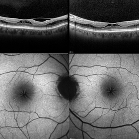
Bilateral ocular coherence tomography and fundus autofluorescence of a 77 year old woman with Macular Foveoschisis. Patient with stable vision since her last appointment (20/30 right eye and 20/25 left eye) with worsening vitreomacular traction in the right eye. Patient is followed routinely for Plaquenil use.
Photographer: Charlotte Jones
Imaging device: Heidelberg Spectralis
Condition/keywords: fundusautofluorescence, macularfoveoschisis, macularretinoschisis, macularstar, ocularcoherencetomography
-
 Old Inferotemporal BRVO with Bullous Retinoschisis
Jan 15 2022 by Dinesh Rungta, MBBS, DNB
Optos image, OS, of an 85-year-old male showing old IT-BRVO with sclerosed vessel and Bullous Retinoschisis.
Photographer: Pratap Dey, Disha Eye Hospital, West Bengal, India.
Imaging device: Optos Daytona
Condition/keywords: branch retinal vein occlusion (BRVO), retinoschisis
-
 Optic Nerve Pit Right Eye
Feb 15 2021 by Kim Barrett
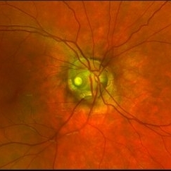
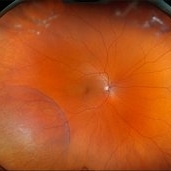
A 14-year-old male presented with vision loss and VF defect. Patient was treated for presumed amblyopia with patching since age 4. He has had neurologic care for post traumatic skull fracture and brain bleed in 2012. IOP's WNL. OD is without retinoschisis or subretinal fluid. Patient is at risk of serous detachment. Current VA OD 20/200+1 PH 20/80.
Photographer: Kim Barrett C.O.A. Retina Specialist of Michigan, Grand Rapids, MI
Imaging device: Optos California
Condition/keywords: amblyopia, hemifield, Humphrey visual field, nerve, optic nerve pit, visual field defect
-
 Outer Retinal Tear in Schisis-Detachment
Mar 25 2016 by Gregory R. Blaha, MD, PhD
Large outer retinal tear in combined retinoschisis-detachment. The retinal vessels are visible going over the retinal break.
Photographer: Janice Neal, Gurley Eye Care Associates
Imaging device: Topcon Mark II
Condition/keywords: retinal tear, retinoschisis
-
 Retinalschisis
Nov 9 2012 by Norman Byer
This 43-year-old woman had the small area of retinalschisis and associated small localized retinal detachment resulting from breaks in both retinal layers when she was first examined. This eye was asymptomatic and has remained exactly the same during two years of observation without treatment. In this picture, the outer layer break is demarcated by the yellow line. The inner layer is seen intact over this hole but has itself two small holes in the upper part of the photograph. Thus, it is evident that even retinoschisis with double layer holes does not necessarily progress to a clinical retinal detachment. The viscous nature of the fluid in the retinoschisis cavity is probably a contributing factor in this non progressive tendency.
Condition/keywords: inner layer holes, outer layer hole, retinoschisis
-
 Retinoschisis
May 1 2015 by Mehul A Shah
A 24-year-old boy presented with diminished vision and found to have retinoschisis with break in internal layer.
Photographer: Mehul Shah, Drashti Netralaya
Imaging device: Zeiss FF450plus
Condition/keywords: bullous retinoschisis
-
 Retinoschisis
Mar 28 2021 by JEFFERSON R SOUSA, Tecg.º (Biomedical Systems Technology)
A 14-year-old male patient was admitted for visual assessment. Visual acuity s/c in the right eye and 20/80 in the left eye. According to family members, he reported low vision since childhood. He had already undergone photocoagulation treatment at another service for which he had a diagnostic hypothesis of Coats' disease. Laboratory tests were requested (HIV, TOXO, TOXOCARIASIS, ACE, VDRL, PPD). In the evaluation, there was significant exudation in the posterior pole, some vascular irregularities in the right eye. In the left eye, there is retinoschisis affecting the entire posterior pole and the nasal region to the optic disc, macula with a characteristic chariot-wheel appearance, well exemplified by OCT-A (Structrure Deep: IPL - 25, OPL - 25).
Photographer: JEFFERSON R SOUSA - Study Center and Ophthalmological Research Dr. Andre M V Gomes, Institute Dr. Suel Abujamra São Paulo-Brazil
Imaging device: Optical coherence tomography system Optical Coherence Tomography system OCT CIRRUS 5000, Line Protocol, HD 21 line. Cirrus 5000 does not do a wide-angle tomographic image. (Structrure Deep: IPL - 25, OPL - 25).
Condition/keywords: Coats' disease, retinoschisis
-
 Retinoschisis
Jun 4 2014 by Henry J. Kaplan, MD
Senile degenerative peripheral retinoschisis with outer wall holes.
Condition/keywords: retinoschisis
-
 Retinoschisis
Feb 26 2025 by Kimberly Wakester
Optomap RGB of a 56-year-old woman with bullous retinoschisis in the right eye. The patient remains stable with very mild progression. Patient is to continue follow up care at 6 month intervals to monitor for worsening progression.
Photographer: Kimberly Wakester, COA
Imaging device: Optos California
Condition/keywords: bullous retinoschisis
-
000---thumb.jpg/image-square;max$300,300.ImageHandler) Retinoschisis in ROP
Retinoschisis in ROP
Jan 22 2013 by Young Hee Yoon, MD, PhD
Images of an 18-year-old male with a history of premature birth (30 weeks).
Photographer: Soohyun Cho, Asan Medical Center, Seoul, Korea
Imaging device: Fluorescein angiography using Optomap, optos
Condition/keywords: retinoschisis
-
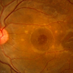
 Stellate Nonhereditary Idiopathic Foveomacular Retinoschisis
Feb 16 2024 by Sayena . Jabbehdari, MD, MPH, MBA candidate
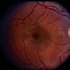
A 67 year old male presented with central vision distortion of right eye.
Photographer: Sayena Jabbehdari MD MPH
Condition/keywords: Stellate nonhereditary idiopathic foveomacular retinoschisis
-
 X - Linked Retinoschisis
Sep 18 2014 by David Callanan, MD
9-year-old male, x - linked retinoschisis.
Condition/keywords: x-linked retinoschisis (XLRS)
-
 X-Linked Retinoschis
Nov 15 2019 by Nelson Chamma Capelanes, MD
SD-OCT of an 28-year-old man with X - linked retinoschisis.
Photographer: Nelson Capelanes, Promedica/Promacula Indaiatuba & UPO Oftalmologia São Paulo
Imaging device: Spectralis
Condition/keywords: x-linked retinoschisis (XLRS)
-
 X-Linked Retinoschisis
Jan 31 2015 by Hamid Ahmadieh, MD
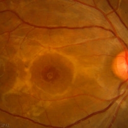
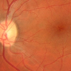
Color fundus photograph of the left eye of a 35-year-old man with x-linked retinoschisis. Please notice peripheral retinoschisis with large inner layer holes and laser scars around an outer layer hole.
Photographer: Shabnam Poureh, Negah Eye Center, Tehran, Iran
Condition/keywords: color fundus photograph, retinoschisis, x-linked retinoschisis (XLRS)
-
 X-Linked Retinoschisis
Jan 31 2015 by Hamid Ahmadieh, MD
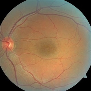
Color fundus photograph of the left eye of a 35-year-old man with x-linked retinoschisis. Please notice the foveal schisis.
Photographer: Shabnam Poureh, Negah Eye Center, Tehran, Iran
Condition/keywords: color fundus photograph, foveal schisis, x-linked retinoschisis (XLRS)
-
 XLJR - X-linked Juvenile Retinoschisis with CME
Jun 25 2020 by Evan N Dunn, MD
40-year-old male with X-linked juvenile retinoschisis. Genetic testing was positive for a pathogenic variant in the RS1 gene.
Photographer: Steve Morris
Condition/keywords: x-linked retinoschisis (XLRS)
-
 Retinoschisis + Retinal Detachment
Mar 9 2017 by Uriel Rubin, MD
Retinal detachment at the posterior edge of a retinoschisis in a 45-year-old male patient.
Condition/keywords: bullous retinoschisis, retinoschisis
-
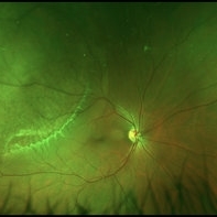
 X-Linked Retinoschisis - Mizuo Nakamura Phenomenon
Apr 30 2020 by Giselle DeOliveira
Fundus photograph montaged of a 19-year-old male with x-linked retinoschisis
Photographer: Giselle DeOliveira
Imaging device: Clarus
Condition/keywords: Mizuo Nakamura Phenomenon, x-linked retinoschisis (XLRS)
 Loading…