Search results (363 results)
-
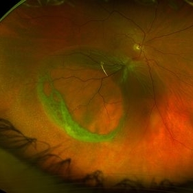
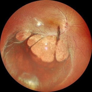
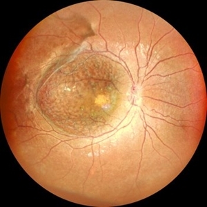 Retinoschisis
Jul 30 2025 by Akansha Sharma
Color fundus photograph of a 2 year old male with both eyes retinoschisis.
Photographer: DR. AKANSHA SHARMA
Condition/keywords: juvenile retinoschisis, RETINOSCHISIS
-
 Retinoschisis
Jul 30 2025 by Akansha Sharma
Color fundus photograph of a 2 year old male with both eyes retinoschisis.
Photographer: DR. AKANSHA SHARMA
Condition/keywords: juvenile retinoschisis, RETINOSCHISIS
-
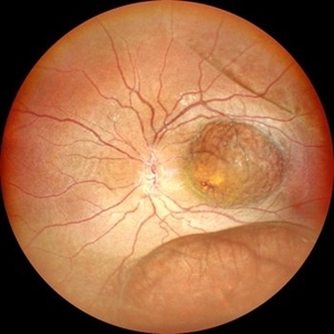
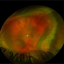
 Retinoschisis with Outer Layer Holes
Jul 18 2025 by Kimberly Wakester
Optomap RGB of an 56-year-old woman with retinoschisis with outer layer holes s/p laser in the left eye. Patient remains stable. Will continue follow up care with dilated exam and optos imaging.
Photographer: Kimberly Wakester, COA, OCT-C
Imaging device: Optos California
Condition/keywords: outer layer hole, retinoschisis
-
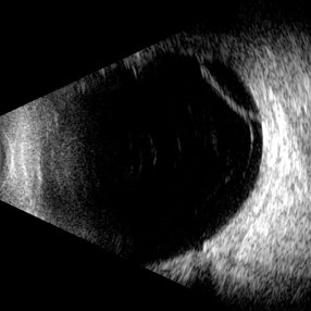
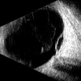 Macular Retinoschisis
Jul 3 2025 by Gustavo Uriel Fonseca Aguirre
This B-mode longitudinal ultrasound scan reveals macular retinoschisis, demonstrating a characteristic splitting of retinal layers with a smooth, dome-shaped elevation. The lesion shows maintained structural integrity of both inner and outer retinal walls without associated subretinal fluid or vitreous traction.
Photographer: Gustavo U. Fonseca Aguirre, Hospital Conde de Valenciana, Ciudad de México
Condition/keywords: macular retinoschisis
-
 Macular Retinoschisis
Jun 26 2025 by rohan jain
Macular retinoschisis
Photographer: Dr. ROHAN JAIN
Condition/keywords: juvenile retinoschisis, RETINOSCHISIS
-
 Macular Retinoschisis
Jun 26 2025 by rohan jain
Macular retinoschisis
Photographer: Dr. ROHAN JAIN
Condition/keywords: inferotemporal retinoschisis, juvenile retinoschisis, Macular retinoschisis, RETINOSCHISIS
-
 Complex Retinal Detachment with PVR and Starfold
Jun 6 2025 by Jenn Geelan
57 year old male with a Complex Retinoschisis related retinal detachment with PVR and a Posterior Star Fold
Photographer: Jenn Geelan, Retina-Vitreous Surgeons of CNY
Imaging device: Optos California
Condition/keywords: proliferative vitreoretinopathy (PVR), rare, Retinal Detachment, retinoschisis, Starfolds, subretinal fluid
-
 Retinoschisis
Apr 21 2025 by Gustavo Uriel Fonseca Aguirre
This B-mode longitudinal ultrasound scan reveals a peripheral temporal retinoschisis, demonstrating a characteristic thin, dome-shaped separation of the retinal layers without associated subretinal fluid or vitreous traction. The lesion shows smooth, convex contours with maintained structural integrity of both retinal layers.
Photographer: Gustavo U. Fonseca Aguirre, Hospital Conde de Valenciana, Ciudad de México
Condition/keywords: retinoschisis
-
 Retinoschisis
Feb 26 2025 by Kimberly Wakester
Optomap RGB of a 56-year-old woman with bullous retinoschisis in the right eye. The patient remains stable with very mild progression. Patient is to continue follow up care at 6 month intervals to monitor for worsening progression.
Photographer: Kimberly Wakester, COA
Imaging device: Optos California
Condition/keywords: bullous retinoschisis
-
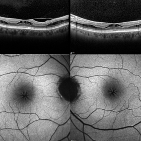
 Foveomacular Retinoschisis
Jan 3 2025 by Drew Mitchell
HD 6x6 OCT-Angiography Structural View of the Deep Inner Retina
Photographer: Drew Mitchell, OCT-C
Imaging device: Zeiss Cirrus 6000
Condition/keywords: foveoschisis, maculoschisis
-
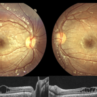
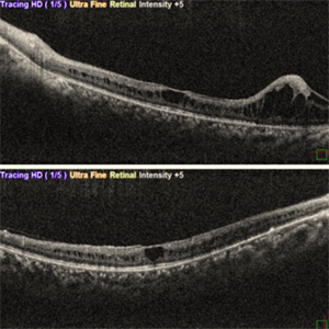 X-Linked Juvenile Retinoschisis ( LE OCT )
Oct 5 2024 by Anand Temkar
This is LE OCT of a 15 year old child with X-linked juvenile retinoschisis.
Photographer: Dr.Anand Temkar- Retina Foundation, Ahmedabad
Imaging device: Mirante
Condition/keywords: OCT, retinoschisis
-
 X-Linked Juvenile Retinoschisis ( RE OCT )
Oct 5 2024 by Anand Temkar
This is RE OCT of a 15 year old child with X-linked juvenile retinoschisis.
Photographer: Dr.Anand Temkar- Retina Foundation, Ahmedabad
Imaging device: Mirante
Condition/keywords: juvenile retinoschisis, OCT, retinoschisis
-
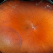
 X-Linked Juvenile Retinoschisis
Oct 5 2024 by Anand Temkar
This is a color photo montage of LE of a 15 year old child with X-linked juvenile retinoschisis.
Photographer: Dr.Anand Temkar- Retina Foundation, Ahmedabad
Imaging device: Mirante
Condition/keywords: x-linked retinoschisis (XLRS)
-
 X-Linked Juvenile Retinoschisis
Oct 5 2024 by Anand Temkar
This is a color photo montage of RE of a 15 year old child with X-linked juvenile retinoschisis.
Photographer: Dr.Anand Temkar- Retina Foundation, Ahmedabad
Imaging device: Mirante
Condition/keywords: x-linked retinoschisis (XLRS)
-
 Branch Retinal Vein Occlusion
Aug 13 2024 by Shakhzod Muratov
Fundus photograph of a 66 year old woman with a BRVO and Bullous retinoschisis.
Photographer: Shakhzod Muratov, S. Fyodorov Eye Microsurgery Federal State Institution
Imaging device: Zeiss Clarus 500
Condition/keywords: BRVO, retinoschisis
-
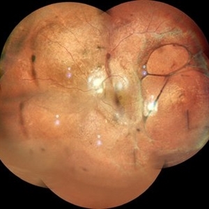
 Juvenile X-linked Retinoschisis
Jul 31 2024 by Tejaswita Verma
Widefield fundus image of a 17 year old boy with BCVA 6/12 showing juvenile X-linked peripheral retinoschisis and foveoschisis bilaterally.
Photographer: DR. TEJASWITA VERMA
Imaging device: MIRANTE
Condition/keywords: juvenile retinoschisis
-
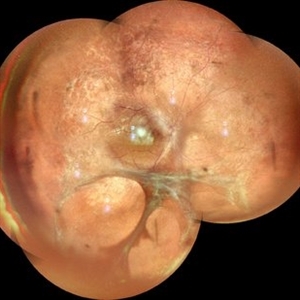
 Juvenile Foveoschisis
Jul 31 2024 by Tejaswita Verma
Central fundus image of a teenage boy with 6/12 vision showing spokewheel pattern in case of juvenile X-linked bilateral foveoschisis.
Photographer: DR. TEJASWITA VERMA
Imaging device: MIRANTE
Condition/keywords: foveoschisis, juvenile retinoschisis
-
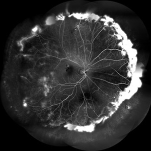 FFA in a Case of Retinoschisis With Fluid
Jul 28 2024 by Prashant K Bawankule, M.S.
Young male of 22 years presented with DOV. Examination showed retinoschisis with fluid in periphery. FFA showed massive leakage in the periphery
Photographer: Prashant Bawankule, Sarakshi Netralaya, Nagpur, Maharashtra , India
Imaging device: Mirante ( by Nidek)
Condition/keywords: retinoschisis
-
 Retinoschisis
Jul 1 2024 by Harsh Hemant Jain, MBBS, DNB
Fundus photograph of a 36-year-old man with a peripheral retinoschisis with outer retinal layer holes.
Photographer: Dr. Muskan Mangal
Imaging device: OPTOS DAYTONA
Condition/keywords: RETINOSCHISIS
-
 JXLR
JXLR
May 26 2024 by shama sharief
12 year old boy with complaints of blurring of vision in both eye for 2 years.
Condition/keywords: juvenile retinoschisis
-
 Stellate Nonhereditary Idiopathic Foveomacular Retinoschisis
Feb 16 2024 by Sayena . Jabbehdari, MD, MPH, MBA
A 67 year old male presented with central vision distortion of right eye.
Photographer: Sayena Jabbehdari MD MPH
Condition/keywords: Stellate nonhereditary idiopathic foveomacular retinoschisis
-
 Dislocated Lens, Posterior OD
Jan 26 2024 by Corey Grant
OPTOS California photo presents a 71 year old male patient with a dislocated lens, posterior in the right eye. Presented on 1/26/24 with posteriorly dislocated SN60WF with a Soemmerring ring. Associated retinal hemorrhage within retinoschisis as well. This will result in a PPV/IOL exchange/SFIOL/STK for the right eye.
Photographer: Corey Grant, Ophthalmic Imager, Retina Specialist of Michigan
Imaging device: OPTOS California
Condition/keywords: color photo, IOL, OD, Optos, OPTOS CALIFORNIA, pars plana vitrectomy (PPV), retina
-
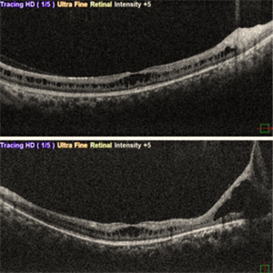
 Macular Foveoschisis
Nov 9 2023 by Charlotte Jones
Bilateral ocular coherence tomography and fundus autofluorescence of a 77 year old woman with Macular Foveoschisis. Patient with stable vision since her last appointment (20/30 right eye and 20/25 left eye) with worsening vitreomacular traction in the right eye. Patient is followed routinely for Plaquenil use.
Photographer: Charlotte Jones
Imaging device: Heidelberg Spectralis
Condition/keywords: fundusautofluorescence, macularfoveoschisis, macularretinoschisis, macularstar, ocularcoherencetomography
-
 Limited Choroidal Hemorrhage S/P Glaucoma Valve Implant OS; Retinoschisis
Aug 21 2023 by Angela Rico
A 52 year old Female presents to office S/P Glaucoma Valve Implant with IOP: 5mmHg OS
Photographer: Angela Rico M.D.
Condition/keywords: choroidal hemorrhage, glaucoma, hypotony, retinoschisis
 Loading…