Search results (111 results)
-
---thumb.jpg/image-square;max$300,300.ImageHandler) Progressive Outer Retinal Necrosis
Progressive Outer Retinal Necrosis
Feb 15 2013 by From the Collections of Thomas M. Aaberg, MD and Thomas M. Aaberg Jr., MD
Color fundus photograph showing extensive confluent retinal whitening, retinal exudation, intraretinal hemorrhage, and sheathing of retinal vessels consistent with infectious retinitis such as progressive outer retinal necrosis (PORN).
Condition/keywords: occlusive retinitis, retinal necrosis
-
 Symptomatic Horseshoe Tear
Nov 9 2012 by Norman Byer
This is a fresh, symptomatic horseshoe tear at the site of a cystic retinal tuft in a 63-year-old man. This is really a double horseshoe tear because the two retinal vessels have resisted the vitreal retinal traction and have preserved an intact bridge of tissue between the tears. Note the prominent vitreous condensation attached to the apex of the upper tear and made much more visible because it is seen superimposed over the dark underlying shadow of scleral indentation.
Condition/keywords: bridge of tissue between tears, cystic retinal tuft, scleral indentation, vitreous condensation
-
 Retinitis pigmentosa AD Slide 1
Oct 22 2012 by Ronald C. Gentile, MD
37 year-old man presents with nyctalopia and tunnel vision. Fundus photo reveals sparing of the fovea with accentuation of the foveal thickness compared to the surrounding atrophic retina. There is waxy pallor of the optic nerve and attenuation of the retinal vessels.
Photographer: The New York Eye & Ear Infirmary Department of Medical Imaging
Condition/keywords: retinitis pigmentosa
-
 Remnant of Hyaloidal Artery
Feb 5 2014 by Gerardo Garcia-Aguirre, MD
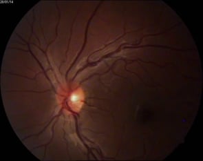
Fundus photograph of the left eye of a 14-year-old asymptomatic female. The photograph is focused on the retina, and a prepapillary vitreous opacity is observed (white arrows). The opacity is attached to the origin of the retinal vessels in the optic nerve head.
Photographer: Gerardo Garcia-Aguirre, MD
Condition/keywords: persistence of the hyaloid artery
-
 Pseudo Retinal Break
Nov 9 2012 by Norman Byer
The next three photographs will illustrate retinal conditions that can easily be mistaken for retinal breaks. For a fourth example of a pseudo retinal break, see slide pair 35. It is important to distinguish these conditions from true retinal breaks. This 49-year-old man was found to have this crescent shaped reddish lesion with a sharp yellow posterior border but without any visible elevated retinal flap. The two blood vessels which traversed this lesion in the presence of a flat retina proved that the retina is intact. This confusing appearance is caused by the presence of white with pressure both behind and in front of the reddish area causing it to resemble a retinal break.
Condition/keywords: pseudo retinal break, reddish lesion, retinal flap, traversing retinal vessels, white with pressure
-
 Combined Hamartoma of the Retinal Pigment Epithelium Case 1
Oct 5 2012 by Ronald C. Gentile, MD
A peripapilary combined hamartoma of the retinal pigment epithelium involving the nasal disc margin. This tumor is slightly elevated, charcoal grey in color with grey-white tissue on it surface. The underlying retinal vessels are obscured.
Photographer: The New York Eye & Ear Infirmary Department of Medical Imaging
Condition/keywords: hamartoma, retinal pigment epithelium
-
 Remnant of Hyaloidal Artery
Feb 5 2014 by Gerardo Garcia-Aguirre, MD
Fundus photograph of the left eye of a 14-year-old asymptomatic female. The photograph is focused on the posterior vitreous where a prepapillary vitreous opacity is observed (white arrows). The opacity is attached to the origin of the retinal vessels in the optic nerve head.
Photographer: Gerardo Garcia-Aguirre, MD
Condition/keywords: persistence of the hyaloid artery
-
---thumb.jpg/image-square;max$300,300.ImageHandler) Primary Hyperoxaluria and Oxalosis
Primary Hyperoxaluria and Oxalosis
Jul 24 2013 by Hamid Ahmadieh, MD
Early phase FA image of the left eye of a 55-year-old man with primary hyperoxaluria and oxalosis, Delayed filling of retinal vessels due to intravascular deposition of calcium oxalate crystals and non-perfusion of the temporal retina are visible.
Photographer: Hanieh Payab, Ophthalmic Research Center, Labbafinejad Medical Center, Tehran
Imaging device: Topcon Fundus Camera
Condition/keywords: ocular manifestation, oxalosis, primary hyperoxaluria
-
 Remnant of Hyaloidal Artery
Feb 5 2014 by Gerardo Garcia-Aguirre, MD
Video of the fundus of the left eye of a 14-year-old asymptomatic female, where a prepapillary vitreous opacity is observed. The opacity is attached to the origin of the retinal vessels in the optic nerve head, and is considered to be a remnant of the hyaloidal artery.
Photographer: Gerardo Garcia-Aguirre, MD
Condition/keywords: persistence of the hyaloid artery
-
 Retina
May 31 2014 by ruth pav
A 32-year-old woman with a history of drug abuse was admitted due to acute manifestation of multiple infarcts, including acute stroke, splenic and renal infarcts, and multiple cutaneous hematomas. Due to decreased vision in her left eye the patient was referred for ophthalmic evaluation. On exam, visual acuity was 6/10 in the right eye and no light perception in her left eye. Ophthalmoscopic examination was normal in the right eye but showed pallor of the optic nerve head with attenuated retinal vessels in the left eye. Fluorescein angiography showed an oval area of hyperfluorescence from from non-perfusion involving the macular center with staining of overlying retinal capillaries.
Photographer: Ruth Pav, Rambam medical center,Hifa Israel.
Imaging device: Zeiss FF4
Condition/keywords: retina
-
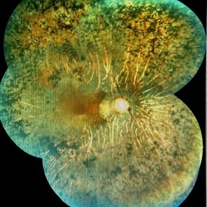
 Optic Atrophy and Attenuated Retinal Vessels Following Endophthalmitis
Jul 12 2014 by Philip J. Polkinghorne, MD
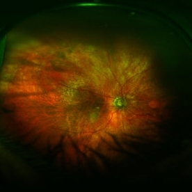
This elderly lady underwent a vitrectomy for post-surgical endophthalmitis. The infection was successfully treated but the functional outcome was poor because of optic atrophy and attenuated retinal vessels.
Photographer: Alex Fraser
Imaging device: Optos Camera
Condition/keywords: attenuated vessels, endophthalmitis, optic atrophy, post-vitrectomy
-
 Intraocular Multiple Cysticercus
Oct 10 2018 by Vishal Agrawal, MD, FRCS,FACS,FASRS
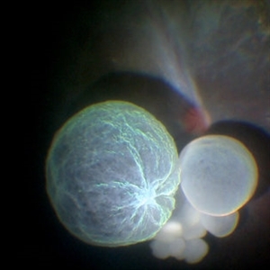
Intraoperative fundus picture of right eye of a 18-year-old boy with complaints of DOV for the past 2 months. There were 12 intravitreal cysts in total with vitritis sclerosis retinal vessels and TRD. To note here, the largest cyst has a flimsy wall and no scolex (possibly ruptured) and the rest of the smaller cysts have a scolex and a taut wall.
Photographer: Vishal Agrawal MD,FRCS
Imaging device: SONY PMW-10 MD HD
Condition/keywords: cysticercosis, scolex
-
---thumb.jpg/image-square;max$300,300.ImageHandler) Peripheral retinal nonperfusion, venous beading and dilatation, retinal microaneurysms, and intraretinal hemorrhage
Peripheral retinal nonperfusion, venous beading and dilatation, retinal microaneurysms, and intraretinal hemorrhage
Feb 15 2013 by From the Collections of Thomas M. Aaberg, MD and Thomas M. Aaberg Jr., MD
Color fundus photograph corresponding to slide titled "staining of retinal vessels, leakage from peripheral retinal neovascularization and peripheral nonperfusion." Shows peripheral retinal nonperfusion, venous beading and dilatation, retinal microaneurysms, and intraretinal hemorrhage.
Condition/keywords: peripheral retinal nonperfusion, proliferative retinopathy, retinal neovascularization
-
 Combined Hamartoma of the Retinal Pigment Epithelium Case 2
Oct 5 2012 by Ronald C. Gentile, MD
Magnified view of the peripapilary combined hamartoma of the retinal pigment epithelium involving the inferior disc margin. This tumor and slightly elevated, charcoal grey to light grey in color with grey-white tissue on it surface. The underlying retinal vessels are obscured with some epiretinal vitreous membranes.
Photographer: The New York Eye & Ear Infirmary Department of Medical Imaging
Condition/keywords: hamartoma, retinal pigment epithelium
-
 Retina
May 31 2014 by ruth pav
A 32-year-old woman with a history of drug abuse was admitted due to acute manifestation of multiple infarcts, including acute stroke, splenic and renal infarcts, and multiple cutaneous hematomas. Due to decreased vision in her left eye the patient was referred for ophthalmic evaluation. On exam, visual acuity was 6/10 in the right eye and no light perception in her left eye. Ophthalmoscopic examination was normal in the right eye but showed pallor of the optic nerve head with attenuated retinal vessels in the left eye. Fluorescein angiography showed an oval area of hyperfluorescence from from non-perfusion involving the macular center with staining of overlying retinal capillaries.
Photographer: Ruth Pav, Rambam Medical Center, Hifa Israel
Imaging device: Zeiss FF4
Condition/keywords: retina
-
 Remnant of Hyaloidal Artery
Feb 5 2014 by Gerardo Garcia-Aguirre, MD
Fundus photograph of the left eye of a 14-year-old asymptomatic female. The photograph is focused on the posterior vitreous where a prepapillary vitreous opacity is observed (see next picture where opacity is marked by arrows). The opacity is attached to the origin of the retinal vessels in the optic nerve head.
Photographer: Gerardo Garcia-Aguirre, MD
Condition/keywords: persistence of the hyaloid artery
-
---thumb.jpg/image-square;max$300,300.ImageHandler) Primary Hyperoxaluria and Oxalosis
Primary Hyperoxaluria and Oxalosis
Jul 24 2013 by Hamid Ahmadieh, MD
Mid phase FA image of the left eye of a 55-year-old man with primary hyperoxaluria and oxalosis, Delayed filling of retinal vessels due to intravascular deposition of calcium oxalate crystals and non-perfusion of the temporal retina are visible.
Photographer: Hanieh Payab, Ophthalmic Research Center, Labbafinejad Medical Center, Tehran
Imaging device: Topcon Fundus Camera
Condition/keywords: ocular manifestation, oxalosis, primary hyperoxaluria
-
 Combined Hamartoma of the Retinal Pigment Epithelium Case 2
Oct 5 2012 by Ronald C. Gentile, MD
A peripapilary combined hamartoma of the retinal pigment epithelium involving the inferior disc margin. This tumor and slightly elevated, charcoal grey to light grey in color with grey-white tissue on it surface. The underlying retinal vessels are obscured with some epiretinal membrane and some striae extending to the inferior nasal macula.
Photographer: The New York Eye & Ear Infirmary Department of Medical Imaging
Condition/keywords: hamartoma, retinal pigment epithelium
-
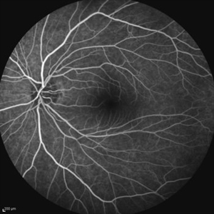
 chronic central serous chorioretinopathy
Oct 31 2012 by Mallika Goyal, MD
Fluorescein angiogram of inferior retina of right eye with chronic CSCR shows dilation of and mild leak from retinal vessels over the inferior serous retinal detachment.
Condition/keywords: central serous chorioretinopathy (CSCR), chronic central serous chorioretinopathy (CSCR), serous retinal detachment
-
 Primary Hyperoxaluria and Oxalosis
Oct 10 2015 by Hamid Ahmadieh, MD
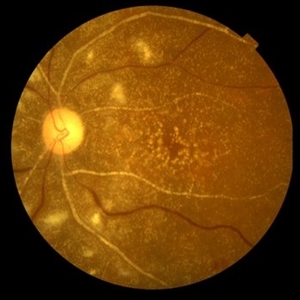
Color fundus photograph of the left eye of a 55-year-old woman with primary hyperoxaluria and oxalosis leading to intraretinal and subretinal deposition of calcium oxalate crystals . In addition, deposition of these crystals in the retinal vessels has led to the occlusion of retinal arterioles and venules leading to multiple cotton wools and dot and blot retinal hemorrhages.
Photographer: Shabnam Pooreh, Negah Eye Center, Tehran, Iran
Condition/keywords: color fundus photograph, oxalosis, primary hyperoxaluria
-
 Asymptomatic Eye in FEVR
Jul 7 2015 by Hamid Ahmadieh, MD
FA image of the asymptomatic left eye of a 28-year-old man with total RD secondary to advanced FEVR in his right eye. Notice straightening of the retinal vessels.
Photographer: Soulmaz Shahmohammad, Negah Eye Center, Tehran, Iran
Imaging device: Specteralis
Condition/keywords: asymptomatic, familial exudative vitreoretinopathy (FEVR)
-
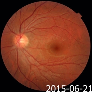
 Pigmentary Paravenous Retinochoroidal Atrophy (Left Eye)
Jun 30 2017 by Navneet Mehrotra, DNB
Fundus photograph of the left eye of a 44-year-old female with bony corpuscle pigmentation along the retinal vessels.
Photographer: Rakesh Juneja, Retina Foundation, Ahmedabad
Condition/keywords: atrophy, pigment, retinochoroidopathy
-
 Asymptomatic Eye in FEVR
Jul 7 2015 by Hamid Ahmadieh, MD
Color fundus photograph of the asymptomatic eye of a patient with FEVR. Notice straightening of the retinal vessels.
Photographer: Soulmaz Shahmohammad, Negah Eye Center, Tehran, Iran
Condition/keywords: color fundus photograph, familial exudative vitreoretinopathy (FEVR)
-
 Retinitis Pigmentosa
Feb 26 2020 by Manuel Ángel Alcántara Delgado, MD
Merged color fundus photograph of a 68-year-old woman with advanced retinitis pigmentosa. It is appreciated bone spicule-shaped pigment deposits, optic disc pallor, retinal atrophy, attenuated retinal vessels and surface wrinkling retinopathy.
Photographer: Manuel Ángel Alcántara Delgado
Condition/keywords: chorioretinal atrophy, choroidal circulation, optic disc pallor, pericentral retinitis pigmentosa, retina, retinitis pigmentosa, retinitis pigmentosa (RP) dystrophy, sector retinitis pigmentosa
-
---thumb.jpg/image-square;max$300,300.ImageHandler) Subfoveal Neovascular Membrane And Bridging Fibrotic Band To The Supertemporal Equator
Subfoveal Neovascular Membrane And Bridging Fibrotic Band To The Supertemporal Equator
Oct 4 2013 by Maurice F. Rabb
Acuity OD of 20/800 and OS 20/20. Manifest refraction shows anisometropia: OD +6.50 and OS + 0.75 spheres. The macula of the right eye has a dense fibrotic cicatricial subfoveal scar with mottling of pigment epithelium in the perifoveal area. There is a bridging fibrotic band that extends in the 10 o'clock meridian to the posterior equator tenting up retinal vessels between the two areas. The fluorescein angiogram done has the frames 20-25 reversed on the positive so that they are all of the right eye. The remainder are correctly oriented and show a subfoveal neovascular membrane with surrounding serous fluid overlying residual exudate. There is the fibrotic band that can be seen in the 10:00 o'clock meridian in the late photographs where retinal vessels under traction from the subretinal band leak somewhat.
Condition/keywords: bridging fibrotic band, subfoveal neovascular membrane, supertemporal equator
 Loading…