 Initializing download.
-
 By Alex P. Hunyor, MD
Retina Associates
Co-author(s): From the slide collection of Dr ABL Hunyor - Uploaded on Jan 11, 2013.
- Last modified by Alex P. Hunyor, MD on Sep 28, 2013.
- Rating
- Appears in
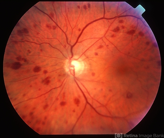
- Ocular ischaemic syndrome
- Condition/keywords
- ocular ischemic syndrome
- Imaging device
- Fundus camera
- Description
- Ocular ischaemic syndrome, left eye - color image, posterior pole. Note: dilated but not tortuous veins, attenuated arteries, and multiple intraretinal haemorrhages.


