Search results (185 results)
-
 Plaquenil Toxicity
Apr 30 2013 by Theodore Leng, MD, MS, FASRS
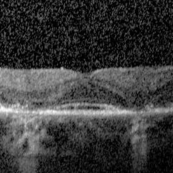
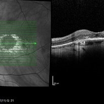
SD-OCT scan from a 44-year-old woman with bilateral plaquenil toxicity. There is damage visible in the outer retina in a perifoveal distribution.
Condition/keywords: hydroxychloroquine toxicity, plaquenil toxicity
-
 Plaquenil Toxicity
Apr 30 2013 by Theodore Leng, MD, MS, FASRS
SD-OCT scan from a 44-year-old woman with bilateral plaquenil toxicity. There is damage visible in the outer retina in a perifoveal distribution.
Condition/keywords: hydroxychloroquine toxicity, plaquenil toxicity
-
 Hollenhorst Plaque
Sep 18 2016 by John T. Thompson, MD
Color photo of Hollenhorst plaque at branch of inferotemporal artery.
Imaging device: Zeiss FF4
Condition/keywords: branch retinal artery occlusion (BRAO), hollenhorst plaque
-
 Plaquenil Toxicity
Apr 30 2013 by Theodore Leng, MD, MS, FASRS
Fundus autofluorescence from a 44-year-old woman with bilateral plaquenil toxicity. There is an area of hyperautofluorescence that corresponds to areas of outer retinal damage.
Condition/keywords: hydroxychloroquine toxicity, plaquenil toxicity
-
 Hollenhorst Plaque in Eye with CRAO
Oct 1 2012 by Jeffrey G. Gross, MD, FASRS
Hollenhorst plaque in eye with CRAO.
Condition/keywords: central retinal artery occlusion (CRAO), hollenhorst plaque, in eye
-
 Kyrieleis arteritis
Feb 15 2013 by From the Collections of Thomas M. Aaberg, MD and Thomas M. Aaberg Jr., MD
Nodular pattern of yellowish intraarterial plaques (aka Kyrieleis arteritis). typically seen in association with toxoplasma chorioretinitis.
Condition/keywords: Kyrieleis arteritis, toxoplasmosis
-
---thumb.jpg/image-square;max$300,300.ImageHandler) Hollenhorst Plaque
Hollenhorst Plaque
-
 Hollenhorst Plaque in Eye with BRAO
Oct 1 2012 by Jeffrey G. Gross, MD, FASRS
Hollenhorst plaque in eye with BRAO.
Condition/keywords: branch retinal artery occlusion (BRAO), hollenhorst plaque
-
 Plaquenil Toxicity
Apr 30 2013 by Theodore Leng, MD, MS, FASRS
Fundus autofluorescence from a 44-year-old woman with bilateral plaquenil toxicity. There is an area of hyperautofluorescence that corresponds to areas of outer retinal damage.
Condition/keywords: hydroxychloroquine toxicity, plaquenil toxicity
-
 Plaquenil Toxicity
Apr 30 2013 by Theodore Leng, MD, MS, FASRS
Right eye 10-2 HVF from a 44-year-old woman with bilateral plaquenil toxicity. A ring scotoma is present.
Condition/keywords: hydroxychloroquine toxicity, plaquenil toxicity
-
 Plaquenil Toxicity OCT
Dec 6 2016 by Courtney Crawford, MD, FACS
70-year-old woman with history of plaquenil use for rheumatoid arthritis.
Condition/keywords: plaquenil toxicity
-
 Hypertensive Choroidopathy - Right Eye
Dec 21 2016 by Maciej Czepita
Fundus photograph and SD-OCT scan as well as fundus autofluorescence image (FAF) of the right eye of a 70-year-old woman with hypertensive choroidopathy. In the fundus image numerous Elschnig's spots are visible. Note the Hollenhorst plaque in the superior temporal artery. In the SD-OCT scan (green line on the fundus image) the RPE layer is uneven. Numerous hypo and hyperautofluorescent patches can be seen in the fundus autofluorescence image.
Photographer: Maciej Czepita, M.D., Ph.D., Pomeranian Medical University, Szczecin, Poland
Imaging device: Heidelberg Spectralis HRA+OCT
Condition/keywords: hypertensive choroidopathy
-
 Plaquenil Toxicity
Apr 30 2013 by Theodore Leng, MD, MS, FASRS
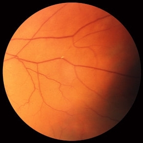
Color fundus photograph from a 44-year-old woman with bilateral plaquenil toxicity. A mild bulls-eye maculopathy is present.
Condition/keywords: hydroxychloroquine toxicity, plaquenil toxicity
-
 Plaquenil Toxicity
Apr 30 2013 by Theodore Leng, MD, MS, FASRS
Left Eye 10-2 HVF from a 44-year-old woman with bilateral plaquenil toxicity. A ring scotoma is present.
Condition/keywords: hydroxychloroquine toxicity, plaquenil toxicity
-
 Exudative Macular Degeneration, Prominent Plaque - FC
Oct 9 2012 by James B. Soque, CRA, OCT-C, COA, FOPS
88 y/o WM with extensive history of EMD and prominent plaque OS. Large heme OS has resolved with a variety of anti-VEGF therapy, and only a small heme, IT to fovea, remains. VA OS cc 20/50. (3) three photos: Color Photo, Early FA, and Late FA enclosed. 50 Degree, no mag.
Photographer: James Soque CRA COA
Imaging device: Topcon TRC 50 EX, with OIS V 10.5.74 Software. 5 MP Camera
Condition/keywords: exudative age-related macular degeneration
-
 Toxoplasmosis Slide 2
Oct 22 2012 by Ronald C. Gentile, MD
One month following treatment with Bactrim, Clindamycin, and oral prednisone the focal area chorioretinitis has coalesced with a decrease in overlying vitreous inflammation. Kyrieleis plaques can be seen along the inferior retinal arteriole.
Photographer: The New York Eye & Ear Infirmary Department of Medical Imaging
Condition/keywords: posterior uveitis, toxoplasmosis
-
 Plaquenil (Hydroxychloroquine) Toxicity Fundus Photo
Feb 26 2016 by Joshua O Mali, MD, FASRS
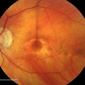
68-year-old female with 15 year history of plaquenil (hydroxychloroquine) use
Condition/keywords: plaquenil toxicity
-
 Plaquenil Toxicity
Apr 30 2013 by Theodore Leng, MD, MS, FASRS
Color fundus photograph from a 44-year-old woman with bilateral plaquenil toxicity. A mild bulls-eye maculopathy is present.
Condition/keywords: hydroxychloroquine toxicity, plaquenil toxicity
-
 Bull's Eye Maculopathy
Jun 29 2013 by Jason S. Calhoun
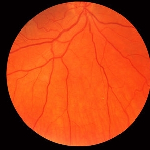
Patient comes in with severe vision loss in her right eye. Patient has been taking plaquenil for about 2 years. Fundus exam shows a bulls eye maculopathy centrally in the right eye.
Photographer: Jason S. Calhoun, Mayo Clinic Jacksonville, Florida
Imaging device: TOPCON TRC 50-EX
Condition/keywords: plaquenil toxicity
-
 Exudative Macular Degeneration, Prominent Plaque - FA early
Oct 9 2012 by James B. Soque, CRA, OCT-C, COA, FOPS
88 y/o WM with extensive history of EMD and prominent plaque OS. Large heme OS has resolved with a variety of anti-VEGF therapy, and only a small heme, IT to fovea, remains. VA OS cc 20/50. (3) three photos: Color Photo, Early FA, and Late FA enclosed. 50 Degree, no mag.
Photographer: James Soque CRA COA
Imaging device: Topcon TRC 50 EX, with OIS V 10.5.74 Software. 5 MP Camera
Condition/keywords: exudative age-related macular degeneration
-
 Central Retinal Artery Occlusion (CRAO)
Dec 27 2016 by Manish Nagpal, MD, FRCS (UK), FASRS
Acute CRAO with hollenhorst plaque.
Photographer: hardik Jain
Condition/keywords: central retinal artery occlusion (CRAO), edema, hollenhorst plaque, retinal infarction
-
 Branch Retinal Artery Occlusion
Oct 2 2013 by Jerald A. Bovino, MD
There is a hollenhorst plaque causing a branch retinal artery occlusion. The patient has scars from prior panretinal laser photocoagulation.
Condition/keywords: branch retinal artery occlusion (BRAO), hollenhorst plaque, pan-retinal photocoagulation (PRP)
-
 Amelanotic Choroidal Melanoma
Apr 12 2019 by David L Kilpatrick, MD
Fundus photograph of a 69-year-old male with an amelanotic choroidal melanoma and corresponding exudative retinal detachment. Transvitreal biopsy was performed at the time of radioactive I-125 plaque placement. The genetic expression profile revealed a Class 1A, PRAME negative tumor.
Photographer: Retina Consultants of Alabama, P. C.
Imaging device: Optos
Condition/keywords: amelanotic melanoma
-
 Choroidal Osteoma 5
Oct 5 2012 by Ronald C. Gentile, MD
B scan ultrasonography with representative A scan of the macular choroidal osteoma. The B scan reveals a characteristic highly reflective plaque consistent with its bone-like calcium composition that persists with low gain. The A scan reveals a large spike.
Photographer: The New York Eye & Ear Infirmary Department of Medical Imaging
Condition/keywords: B scan ultrasound, choroidal tumor, macular choroidal osteoma
-
 Plaquenil (Hydroxychloroquine) Toxicity OCT
Feb 26 2016 by Joshua O Mali, MD, FASRS
68-year-old female with 15 year history of plaquenil (hydroxychloroquine) use.
Condition/keywords: plaquenil toxicity
 Loading…