Search results (185 results)
-
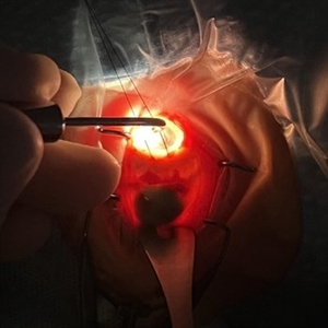 Intraoperative Transillumination of Choroidal Melanoma
Apr 18 2025 by Virginia Gebhart
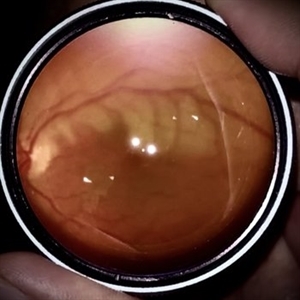
Intraoperative photo of transillumination of choroidal melanoma before plaque placement in 36 year old female.
Photographer: Chris Bergstrom, MD, OD
Imaging device: iphone
Condition/keywords: choroidal melanoma, intraoperative, transillumination
-
 Treated Melanoma with Iluvien Implant
Apr 9 2025 by Virginia Gebhart
62 year old female 4 mo s/p brachytherapy for amelanotic choroidal melanoma. Iluvien implant given 4 wks s/p plaque removal, lesion is stable with resolved exudative detachment and subretinal fluid
Photographer: Virginia Gebhart, Retina Consultants of Carolina
Imaging device: Optos California
Condition/keywords: amelanotic melanoma, brachytherapy, choroidal melanoma, Iluvien, melanoma
-
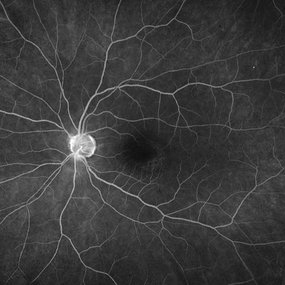
 Retinal Vasculitis
Mar 26 2025 by Korey Starkey
41 year-old patient presents with vascular FA findings of occlusive vasculitis with four quadrant Kyrieleis plaques OU showcases a possibly rare but reported atypical presentation of Behcet's Syndrome.
Photographer: Korey Starkey
Imaging device: Optos
Condition/keywords: FA early phase, Fundus Fluorescein Angiography, ischemia, Optos, retinal vasculitis, ultra-wide field imaging, venous beading
-
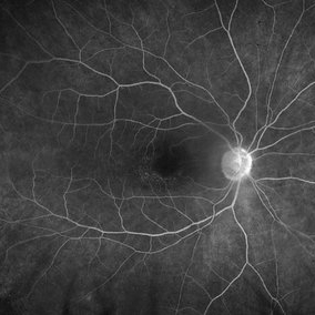
 Retinal Vasculitis
Mar 26 2025 by Korey Starkey
41 year-old patient presents with vascular FA findings of occlusive vasculitis with four quadrant Kyrieleis plaques OU showcases a possibly rare but reported atypical presentation of Behcet's Syndrome.
Photographer: Korey Starkey
Imaging device: Optos
Condition/keywords: Behcet's Disease, FA early phase, Fundus Fluorescein Angiography, Optos, retinal vasculitis, ultra-wide field imaging, venous beading
-
 Regressing Choroidal Melanoma
Mar 10 2025 by Virginia Gebhart
56 year old male 4 months s/p plaque brachytherapy for choroidal melanoma. Tumor is regressing, there is an exudative detachment with worsening SRF. Treated with Avastin to promote hopeful improvement of the SRF
Photographer: Virginia Gebhart, Retina Consultants of Carolina
Imaging device: Optos California
Condition/keywords: brachytherapy, Choroidal melanoma, exudative detachment, melanoma
-
 Choroidal Melanoma
Mar 10 2025 by Virginia Gebhart
56 year old female with new choroidal melanoma. Pt states they have a "freckle" that had been monitored for 26 years, last CEE was over 2 years ago. Clinical exam and ancillary testing consistent with uveal melanoma. Pt scheduled for plaque brachytherapy with transretinal biopsy of the tumor for genetic testing. Pt also scheduled for CT scan of chest/abdomen to rule out metastatic disease.
Photographer: Virginia Gebhart, Retina Consultants of Carolina
Imaging device: Optos California
-
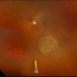
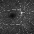
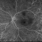
 Choroidal Melanoma 3 Ways
Jan 16 2025 by Virginia Gebhart
RGB/FA/ICG of 76 year old female with a new choroidal melanoma. Pt scheduled for plaque radiation. BCVA 20/400
Photographer: Virginia Gebhart, Retina Consultants of Carolina
Imaging device: Optos California
Condition/keywords: fluorescein angiogram (FA), indocyanine green (ICG) angiography, OPTOS CALIFORNIA RGB
-
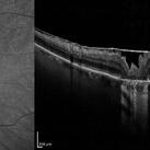
 NIR and OCT Right Eye Early Parafoveal Changes Plaquenil
Jan 14 2025 by Kyle D Kovacs, MD
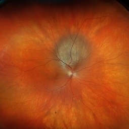
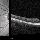
58 year old woman with 11 year history of plaquenil use with early parafoveal outer retinal attenuation and bull's eye on near-infrared imaging. Right eye.
Condition/keywords: OCT, plaquenil toxicity
-
 NIR and OCT Left Eye Early Parafoveal Changes Plaquenil
Jan 14 2025 by Kyle D Kovacs, MD
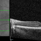
58 year old woman with 11 year history of plaquenil use with early parafoveal outer retinal attenuation and bull's eye on near-infrared imaging. Left eye
Condition/keywords: OCT, plaquenil toxicity
-
 New Iris Melanoma
Oct 10 2024 by Virginia Gebhart
56 year old male with new amelanotic melanoma emanating from the ciliary body through the posterior iris epithelium. CT scan showed no evidence of metastatic disease. Pt scheduled for radioactive plaque and tumor biopsy
Photographer: Virginia Gebhart, Retina Consultants of Carolina
Imaging device: Samsung Galaxy
Condition/keywords: amelanotic melanoma, iris melanoma
-
 Occlusive Retinal Vasculitis
Oct 3 2024 by Logan ryzenga
4 view ultra-widefield Optos fluorescein angiogram in the left eye of a 39 year old woman occlusive retinal vasculitis with four quadrant Kyrieleis plaques. This is a showcase of a suspected, rarely reported, and atypical presentation of Behcet's Syndrome.
Photographer: Logan Ryzenga
Imaging device: Optos California
Condition/keywords: Behcet's Disease, Behcet's uveitis, Fluorescein angiography, fluorescein leakage, kyrieleis plaques, non-perfusion, OPTOS, OPTOS CALIFORNIA, ultra-wide field imaging, Uveitis
-
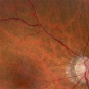
 Branch Retinal Artery Occlusion
Oct 1 2024 by Angel Enrique Flores Pineda
Fundus photograph of a 78-year-old woman with poorly controlled systemic arterial hypertension and dyslipidemia. Hollenhorst plaque can be observed.
Photographer: Angel Enrique Flores Pineda, Hospital General de Zona #20
Imaging device: Smartphone (IPhone 15 plus)
Condition/keywords: branch retinal artery occlusion (BRAO)
-
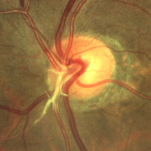
 Hollenhorst Plaque
Jun 25 2024 by Virginia Gebhart
75 year female with complaint of shadow in the bottom of her vision for many years. Hollenhorst plaque on superior pole of the disc and sclerotic superotemporal arteriole. Also DBHs superiorly most likely due to combined BRAO/BRVO.
Photographer: Virginia Gebhart
Imaging device: Topcon 50DX
Condition/keywords: branch retinal artery occlusion (BRAO), branch retinal vein occlusion (BRVO), hollenhorst plaque, sclerotic arteriole
-
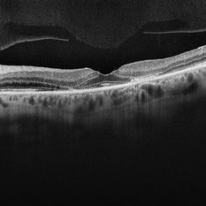
 Bullseye Maculopathy
Jan 22 2024 by Kali Jend
Optical coherence tomography of a 73-year-old female with Bullseye Macular Changes affecting her left eye. Patient reports having a family history of this condition and denies prior Plaquenil or Elmiron use. Compared to previous imaging, the patient's condition progressed in the left eye from 2020 to 2023. Patient has a history of fluctuating Diabetic Macular Edema and a current Epiretinal Membrane as well. Patient's vision was Ncc20/60 at the time the image was taken.
Photographer: Kali Jend
Imaging device: Heidelberg Spectralis
Condition/keywords: bullseye maculopathy, epiretinal membrane (ERM), heidelberg spectralis, left eye, macular pucker, OCT, optical coherence tomography (OCT)
-
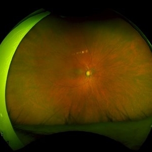
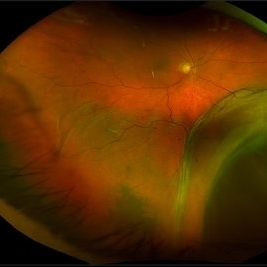 New Choroidal Melanoma
Jan 4 2024 by Virginia Gebhart
77 year old male with a bilobed pigmented mass with exudative RD, and trace inflammation present in AV consistent with choroidal melanoma. Mass extends into ciliary body. Pt scheduled for MRI prior to plaque radiation to rule out metastasis.
Photographer: Virginia Gebhart
Imaging device: Optos California
Condition/keywords: ciliary body melanoma, exudative retinal detachment
-
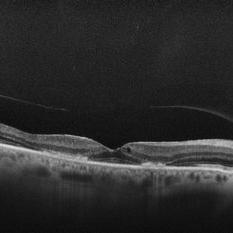
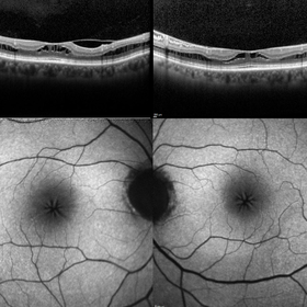 Macular Foveoschisis
Nov 9 2023 by Charlotte Jones
Bilateral ocular coherence tomography and fundus autofluorescence of a 77 year old woman with Macular Foveoschisis. Patient with stable vision since her last appointment (20/30 right eye and 20/25 left eye) with worsening vitreomacular traction in the right eye. Patient is followed routinely for Plaquenil use.
Photographer: Charlotte Jones
Imaging device: Heidelberg Spectralis
Condition/keywords: fundusautofluorescence, macularfoveoschisis, macularretinoschisis, macularstar, ocularcoherencetomography
-
 Hollenhorst Plaque
Sep 21 2023 by Ben Serar
Fundus photograph showing multiple whitish intra-arterial dot like deposits suggestive of Hollenhorst plaques.
Condition/keywords: hollenhorst plaque
-
 A Glow in the Darkness: Hollenhorst Plaque
Aug 22 2023 by Harsh Vardhan Singh, MS
82 year-old-female with a history of some disturbance of vision in the left eye with the finding of hollenhorst plaque in one of the branches of central retinal artery
Photographer: Dr Harsh Vardhan Singh
Condition/keywords: hollenhorst plaque
-
 A Glow in the Darkness : Hollenhorst Plaque
Aug 21 2023 by Harsh Vardhan Singh, MS
82-year-old-female with a history of some disturbance of vision in the left eye with the finding of hollenhorst plaque in one of the branches of central retinal artery
Photographer: Dr Harsh Vardhan Singh
Condition/keywords: hollenhorst plaque
-
 Hydroxychloroquine Maculopathy
Jul 23 2023 by Ahmad B. Tarabishy, MD
62 year old female with rheumatoid arthritis, treated with hydroxychloroquine 200 mg BID for the past 6-8 years. She presents with blurred vision, difficulty reading, and difficulty transitions from dark to light conditions since 4 months.
Photographer: Dr. Angela Rico
Condition/keywords: hydroxychloroquine toxicity, plaquenil toxicity, toxic maculopathy
 Loading…