Search results (185 results)
-
 New Iris Melanoma
Oct 10 2024 by Virginia Gebhart
56 year old male with new amelanotic melanoma emanating from the ciliary body through the posterior iris epithelium. CT scan showed no evidence of metastatic disease. Pt scheduled for radioactive plaque and tumor biopsy
Photographer: Virginia Gebhart, Retina Consultants of Carolina
Imaging device: Samsung Galaxy
Condition/keywords: amelanotic melanoma, iris melanoma
-
 Plaquenil Toxicity
Apr 30 2013 by Theodore Leng, MD, MS, FASRS
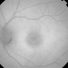
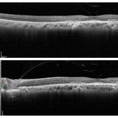
SD-OCT scan from a 44-year-old woman with bilateral plaquenil toxicity. There is damage visible in the outer retina in a perifoveal distribution.
Condition/keywords: hydroxychloroquine toxicity, plaquenil toxicity
-
 Bear Tracks
Nov 10 2020 by Ronald Coriasso
Fundus photo of 68-year-old female with history of plaquenil use. Her findings are most consistent with bear tracks, however these kinds of lesions can be indicative of familial adenomatous polyposis (FAP).
Photographer: Ronald Coriasso
Imaging device: OPTOS
Condition/keywords: bear tracks, familial adenomatous polyposis
-
 HHPlaqueON
Aug 13 2021 by Jeffrey Barker
Hollenhorst Plaque
Photographer: Jeffrey P. Barker, B.S. Retina Vitreous Surgeons of C.N.Y.
Condition/keywords: hollenhorst plaque, optic nerve
-
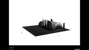 3D OCT of juxtapapillary melanoma
May 15 2020 by Sophia El Hamichi, MD
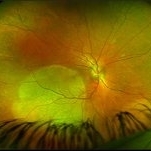
A 63-year-old male with juxtapapillary melanoma of the right eye. Visual acuity at presentation was 20/25 OD. Patient treated with brachytherapy Iodine125 plaque
Photographer: Belinda Rodriguez
Condition/keywords: optical coherence tomography (OCT)
-
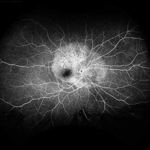 Acute Syphilitic Posterior Placoid Chorioretinitis
May 4 2021 by RAFAEL REIS PEREIRA, MD
A 31-year-old patient with a complaint of photophobia and low visual acuity OD in the previous three weeks. BCVA was 20/60 and 20/20 The fundus examination revealed a placoid white lesion in the posterior pole and vitreous cells in the right eye. The left eye was unremarkable. Fluorescein angiography reveals hyperfluorescent plaque with distinctive “leopard spots” hypofluorescence.
Imaging device: Opto California
Condition/keywords: acute syphilitic posterior placoid chorioretinitis
-
 Branch Retinal Artery Occlusion
Sep 9 2018 by Gabriela Lopezcarasa Hernandez, MD
88-year-old female patient with sudden decrease in visual acuity and scotoma in left eye, please notice the widening retina due to retinal edema of branch occlusion with hollenhorst plaque in the artery and the optic nerve.
Photographer: Araceli Rojas
Imaging device: Zeiss FF4
Condition/keywords: branch retinal artery occlusion (BRAO)
-
 Branch Retinal Artery Occlusion
Sep 9 2018 by Gabriela Lopezcarasa Hernandez, MD
88-year-old female patient with sudden decrease in visual acuity and scotoma in left eye, please notice the branch occlusion with hollenhorst plaque and the delay perfusion in the involved arteria.
Photographer: Araceli Rojas
Imaging device: Zeiss FF4
Condition/keywords: branch retinal artery occlusion (BRAO)
-
 Branch Retinal Artery Occlusion
Mar 27 2018 by Nichole Lewis
Branch retinal artery occlusion with a Hollenhorst Plaque.
Photographer: Nichole Lewis
Condition/keywords: branch retinal artery occlusion (BRAO), hollenhorst plaque
-
 Central Retinal Artery Occlusion (CRAO)
Dec 27 2016 by Manish Nagpal, MD, FRCS (UK), FASRS
Acute CRAO with hollenhorst plaque.
Photographer: hardik Jain
Condition/keywords: central retinal artery occlusion (CRAO), edema, hollenhorst plaque, retinal infarction
-
 Choroidal Melanoma 3 Ways
Jan 16 2025 by Virginia Gebhart
RGB/FA/ICG of 76 year old female with a new choroidal melanoma. Pt scheduled for plaque radiation. BCVA 20/400
Photographer: Virginia Gebhart, Retina Consultants of Carolina
Imaging device: Optos California
Condition/keywords: fluorescein angiogram (FA), indocyanine green (ICG) angiography, OPTOS CALIFORNIA RGB
-
 Hydroxychloroquine Maculopathy
Jul 23 2023 by Ahmad B. Tarabishy, MD
62 year old female with rheumatoid arthritis, treated with hydroxychloroquine 200 mg BID for the past 6-8 years. She presents with blurred vision, difficulty reading, and difficulty transitions from dark to light conditions since 4 months.
Photographer: Dr. Angela Rico
Condition/keywords: hydroxychloroquine toxicity, plaquenil toxicity, toxic maculopathy
-
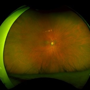
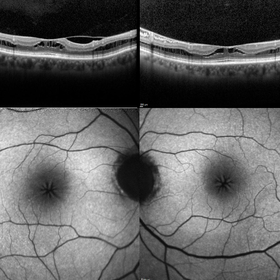 Macular Foveoschisis
Nov 9 2023 by Charlotte Jones
Bilateral ocular coherence tomography and fundus autofluorescence of a 77 year old woman with Macular Foveoschisis. Patient with stable vision since her last appointment (20/30 right eye and 20/25 left eye) with worsening vitreomacular traction in the right eye. Patient is followed routinely for Plaquenil use.
Photographer: Charlotte Jones
Imaging device: Heidelberg Spectralis
Condition/keywords: fundusautofluorescence, macularfoveoschisis, macularretinoschisis, macularstar, ocularcoherencetomography
-
 Occlusive Retinal Vasculitis
Oct 3 2024 by Logan ryzenga
4 view ultra-widefield Optos fluorescein angiogram in the left eye of a 39 year old woman occlusive retinal vasculitis with four quadrant Kyrieleis plaques. This is a showcase of a suspected, rarely reported, and atypical presentation of Behcet's Syndrome.
Photographer: Logan Ryzenga
Imaging device: Optos California
Condition/keywords: Behcet's Disease, Behcet's uveitis, Fluorescein angiography, fluorescein leakage, kyrieleis plaques, non-perfusion, OPTOS, OPTOS CALIFORNIA, ultra-wide field imaging, Uveitis
-
 Plaquenil Toxicity
Jul 11 2018 by Sarah Oelrich
Plaquenil Toxicity
Photographer: Sarah Oelrich CRA, Southeastern Retina Associates, Knoxville TN
Condition/keywords: plaquenil toxicity
-
 Plaquenil Toxicity
Apr 30 2013 by Theodore Leng, MD, MS, FASRS
SD-OCT scan from a 44-year-old woman with bilateral plaquenil toxicity. There is damage visible in the outer retina in a perifoveal distribution.
Condition/keywords: hydroxychloroquine toxicity, plaquenil toxicity
-
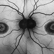
 Plaquenil Toxicity
Apr 30 2013 by Theodore Leng, MD, MS, FASRS
Fundus autofluorescence from a 44-year-old woman with bilateral plaquenil toxicity. There is an area of hyperautofluorescence that corresponds to areas of outer retinal damage.
Condition/keywords: hydroxychloroquine toxicity, plaquenil toxicity
-
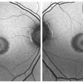 Plaquenil Toxicity
Feb 25 2021 by Niloofar Piri, MD
Bilateral fundus autofluorescence of a patient with hydroxycholoroquine (plaquenil) toxicity demonstrating classic bull's eye pattern.
Condition/keywords: bull's eye maculopathy, hydroxychloroquine toxicity, plaquenil toxicity
-
 Plaquenil Toxicity
Jun 8 2016 by John S. King, MD
AF 6 years med.
Condition/keywords: bull's eye maculopathy, plaquenil toxicity
-
 Plaquenil Toxicity: "Flying Saucer"
Feb 25 2021 by Niloofar Piri, MD
SD- OCT of the same patient with hydroxychloroquine (Plaquenil) toxicity, demonstrating classic "Flying Saucer" pattern secondary to parafoveal RPE atrophy, and outer retinal layer disruption including EZ loss, and outer nuclear layer loss.
Condition/keywords: bull's eye maculopathy, hydroxychloroquine toxicity, plaquenil toxicity
-
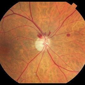
 Posterior Placoid Chorioretinopathy
Dec 19 2020 by John S. King, MD
44-year-old white female seen over the weekend complaining of a "spot" in her vision centrally OD for three days. She was referred over by another eye doctor who was concerned about a possible retinal detachment vs ARN in that eye. Her past medical history includes adrenal insufficiency for which she takes a low dose of hydrocortisone, thyroxine (post thyroidectomy), Plaquenil (inflammatory arthritis). She is divorced with one partner and denies any IVDU. Va 20/200 OD and 20/20 OS, IOP 12 OU, pupils mydriatic post gtts (light desaturation OD). There was 1+ A/C cell OD, O/W unremarkable anterior segment OU; in the posterior segment OD there was 1+ vitritis with a diffusely swollen optic disc and a large yellowish placoid lesion in the macula with yellowish border and extended out past the arcades inferiorly, as well as another lesion smaller in the IN periphery, and two possible smaller spots SN (See Photo above). There was a trace vitreous cell OS with a large, granular placoid lesion nasally. The OCT showed mild subfoveal fluid with nodular areas in the RPE and some overlying irregular architecture of the outer retina. Syphilis was a concern at this point. She denied any hand or foot rash, and said that she was recently working on the house, and her hands were dried out. There did appear to be a rash on the hand, and later learned that she had a rash on the soles of her feet. She was sent to ED for a work-up and her syphilis IgG was positive and VDRL 1:128, and negative for HIV. She was started on a course IV Penicillin (40mg PO steroid two days after tx started). She has responded well. A few days after treatment her visual acuity has improved to 20/60 OD; there was no anterior segment inflammation OU, and decreased vitreous cell OU. Disc edema was improved. The large placoid lesion in the macula of the right eye was slightly enlarged, but more granular in appearance without a distinct yellowish border, and the smaller lesions SN had dissipated. OCT showed resolution of the subfoveal fluid and an improved appearance of the outer retina and RPE layer.
Imaging device: Optos CA
Condition/keywords: acute syphilitic posterior placoid chorioretinitis, syphilis
-
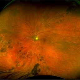
 Amelanotic Choroidal Melanoma
Apr 12 2019 by David L Kilpatrick, MD
Fundus photograph of a 69-year-old male with an amelanotic choroidal melanoma and corresponding exudative retinal detachment. Transvitreal biopsy was performed at the time of radioactive I-125 plaque placement. The genetic expression profile revealed a Class 1A, PRAME negative tumor.
Photographer: Retina Consultants of Alabama, P. C.
Imaging device: Optos
Condition/keywords: amelanotic melanoma
-
 Choroidal Melanoma
Jan 8 2016 by Jared Watson
42-year-old white female, S/P I-125 plaque brachytherapy.
Photographer: Jared Watson COT/CRA
Imaging device: Topcon 50EX, OIS-Merge
-
 Exudative Macular Degeneration, Prominent Plaque - FA early
Oct 9 2012 by James B. Soque, CRA, OCT-C, COA, FOPS
88 y/o WM with extensive history of EMD and prominent plaque OS. Large heme OS has resolved with a variety of anti-VEGF therapy, and only a small heme, IT to fovea, remains. VA OS cc 20/50. (3) three photos: Color Photo, Early FA, and Late FA enclosed. 50 Degree, no mag.
Photographer: James Soque CRA COA
Imaging device: Topcon TRC 50 EX, with OIS V 10.5.74 Software. 5 MP Camera
Condition/keywords: exudative age-related macular degeneration
 Loading…