Search results (19 results)
-
---thumb.jpg/image-square;max$300,300.ImageHandler) Primary Hyperoxaluria and Oxalosis
Primary Hyperoxaluria and Oxalosis
Jul 24 2013 by Hamid Ahmadieh, MD
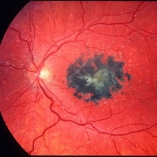
Color fundus photograph of the right eye of a 55-year-old man with primary hyperoxaluria and oxalosis. Characteristic crystalline retinopathy (flecked retina), black geographic maculopathy, and partial optic atrophy are visible. In addition, occluded branches of central retinal artery due to calcium oxalate deposition are visible.
Photographer: Hanieh Payab, Ophthalmic Research Center, Labbafinejad Medical Center, Tehran
Imaging device: Topcon Fundus Camera
Condition/keywords: oxalosis, primary hyperoxaluria
-
 Oxalosis
May 2 2013 by Henry J. Kaplan, MD
Crystalline retinopathy in oxalosis as a result of calcium oxalate deposits in the retina ; also deposition in RPE which causes fleck retina as pigmentary lesion in the center.
Condition/keywords: crystalline retinopathy, oxalosis
-
---thumb.jpg/image-square;max$300,300.ImageHandler) Primary Hyperoxaluria and Oxalosis
Primary Hyperoxaluria and Oxalosis
Jul 24 2013 by Hamid Ahmadieh, MD
Red-free image of the left eye of a 55-year-old man with primary hyperoxaluria and oxalosis. Extensive deposition of calcium oxalate crystals are demonstrated in the RPE . Deposition of crystals in retinal arteries as well as venous caliber abnormality are also visible.
Photographer: Hanieh Payab, Ophthalmic Research Center, Labbafinejad Medical Center, Tehran
Imaging device: Topcon Fundus Camera
Condition/keywords: ocular manifestation, oxalosis, primary hyperoxaluria, red-free
-
---thumb.jpg/image-square;max$300,300.ImageHandler) Primary Hyperoxaluria and Oxalosis
Primary Hyperoxaluria and Oxalosis
Jul 24 2013 by Hamid Ahmadieh, MD
Color fundus photograph of the right eye of a 55-year-old man with primary hyperoxaluria and oxalosis. Characteristic crystalline retinopathy (flecked retina), black geographic maculopathy, and partial optic atrophy are visible. In addition, occluded branches of central retinal artery due to calcium oxalate deposition are visible.
Photographer: Hanieh Payab, Ophthalmic Research Center, Labbafinejad Medical Center, Tehran
Imaging device: Topcon Fundus Camera
Condition/keywords: oxalosis, primary hyperoxaluria
-
---thumb.jpg/image-square;max$300,300.ImageHandler) Primary Hyperoxaluria and Oxalosis
Primary Hyperoxaluria and Oxalosis
Jul 24 2013 by Hamid Ahmadieh, MD
Early phase FA image of the left eye of a 55-year-old man with primary hyperoxaluria and oxalosis, Delayed filling of retinal vessels due to intravascular deposition of calcium oxalate crystals and non-perfusion of the temporal retina are visible.
Photographer: Hanieh Payab, Ophthalmic Research Center, Labbafinejad Medical Center, Tehran
Imaging device: Topcon Fundus Camera
Condition/keywords: ocular manifestation, oxalosis, primary hyperoxaluria
-
---thumb.jpg/image-square;max$300,300.ImageHandler) Primary Hyperoxaluria and Oxalosis
Primary Hyperoxaluria and Oxalosis
Jul 24 2013 by Hamid Ahmadieh, MD
Red-free image of the left eye of a 55-year-old man with primary hyperoxaluria and oxalosis. Extensive deposition of calcium oxalate crystals are demonstrated in the RPE . Deposition of crystals in retinal arteries as well as venous caliber abnormality are also visible.
Photographer: Hanieh Payab, Ophthalmic Research Center, Labbafinejad Medical Center, Tehran
Imaging device: Topcon Fundus Camera
Condition/keywords: oxalosis, primary hyperoxaluria, red-free
-
---thumb.jpg/image-square;max$300,300.ImageHandler) Primary Hyperoxaluria and Oxalosis
Primary Hyperoxaluria and Oxalosis
Jul 24 2013 by Hamid Ahmadieh, MD
Red-free image of the left eye of a 55-year-old man with primary hyperoxaluria and oxalosis. Extensive deposition of calcium oxalate crystals are demonstrated in the RPE . Deposition of crystals in retinal arteries as well as venous caliber abnormality are also visible.
Photographer: Hanieh Payab, Ophthalmic Research Center, Labbafinejad Medical Center, Tehran
Imaging device: Topcon Fundus Camera
Condition/keywords: ocular manifestation, oxalosis, primary hyperoxaluria, red-free
-
---thumb.jpg/image-square;max$300,300.ImageHandler) Primary Hyperoxaluria and Oxalosis
Primary Hyperoxaluria and Oxalosis
Jul 24 2013 by Hamid Ahmadieh, MD
Late phase FA image of the left eye of a 55-year-old man with primary hyperoxaluria and oxalosis. Profound leakage from disc due to NVD is visible. Vasoproliferative retinopathy has occurred secondary to retinal ischemia due to intravascular deposition of calcium oxalate crystals.
Photographer: Hanieh Payab, Ophthalmic Research Center, Tehran
Imaging device: Topcon Fundus Camera
Condition/keywords: oxalosis, primary hyperoxaluria, vasoproliferative retinopathy
-
---thumb.jpg/image-square;max$300,300.ImageHandler) Primary Hyperoxaluria and Oxalosis
Primary Hyperoxaluria and Oxalosis
Jul 24 2013 by Hamid Ahmadieh, MD
Color fundus photograph of the left eye of a 55-year-old man with primary hyperoxaluria and oxalosis. Vitreous hemorrhage originating from NVD due to vasoproliferative retinopathy is seen.
Photographer: Hanieh Payab, Ophthalmic Research Center, Tehran
Imaging device: Topcon Fundus Camera
Condition/keywords: neovascularization of the disc (NVD), oxalosis, primary hyperoxaluria, vasoproliferative retinopathy
-
---thumb.jpg/image-square;max$300,300.ImageHandler) Primary Hyperoxaluria and Oxalosis
Primary Hyperoxaluria and Oxalosis
Jul 24 2013 by Hamid Ahmadieh, MD
Red-free image of the left eye of a 55-year-old man with primary hyperoxaluria and oxalosis. Extensive deposition of calcium oxalate crystals are demonstrated in the RPE . Deposition of crystals in retinal arteries as well as venous caliber abnormality are also visible.
Photographer: Hanieh Payab, Ophthalmic Research Center, Labbafinejad Medical Center, Tehran
Imaging device: Topcon Fundus Camera
Condition/keywords: ocular manifestation, oxalosis, primary hyperoxaluria, red-free
-
---thumb.jpg/image-square;max$300,300.ImageHandler) Primary Hyperoxaluria and Oxalosis
Primary Hyperoxaluria and Oxalosis
Jul 24 2013 by Hamid Ahmadieh, MD
Mid phase FA image of the left eye of a 55-year-old man with primary hyperoxaluria and oxalosis, Delayed filling of retinal vessels due to intravascular deposition of calcium oxalate crystals and non-perfusion of the temporal retina are visible.
Photographer: Hanieh Payab, Ophthalmic Research Center, Labbafinejad Medical Center, Tehran
Imaging device: Topcon Fundus Camera
Condition/keywords: ocular manifestation, oxalosis, primary hyperoxaluria
-
 Primary Hyperoxaluria and Oxalosis
Oct 10 2015 by Hamid Ahmadieh, MD
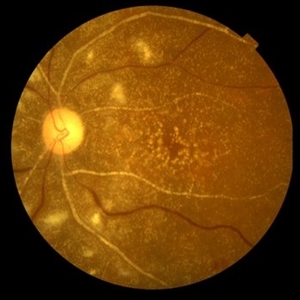
Color fundus photograph of the left eye of a 55-year-old woman with primary hyperoxaluria and oxalosis leading to intraretinal and subretinal deposition of calcium oxalate crystals . In addition, deposition of these crystals in the retinal vessels has led to the occlusion of retinal arterioles and venules leading to multiple cotton wools and dot and blot retinal hemorrhages.
Photographer: Shabnam Pooreh, Negah Eye Center, Tehran, Iran
Condition/keywords: color fundus photograph, oxalosis, primary hyperoxaluria
-
 Oxalosis / Methoxyflurane
-
 Primary Hyperoxaluria and Oxalosis
Oct 10 2015 by Hamid Ahmadieh, MD
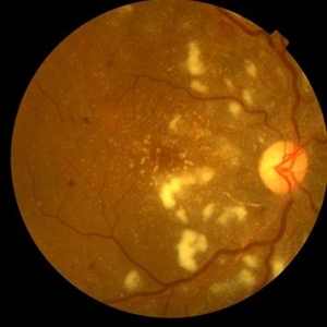
Color fundus photograph of the right eye of a 55-year-old woman with primary hyperoxaluria and oxalosis leading to intraretinal and subretinal deposition of calcium oxalate crystals . In addition, deposition of these crystals in the retinal vessels has led to the occlusion of retinal arterioles and venules leading to multiple cotton wools and dot and blot retinal hemorrhages.
Photographer: shabnam Pooreh, Negah Eye Center, Tehran, Iran
Condition/keywords: color fundus photograph, oxalosis, primary hyperoxaluria
-
 Primary Hyperoxaluria and Oxalosis
Oct 10 2015 by Hamid Ahmadieh, MD
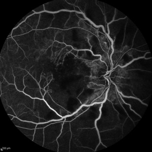
FA image of the left eye of a 55-year-old woman with primary hyperoxaluria and oxalosis showing occlusion of the retinal arteries. Staining of calcium oxalate crystals is visible.
Photographer: shabnam Pooreh, Negah Eye Center, Tehran, Iran
Condition/keywords: oxalosis, primary hyperoxaluria
-
 Primary Hyperoxaluria and Oxalosis
Oct 10 2015 by Hamid Ahmadieh, MD
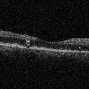
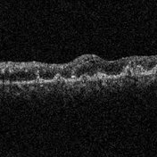
OCT image of the right eye of a 55- year- old woman with primary hyperoxaluria and oxalosis. Intraretinal and sub- retinal deposition of calcium oxalate crystals are visible .
Photographer: Shabnam Pooreh, Negah Eye Center, Tehran, Iran
Condition/keywords: optical coherence tomography (OCT), oxalosis, primary hyperoxaluria
-
 Primary Hyperoxaluria and Oxalosis
Oct 10 2015 by Hamid Ahmadieh, MD
Late venous phase FA image of the right of a 55-year-old woman with primary hyperoxaluria and oxalosis . Notice macular infarction and areas of capillary non -perfusion in retinal mid periphery due to the occlusion of retinal arterioles.
Photographer: Shabnam Pooreh, Negah Eye Center, Tehran, Iran
Condition/keywords: oxalosis, primary hyperoxaluria
-
 Oxalosis / Methoxyflurane
-
 Primary Hyperoxaluria and Oxalosis
Oct 10 2015 by Hamid Ahmadieh, MD
OCT image of the left eye of a 55- year- old woman with oxalosis. Intraretinal and sub -retinal deposition of calcium oxalate crystals are seen.
Photographer: Shabnam Pooreh, Negah Eye Center, Tehran, Iran
Condition/keywords: optical coherence tomography (OCT), oxalosis
 Loading…