Search results (1761 results)
-
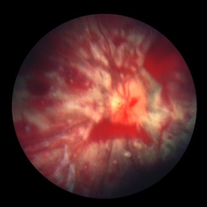
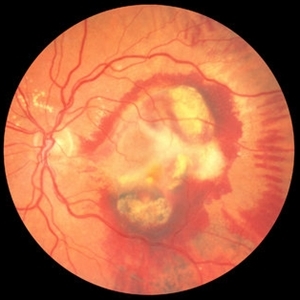
 Subhyaloid Hemorrhage
Oct 8 2012 by Jeffrey G. Gross, MD, FASRS
Subhyaloid hemorrhage, layered, with surrounding subretinal hemorrhage.
Condition/keywords: subhyaloid hemorrhage, subretinal hemorrhage
-
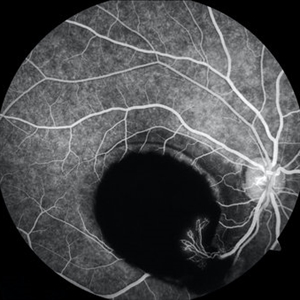 Sub-ILM Hemorrhage with Neovessels
Apr 30 2020 by Saurabh Deshmukh, MBBS, DNB, FVRS, MNAMS
Late arteriovenous phase FA showing a large sub-internal limiting membrane hemorrhage with overlying neovessels. This hypertensive patient presented with a visual acuity of counting fingers at 2 meters. The patient was advised intravitreal anti-VEGF injection, Nd: YAG Membranotomy, and systemic control of hypertension.
Photographer: Saurabh Deshmukh, Sri Sankaradeva Nethralaya, Guwahati, India
Imaging device: Topcon TRC-50 DX
Condition/keywords: hypertensive retinopathy, neovascularization elsewhere (NVE), subILM hemorrhage
-
---thumb.jpg/image-square;max$300,300.ImageHandler) Roth Spot
Roth Spot
Feb 27 2013 by Henry J. Kaplan, MD
Roth spots due to subacute bacterial endocardiris in a patient with the diagnosis of AIDS .
Condition/keywords: AIDS, subacute bacterial endocardiris, white centered retinal hemorrhage (Roth Spot)
-
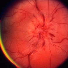 Papillitis
May 2 2013 by Henry J. Kaplan, MD
Anterior optic neuropathy or papillitis in the right eye; notice the blurred optic disc margin, engorged capillaries and flame shaped hemorrhages at the margin.
Condition/keywords: optic disc edema, optic disc swelling, papillitis
-
 Retinal Vasculitis with Hemorrhages and Cotton Wool Spots
Oct 16 2012 by Jeffrey G. Gross, MD, FASRS
Retinal vasculitis with hemorrhages and cotton wool spots.
Condition/keywords: cotton wool spots, retinal vasculitis
-
 Ocular Manifestation of Acute Leukemia
Sep 8 2012 by Hamid Ahmadieh, MD
Color fundus photograph of a 26-year-old man with acute leukemia.
Photographer: Hamid Ahmadieh, MD, Ophthalmic Research Center, Labbafinejad Medical Center, Shahid Beheshti University of Medical Sciences , Tehran
Imaging device: Topcon Fundus Camera
Condition/keywords: acute leukemia, white centered retinal hemorrhage (Roth Spot)
-
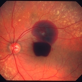
 "Boat-Shaped" Preretinal Hemorrhage
Feb 21 2019 by Mitzy E Torres Soriano, MD
Color fundus photograph showing preretinal (subhyaloid) hemorrhage in a diabetic patient with proliferative diabetic retinopathy.
Photographer: Andrea Vitale, MD
Condition/keywords: preretinal hemorrhage, proliferative diabetic retinopathy (PDR), subhyaloid hemorrhage
-
 Shaken Baby Syndrome
Sep 20 2012 by Jeffrey G. Gross, MD, FASRS
Shaken Baby Syndrome with many hemorrhages and cotton wool spots
Condition/keywords: cotton wool spots, shaken baby syndrome
-
 Subfoveal Subretinal Hemorrhage
Aug 28 2012 by Sharon Fekrat, MD FACS FASRS
subfoveal subretinal hemorrhage, right eye.
Photographer: Michael P. Kelly, FOPS Director, Duke Eye labs Duke University Eye Center Durham, NC
Imaging device: Zeiss FF40
Condition/keywords: subretinal hemorrhage
-
 Lyme Disease
Feb 13 2013 by From the Collections of Thomas M. Aaberg, MD and Thomas M. Aaberg Jr., MD
Papilledema, intra-retinal hemorrhage, periopticneuritis.
Condition/keywords: intraretinal hemorrhage, Lyme disease, periopticneuritis
-
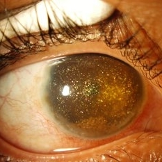 Synchysis Scintillans
Sep 17 2015 by Jessica G Lee, MD
24-year-old male with history of chronic retinal detachment.
Photographer: Bob Masini
Condition/keywords: cholesterol crystals, refractile bodies, synchysis scintillans, trauma, vitreous hemorrhage
-
 CRVO with Flame Hemorrhages
Oct 1 2012 by Jeffrey G. Gross, MD, FASRS
CRVO with flame hemorrhages and cotton wool spots 20/80.
Condition/keywords: 20/80, central retinal vein occlusion (CRVO), cotton wool spots
-
 Sickle Salmon-Patch Hemorrhage
Oct 23 2012 by Larry Halperin, MD
Salmon-patch hemorrhage in sickle cell
Condition/keywords: salmon patch, sickle cell retinopathy
-
 Optic Disc Edema and Hemorrhages with Subdural Hematoma
Oct 1 2012 by Jeffrey G. Gross, MD, FASRS
Optic disc edema and hemorrhages with subdural hematoma.
Condition/keywords: optic disc edema, subdural hematoma
-
 Subhyaloid Hemorrhage
Oct 8 2012 by Jeffrey G. Gross, MD, FASRS
Subhyaloid hemorrhage, layered, with surrounding subretinal partial reabsorbed hemmorrhage, s/p YAG, hyaloidotomy.
Condition/keywords: hyaloidotomy, subhyaloid hemorrhage, subretinal hemorrhage
-
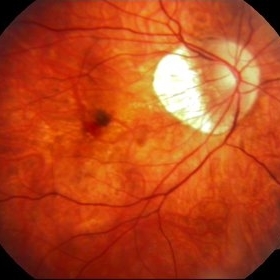 Myopic Choroidal Neovascular Membrane
Mar 25 2013 by Ratimir Lazic, MD, PhD
Color fundus photography of a 33-year-old female. In macular area subretinal hemorrhage can be seen. Area of atrophy temporal from PNO. Myopic changes of posterior pole and mid periphery can be noticed. The patient has been treated with 2 consecutive ranibizumab intravitreal injections. BCVA at baseline was 0,05 (Snellen lines) and 0,3 (Snellen lines) 2 months after.
Photographer: Marko Lukic, MD
Imaging device: Zeis Visucam Lite 2
Condition/keywords: high myopia, myopic choroidal neovascularization (CNV), ranibizumab
-
 ARMD with Disciform Scar
Oct 16 2012 by Jeffrey G. Gross, MD, FASRS
ARMD with disciform scar, RPE contracture, and subretinal hemorrhage, CF.
Condition/keywords: disciform scar, retinal pigment epithelium (RPE) contracture, subretinal hemorrhage
-
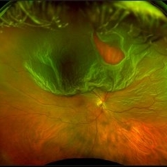 Optos Giant Tear within Retinal Detachment
Apr 30 2019 by Lauren Whaley
Noticed an inferior visual field defect on a patient with history of vitreous hemorrhage. Decided to take an Optos image and this is what we found. Doctor performed pneumatic retinopexy in office and patient recovering well.
Photographer: Lauren R. Whaley
Imaging device: Optos
Condition/keywords: Optos, retinal tear, subretinal fluid
-
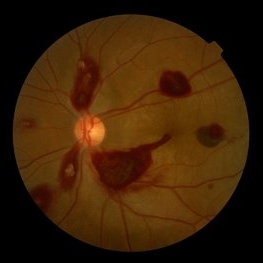
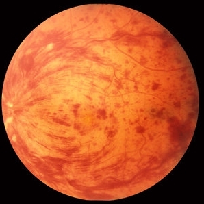
 Leukemic Retinopathy
Oct 9 2012 by Sharon Fekrat, MD FACS FASRS
22-year-old female with new diagnosis of acute myelogenous leukemia. White blood cell count was 35,000,000,000 cells/L. Note Roth Spots.
Photographer: Tiffanie Keaton, Duke Eye Imaging, Durham, NC
Condition/keywords: acute leukemia, white centered retinal hemorrhage (Roth Spot)
-
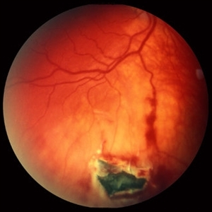 Intraocular Foreign Body, Metallic, in Inferior Retina with Hemorrhage
Oct 1 2012 by Jeffrey G. Gross, MD, FASRS
IOFB, metallic, in inferior retina with hemorrhage.
Condition/keywords: inferior retina, intraocular foreign body
-
 Roth Spots
Jul 11 2013 by Jerald A. Bovino, MD
No history, part of stereo pair.
Condition/keywords: stereo pair, white centered retinal hemorrhage (Roth Spot)
-
 PDR with Active NVD
Oct 8 2012 by Jeffrey G. Gross, MD, FASRS
PDR with active NVD and preretinal hemorrhage, mild VH and partial PRP.
Condition/keywords: neovascularization of the disc (NVD), preretinal hemorrhage, scatter laser photocoagulation, vitreous hemorrhage
-
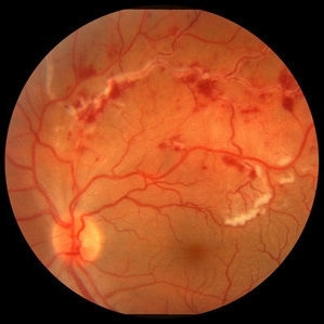
 Diabetic Retinal Hemorrhages in Proliferative Diabetes
Sep 10 2012 by James B. Soque, CRA, OCT-C, COA, FOPS
Fundus Photo of Severe Proliferative Diabetic with Retinal Hemorrhages, Left eye, scattered laser treatment. View: 50 Degrees
Photographer: James Soque, CRA, COA, Island Retina, Shirley, NY
Imaging device: Topcon TRC 50 DX
Condition/keywords: proliferative diabetic retinopathy (PDR)
-
 Proliferative Diabetic Retinopathy
Sep 17 2012 by Michael P. Kelly, FOPS
Retinal fundus photograph of a patient with PDR and NVD.
Photographer: Michael P. Kelly, FOPS Director, Duke Eye Labs, Duke University Hospital, Duke Eye Center
Imaging device: Topcon
Condition/keywords: blot hemorrhages, neovascularization of the disc (NVD)
-
 Ruptured retinal arterial macroaneurysm
Jan 11 2013 by Alex P. Hunyor, MD
Retinal arterial macroaneurysm with subretinal and preretinal hemorrhage
Condition/keywords: retinal arterial macroaneurysm
 Loading…