Search results (1761 results)
-
 Angiographic Storm: Fluorescein Leakage in Retinal Vasculitis
Nov 17 2025 by SHRADDHA RAJ SHRIVASTAVA
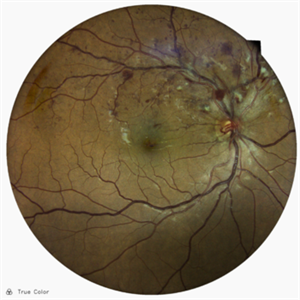
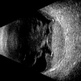
This left eye montage fundus fluorescein angiography (FFA) image of a 19 year old male with idiopathic retinal vasculitis, having skip vasculitic lesions predominantly involving retinal veins. There are areas of blocked fluorescence due to intraretinal hemorrhages, the involved veins have filling defects and occlusions, leading to formation of numerous collateral channels. The inflamed vessels also show perivascular fuzzy hyperfluorescent stain due to leakage of dye. We can also see multiple peripheral capillary non perfusion (CNP) areas, with a 'hot disc', suggestive of ongoing inflammation.
Photographer: Dr. Shraddha Raj Shrivastava
Imaging device: Nidek Mirante SLO/OCT (Confocal scanning/Spectral domain OCT)
Condition/keywords: FA late phase leakage, Fundus Fluorescein Angiography, idiopathic retinal vasculitis, optic disc leakage, VASCULITIS
-
 Bleeding Laser
Nov 8 2025 by Andrea Arriola-Lopez, MD MSc
26 year-old male, reports seeing a red dot with his left eye after staring at a projector light. BCVA 0.7 logMar.
Photographer: Dr. Waldemar Godoy, Clínica Godoy. Jalapa, Guatemala.
Imaging device: OPTOPOL SD-OCT
Condition/keywords: subhyaloid blood, subhyaloid hemorrhage
-
 Neovascular Medusa: A Bad Hair Day at the Optic Disc
Nov 4 2025 by SHRADDHA RAJ SHRIVASTAVA
Left eye pseudocolor fundus photo of 67 year old male, diagnosed with both eyes proliferative diabetic retinopathy, showing hair-like fronds of active neovascularisation at the disc (NVD) extending into the vitreous, giving the medusa-head appearance. There is a band of fibrovascular proliferation nasal to the disc, with presence of hard exudates and dot hemorrhages at the macula.
Photographer: Dr. Shraddha Raj Shrivastava
Imaging device: Nidek Mirante SLO/OCT (Confocal scanning/Spectral domain OCT
Condition/keywords: Diabetic Retinopathy, fibrovascular proliferation, Neovascularisation at the Disc (NVD), proliferative diabetic retinopathy (PDR)
-
 Multilayer Trauma
Nov 3 2025 by Malvika Singh
Fundus photograph of a 34 year old following trauma showing a choroidal rupture, a sub RPE and sub retinal bleed.
Photographer: Dr Malvika Singh, Retina Foundation, Ahmedabad, India
Imaging device: Mirante SLO/OCT
Condition/keywords: Choroidal Rupture, subretinal hemorrhage
-
 The Great Vascular Traffic Jam: Combined Retinal Vein and Artery Occlusion
Oct 29 2025 by SHRADDHA RAJ SHRIVASTAVA
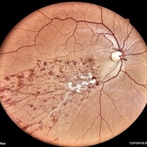
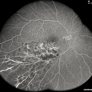
57 year old female, recently diagnosed with accelerated hypertension, developed Right eye Combined Retinal Vein and Artery Occlusion. Posterior pole image showed severe disc edema with peri-papillary haemorrhages. There is significant retinal whitening suggestive of edema leading to the classic cherry red spot at the macula. We can also see segmented flow of blood in retinal arterioles, which is the characteristic cattle-trucking seen in central retinal artery occlusion (CRAO). Widefield image revealed multiple intra-retinal blot hemorrhages in all quadrants with tortuous dilated vessels suggestive of central retinal vein occlusion (CRVO).
Photographer: Dr. Shraddha Raj Shrivastava
Imaging device: Nidek Mirante SLO/OCT (Confocal scanning/Spectral domain OCT)
Condition/keywords: central retinal vascular obstruction, central retinal vein occlusion (CRVO), CRVO with macular edema
-
 The Great Vascular Traffic Jam: Combined Retinal Vein and Artery Occlusion
Oct 29 2025 by SHRADDHA RAJ SHRIVASTAVA
57 year old female, recently diagnosed with accelerated hypertension, developed Right eye Combined Retinal Vein and Artery Occlusion. Posterior pole image showed severe disc edema with peri-papillary hemorrhages. There is significant retinal whitening suggestive of edema leading to the classic cherry red spot at the macula. We can also see segmented flow of blood in retinal arterioles, which is the characteristic cattle-trucking seen in central retinal artery occlusion (CRAO). Widefield image revealed multiple intra-retinal blot hemorrhages in all quadrants with tortuous dilated vessels suggestive of central retinal vein occlusion (CRVO).
Photographer: Dr. Shraddha Raj Shrivastava
Imaging device: Nidek Mirante SLO/OCT (Confocal scanning/Spectral domain OCT)
Condition/keywords: central retinal artery occlusion (CRAO), central retinal vascular obstruction, central retinal vein occlusion (CRVO), CRVO with macular edema
-
 Proliferative Ring of Fire
Oct 29 2025 by SHRADDHA RAJ SHRIVASTAVA
Right eye color fundus photo of 57 year old male, diagnosed with both eyes high risk proliferative diabetic retinopathy (PDR). Posterior pole reveals Neovascularization of disc (NVD) with extensive fibrovascular proliferations (FVPs) overlying the disc and along the arcades. We can also see a florid network of neovascularization (NVEs), with veins showing looping and beading changes. Hard exudates and dot-blot hemorrhages were seen at the macula.
Photographer: Dr. Shraddha Raj Shrivastava
Imaging device: Nidek Mirante SLO/OCT (Confocal scanning/Spectral domain OCT)
Condition/keywords: fibrovascular proliferation, Neovascularisation elsewhere (NVE), NVE, proliferative diabetic retinopathy (PDR), venous beading
-
 Superior Temporal Venous Branch Occlusion
Oct 23 2025 by Vicente Nicanor Mancilla Guerrero
Confocal laser retinography of the right eye of a 45-year-old female patient with hypertension (+). Venous dilation is evidenced together with the presence of flame hemorrhages and cottony spots in the upper temporal arch.
Photographer: Vicente Mancilla G, Medical Technologist in Ophthalmology
Imaging device: Compass CenterVue
Condition/keywords: branch retinal vein occlusion (BRVO)
-
 Severe Papilledema
Oct 13 2025 by Virginia Gebhart
26 year old female with severe optic nerve edema OU with nerve fiber layer hemorrhages. Pt reports blurry vision, intermittent vision loss, and ongoing headaches. VF testing showed enlarged blind spots and possible early acuate defects OU. Pt sent to ER for MRI of brain to rule out tumor. Results pending.
Photographer: Virginia Gebhart, Retina Consultants of Carolina
Imaging device: Topcon TRC 50DX
Condition/keywords: idiopathic intracranial hypertension, optic nerve edema, papilledema
-
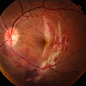
 Intricate Dance of Hemorrhage: BRVO with SHH with VH
Oct 11 2025 by Aditya S Kelkar, MS, FRCS, FASRS,FRCOphth
Retinal image of a 31-year-old male diagnosed with Branch Retinal Vein Occlusion (BRVO) alongside a subhyaloid hemorrhage and vitreous hemorrhage. The BRVO is evident by the disrupted blood flow in the retinal veins, leading to fluid leakage and hemorrhages. The blood leakage here shows the intricate pattern of hemorrhage revealing the hidden secrets of ocular health.
Photographer: Rhishita
Imaging device: optos daytona
Condition/keywords: branch retinal vein occlusion (BRVO), Hemorraghe, Retinal Vein Occlusion, Sub hyaloid haemorrhage, Vitreous hemorrhage
-
 Subhyaloid Hemorrhage
Oct 4 2025 by NIDHI PANWAR, MD FRCS Glasgow FNB FICO
Fully resolved subhyaloid hemorrhage post yag hyaloidotomy, 4 week later
Photographer: Ms Ola
Imaging device: optos
Condition/keywords: subhyaloid hemorrhage
-
 Subhyaloid Hemorrhage
Oct 4 2025 by NIDHI PANWAR, MD FRCS Glasgow FNB FICO
3 days post yag hyaloidotomy ( clotted blood can be seen just below hyaloidotomy site )
Photographer: Ms Ola
Imaging device: OPTOS
Condition/keywords: subhyaloid hemorrhage
-
 Subhyaloid Hemorrhage
Oct 4 2025 by NIDHI PANWAR, MD FRCS Glasgow FNB FICO
Immediately post yag hyaloidotomy
Photographer: Ms Ola
Imaging device: OPTOS
Condition/keywords: subhyaloid hemorrhage
-
 Subhyaloid Hemorrhage
Oct 4 2025 by NIDHI PANWAR, MD FRCS Glasgow FNB FICO
Pitting of ILM seen using green laser ( just before Yag hyaloidotomy)
Photographer: Ms Ola
Imaging device: OPTOS
Condition/keywords: Subhyaloid Hemorrhage
-
 Subhyaloid Hemorrhage
Oct 4 2025 by NIDHI PANWAR, MD FRCS Glasgow FNB FICO
33 Year old asymptomatic male with history of sudden blurring of right eye vision, after 5 days of observation by patient, vision getting more worse.
Photographer: Ms Ola
Imaging device: OPTOS
Condition/keywords: subhyaloid hemorrhage
-
 Subhyaloid Hemorrhage
Oct 4 2025 by NIDHI PANWAR, MD FRCS Glasgow FNB FICO
33 Year old asymptomatic male with history of sudden blurring of right eye vision since 7 days ,
Photographer: Ms Ola
Imaging device: OPTOS
Condition/keywords: SUBHYALOID HEMORRHAGE
-
 Choroidal Rupture
Sep 30 2025 by César Adrián Gomez Valdivia, MD
This fundus photograph shows curvilinear streaks of choroidal rupture radiating from the fovea, associated with subretinal hemorrhage. The rupture lines appear as crescent-shaped, whitish streaks representing a break in Bruch’s membrane, choriocapillaris, and retinal pigment epithelium.
Photographer: @eyemissu2
Imaging device: TOPCON TRX
Condition/keywords: Choroidal, Rupture
-
 Strained Retina
Sep 27 2025 by Malvika Singh
Fundus photograph of a 44 year old male showing hemorrhages at different layers.
Photographer: Dr Malvika Singh, Retina Foundation, Ahmedabad, India
Imaging device: Mirante SLO/OCT
Condition/keywords: valsalva retinopathy
-
 Strained Retina
Sep 27 2025 by Malvika Singh
Fundus photograph of a 44 year old male showing hemorrhages at different layers.
Photographer: Dr Malvika Singh, Retina Foundation, Ahmedabad, India
Imaging device: Mirante SLO/OCT
Condition/keywords: valsalva retinopathy
-
 Type 1 Aneurysmal Neovascularization
Sep 22 2025 by Gustavo Uriel Fonseca Aguirre
This transverse B-scan demonstrates vitreous hemorrhage, a bullous retinal detachment involving the macula, and dense subretinal hemorrhage, consistent with type 1 aneurysmal neovascularization. The scan reveals significant exudative activity with multi-level bleeding.
Photographer: Gustavo U. Fonseca Aguirre, Hospital Conde de Valenciana, Ciudad de México
Condition/keywords: polypoidal choroidal vasculopathy (PCV), Type 1 Aneurysmal Neovascularization
-
 Lower Temporal Branch Retinal Vein Occlusion
Sep 16 2025 by Seif Allah Anwar
Fundus photograph of a 46-year old hypertensive male patient showing sheathed lower temporal retinal vein with whitish cotton wool spots and hemorrhages ( dots, blots and flame shaped) along the area drained by the obstructed vein with vein.
Photographer: Dr Seif Anwar. FRCSEd
Imaging device: CENTERVUE EIDON
Condition/keywords: Lower temporal branch retinal vein occlusion
-
 Lower Temporal Branch Retinal Vein Occlusion
Sep 16 2025 by Seif Allah Anwar
FFA of A 46-year old hypertensive male patient showing blocked fluorescence by retinal hemorrhages along the area drained by the obstructed vein with areas of retinal capillary drop-out.
Photographer: Dr Seif Anwar , FRCSEd
Imaging device: CENTERVUE EIDON
Condition/keywords: Lower temporal branch retinal vein occlusion
-
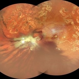 Table Top Tractional Retinal Detachment With Vitreous Hemorrhage in a Case of Proliferative Diabetic Retinopathy
Sep 12 2025 by Akansha Sharma
Color fundus photograph of a 56 year old male with table top tractional retinal detachment with vitreous hemorrhage in a case of proliferative diabetic retinopathy.
Photographer: DR. AKANSHA SHARMA
Condition/keywords: pan-retinal photocoagulation (PRP), PDR, proliferative diabetic retinopathy (PDR), PRP, TABLE TOP TRD, tractional retinal detachment, TRD, VH, vitreous hemorrhage
-
 Subhyaloid Hemorrhage With Vitreous Hemorrhage
Sep 12 2025 by Akansha Sharma
Color fundus photograph of a 56 year old hypertensive and diabetic female who presented with subhyaloid hemorrhage along with vitreous hemorrhage after being administered high dose anti-platelet therapy pre- and post a cardiac procedure.
Photographer: DR. AKANSHA SHARMA
Condition/keywords: SHH, sub ILM hemorrhage, subhyaloid hemorrhage, VH, vitreous hemorrhage
-
 Macular Tributary Retinal Venous Occlusion
Sep 7 2025 by Anand Temkar
A 55 yrs old female, k/c/o DM ( type II ) since past 5 yrs ( on medication ). Her vision was 6/6 in RE and 6/24 in her LE. IOP was 16 in both eyes. On examination, RE was WNL, and in LE ( color photo ) we noticed exudates, small hemorrhages, edema and sclerosed vessel ( depicted by black arrow. OCT LE shows altered foveal contour with cystic spaces and intraretinal hyperreflective material ( IRHRM ).
Photographer: Dr.Anand Temkar- Vasan Eye Hospital, Tiruchirappalli
Imaging device: Opticon
Condition/keywords: macular branch retinal vein occlusion (BRVO), venous occlusion
 Loading…