Search results (77 results)
-
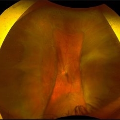
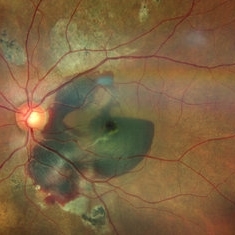
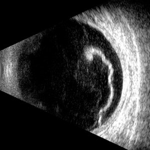 Giant Retinal Tear
Jul 5 2025 by Gustavo Uriel Fonseca Aguirre
This B-mode longitudinal ultrasound scan reveals a giant retinal tear, demonstrating a circumferential retinal flap with rolled edges extending over M-X to M-I. The vitreous shows diffuse hemorrhage and anterior-posterior traction strands inserting at the tear margins. The remaining retina appears attached without subretinal fluid.
Photographer: Gustavo U. Fonseca Aguirre, Hospital Conde de Valenciana, Ciudad de México
Condition/keywords: giant retinal tear
-
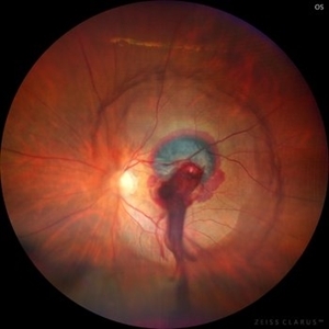
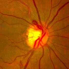 Prepapillary Vascular Loop
Jul 4 2025 by KANWALJEET HARJOT MADAN, M.S. (Ophthalmology); FAICO (Vitreous - Retina)
This is the fundus picture of right eye of a young 32 years female depicting pre papillary vascular loop. A prepapillary vascular loop is a congenital anomaly of the optic disc that presents as an elevated and twisted bundle of vessels projecting into the vitreous cavity. It is a benign condition, usually unilateral but can be bilateral. It is asymptomatic and discovered during routine eye examination. This anomaly can sometimes cause complications like branch retinal artery occlusion, vitreous hemorrhage, or sub retinal hemorrhage.
Photographer: Dr. Kanwaljeet Harjot Madan, Thind Eye Hospital, Jalandhar City (Punjab) INDIA.
Imaging device: Zeiss Fundus Camera
Condition/keywords: branch retinal artery occlusion (BRAO), optic disc, Prepapillary Vascular Loop, SUB RETINAL HEMORRHAGE, Vitreous hemorrhage
-
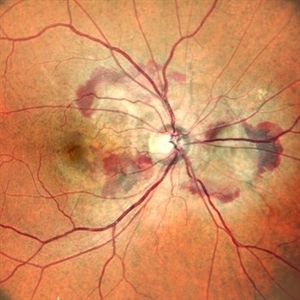 Right Eye Color Photo With Hemorrhages in Case of CNVM With Angioid Streaks
Nov 29 2024 by Anand Temkar
A 45 year old male came with chief complaint of blurring vision in right eyes since past 4 days. His vision is 6/12 in right eye and 6/9 in left eye. His vision was 14 mmHg in right eye and 16 mmHg in left eye. He was diagnosed with Angioid Streaks in both eyes about a year ago, then he developed choroidal neovascularization in his left eye 8 months ago, for which he received AntiVEGF injections x 3. Left eye is a stable eye now. Patient presented with right eye choroidal neovascularization in a case of Angioid Streaks on recent follow up. We have advised him right eye AntiVEGF injections x 3. In this image, the right eye color photo shows bleed from CNVM in case of angioid streaks.
Photographer: Dr.Anand Temkar- Retina Foundation, Ahmedabad
Imaging device: Mirante
Condition/keywords: Angioid Streaks, choroidal neovascular membrane (CNVM)
-
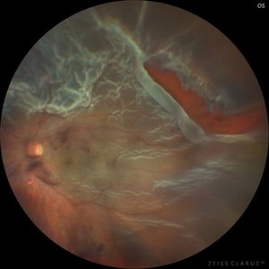
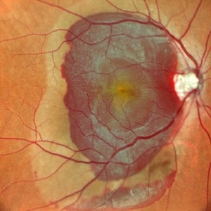
 Large Retinal Tear from a Shuttlecock Injury
Oct 11 2024 by Ahmad B. Tarabishy, MD
27 year old woman presenting with floaters and a shadow in her temporal visual field OS. Approximately one week earlier, she was injured in her left eye by a shuttlecock while playing badminton. Fundus exam reveals mild vitreous hemorrhage and a large retinal tear with a small cuff of surrounding SRF.
Photographer: Angela Rico, M.D.
Imaging device: Optos
Condition/keywords: blunt trauma, ocular trauma, retinal tear
-
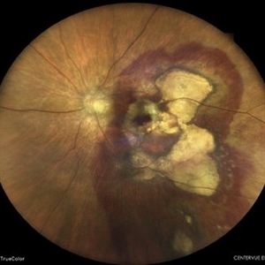
 Choroidal Fracture
Oct 27 2024 by César Adrián Gómez Valdivia, MD
Fundus photograph of a traumatic choroidal fracture & extra-macular sub-retinal hemorrhage.
Photographer: @eyemissu2
Imaging device: TOPCON TRC-50DX
Condition/keywords: Choroidal Fracture
-
 Suprachoroidal Hemorrhage
Dec 3 2024 by Dibya Prabha
Colour Fundus photograph of 62 Year old female patient with Suprachoroidal hemorrhage post trauma
Photographer: Dibya Prabha, LV Prasad eye Institute, Hyderabad
Condition/keywords: suprachoroidal hemorrhage
-
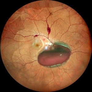
 Ruptured Retinal Artery Macroaneurysm
Jun 18 2024 by KANWALJEET HARJOT MADAN, M.S. (Ophthalmology); FAICO (Vitreous - Retina)
This is a fundus photo depicting ruptured Retinal Artery Macroaneurysm (RAM) in the left eye of a 63 years old female. RAM is an acquired saccular or fusiform dilatation of the retinal arterioles that usually occur within the first three orders of bifurcation. The Superotemporal artery is the most common location. RAM may be asymptomatic or cause a number of complications such as macular edema, serous macular detachment, and hemorrhages.
Photographer: Dr Kanwaljeet Harjot Madan
Condition/keywords: Haemorrhage, macroaneurysm, retinal arteriole
-
 Post Combined Surgery of Cataract, TRD & Vitreous Hemorrhage
Jun 27 2024 by Sanauddin Samejo , Diploma (Ophthalmic Technician Training Course)
A 27 year-old diabetic female visited the clinic one week after combined surgery of cataract, tractional retinal detachment and vitreous hemorrhage.
Photographer: Sanauddin Samejo, Burjeel Hospital, Abu Dhabi, UAE
Imaging device: Silver Stone Optos
Condition/keywords: Combined Surgery Cataract Tractional Retinal Detachment Vitreous Hemorrhage, POST SURGERY, Retinal Detachment, TRD
-
 Dislocated Lens, Posterior OD
Jan 26 2024 by Corey Grant
OPTOS California photo presents a 71 year old male patient with a dislocated lens, posterior in the right eye. Presented on 1/26/24 with posteriorly dislocated SN60WF with a Soemmerring ring. Associated retinal hemorrhage within retinoschisis as well. This will result in a PPV/IOL exchange/SFIOL/STK for the right eye.
Photographer: Corey Grant, Ophthalmic Imager, Retina Specialist of Michigan
Imaging device: OPTOS California
Condition/keywords: color photo, IOL, OD, Optos, OPTOS CALIFORNIA, pars plana vitrectomy (PPV), retina
-
 Total Rhegmatogenous retinal detachment with lattice degeneration & Vitreous haemorrhage
Jul 31 2023 by Harsh Vardhan Singh, MS
72-year male presented PVD induced total retinal detachment with vitreous hemorrhage
Photographer: Dr Harsh Vardhan Singh, AIIMS, Guwahati
Imaging device: Zeiss Clarus 700
Condition/keywords: chronic retinal detachment, hemorrhage, rrd
-
 Candy Stripe Sign
Mar 30 2023 by pedro fernandes souza neto
Candy Stripe Sign, patient with proliferative diabetic retinopathy progressing to vitreous hemorrhage and subsequently to ghost cell glaucoma.
Photographer: Marlos Henrique Oliveira Junior, Federal University of Bahia.
Condition/keywords: dehemoglobinized hemorrhage, diabetes, diabetic glaucoma
-
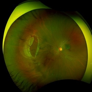
 PEHCR (Peripheral Exudative Hemorrhagic Chorioretinopathy)
May 12 2023 by Niloofar Piri, MD
Ultrawide fundus photograph of the left eye demonstrating extensive peripheral hemorrhagic exudative detachment in a 79 yo Caucasian female with prior history of non-exudative AMD. Recent diagnosis of Acute myeloid leukemia with low platelet count which might have contributed to the above presentatuon. Please note the temporal subretinal hemorrhage as well as RPE atrophy and hyperplasia in the macula.
Photographer: Rocio Bentivegna, MD, Saint Louis University; Jessica Maddox, COA, Saint Louis University
Condition/keywords: peripheral exudative hemorrhagic chorioretinopathy (PEHCR)
-
 Subretinal Hemorrhage
Feb 28 2023 by Akansha Sharma
Color fundus photograph of an 84-year old male with subretinal hemorrhage associated with areas of scarring.
Photographer: Dr. Urmil Shah, Dr. Denish Patel, Dr. Akansha Sharma, Bharati Eye Hospital, Ahmedabad, Gujarat
Condition/keywords: choroidal neovascularization (CNV), subretinal hemorrhage
-
 Lady in a dress
Feb 9 2023 by Shelby Helton
Fluorescein Angiography on a 67-year-old male with significant RPE changes secondary to a severe subretinal hemorrhage that required a vitrectomy with subretinal TPA in 2013.
Photographer: Shelby Helton
Imaging device: Heidelberg Spectralis
Condition/keywords: wet age-related macular degeneration (wet AMD)
-
 High risk Proliferative Diabetic Retinopathy treated with Pan Retinal Photocoagulation
Nov 5 2022 by Somnath Chakraborty, MD
A Fundus Photo Montage of 43 year old Asian Male with Type 2 Diabetes Mellitus since 7 years who presented with sudden onset diminition of vision in his Left eye. BCVA OS was 20/200. He was diagnosed to have Pre retinal bleed due to Proliferative Diabetic Retinopathy and was treated with Pan Retinal Photocoagulation. This image shows a large neo-cascular frond at the disc and superior to it with Pre-retinal bleed and Fresh laser marks along
Photographer: Pulak Roy
Condition/keywords: diabetic blindness, diabetic retinopathy vitrectomy study (DRVS), fresh laser burns, laser photocoagulation, preretinal hemorrhage, proliferative diabetic retinopathy (PDR)
-
 Choroidal Melanoma
Nov 3 2022 by pedro fernandes souza neto
Transillumination of Enucleation specimen of Choroidal Melanoma: anterior chamber is closed. Total secondary retinal detachment with subretinal serous fluid and some subretinal hemorrhages are present.
Photographer: Eduardo Marback, Federal University of Bahia, Brazil
Condition/keywords: enucleation, melanoma
-
 Subretinal BSS and air
Apr 12 2022 by Nassim Alejandro Abreu Arbaje, MD
67 year old female who presented with complaints of 5 days of decreased vision of her left eye. She underwent PPV + BSS and Air injection in the subretinal space
Photographer: Nassim Abreu, Dr. Elias Santana Hospital
Imaging device: Ngenuity 3D system screenshot
Condition/keywords: subretinal hemorrhage
-
 Submacular Hemorrhage PCV
May 6 2022 by Shobhit Chawla, M.S.
Submacular hemorrhage in a 38 years old female patient cause polyp bleed in PCV.
Photographer: Shobhit Chawla
Imaging device: Zeiss Clarus 500
Condition/keywords: polypoidal choroidal vasculopathy (PCV), submacular hemorrhage
-
 Subretinal Bleed
Jul 12 2022 by Akansha Sharma
73 year old diabetic and hypertensive female presented with sub-retinal hemorrhage for which she was operated with pars-plana vitrectomy with intra-vitreal anti-VEGF
Photographer: Dr. Akansha Sharma-Retina Foundation, Ahmedabad
Condition/keywords: subretinal hemorrhage, subretinal blood
-
 Macular Hemorrhage Secondary to Anemic Retinopathy
Apr 18 2022 by Deepak Bhojwani, MS
Fundus image of a young 28 year old patient who has been diagnosed as 'PRIMARY BONE MARROW APLASIA' by hematologist showing large macular hemorrhage (sub -ILM Heme mound). Few Roth spots were also seen in midperiphery suggesting 'ANEMIC RETINOPATHY'.
Photographer: DEEPAK BHOJWANI
Condition/keywords: anaemic retinopathy, BONE MARROW APLASIA
-
 JXT and Proliferative Diabetic Retinopathy
Jan 13 2022 by ASRS Staff
Wide field photograph of 50 year-old woman, known case of JXT in both eyes and known diabetic, after 9 months of PPV for subhyaloid hemorrhage.
Imaging device: Nidek Mirante
Condition/keywords: florid type PDR, JXT, pars plana vitrectomy (PPV)
-
 Perforating Ocular Trauma and Choroidal Rupture due to Shotgun Pellet
Mar 31 2022 by Franco Benvenuto, MD
Fundus photograph of a 17-year-old with shotgun injuries with numerous metal pellets in the chest, neck, brain, and face. Fundus exploration showed the left globe with posterior-inferior eye rupture, vitreous hemorrhages and choroidal rupture.
Photographer: Franco Benvenuto, Universidad de Buenos Aires, Argentina. Universidad de Guadalajara, México.
Condition/keywords: choroidal rupture, penetrating trauma, shotgun
-
 Valsalva Retinopathy
Dec 20 2021 by Unnati Vishwanath Shukla, M. S. ,DNB, FVRS FNERF, MNAMS,PhD Scholar(Retina)
26-year-old male with Valsalva Retinopathy. History of severe cough for 3 days. All hematological investigations were within normal limits.
Photographer: Dr. Unnati Shukla, Consultant, Retina Foundation, Ahmedabad
Imaging device: Nidek Mirante
Condition/keywords: subhyaloid hemorrhage, subretinal hemorrhage, valsalva retinopathy
-
 Subhyaloid Hemorrhage
Mar 1 2021 by Narciso F. Atienza, MD, MBA, FASRS, FPCS, FPAO.
Fundus photo of the left eye, 29-year-old male patient, with previous history of 6 cycles of chemotherapy from Hodgkins lymphoma. Photograph, however looks more like leukemic subhyaloid hemorrhage.
Photographer: Narciso Atienza, Jr. MD, MBA. Legazpi Eye Center
Condition/keywords: Hodgkins lymphoma, subhyaloid hemorrhage
-
 T-Rex Hemorrhage
Jul 2 2021 by Jeffrey Barker
T-Rex hemorrhage.
Photographer: Jeffrey P. Barker
Condition/keywords: hemorrhage
 Loading…