Search results (309 results)
-
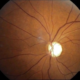
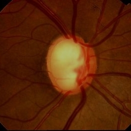
 Normal Fundus image
Aug 30 2018 by Timothy Adeyemo
Fundus photograph of a healthy 31-year-old lady being examined for glaucoma.
Photographer: Adeyemo Timothy, National Eye Centre, Kaduna, Nigeria.
Imaging device: Topcon's TRC- NW8 plus
Condition/keywords: normal retina
-
 Gonioscopy: Pigment Dispersion Glaucoma
Jul 8 2013 by Jason S. Calhoun
Patient with no family history of glaucoma, came in with elevated IOP. During gonioscopy exam. brown pigment overlying the trabecular meshwork. Also, trans-illumination defects on the iris.
Photographer: Jason S. Calhoun, Department of Ophthalmology, Mayo Clinic Jacksonville, Florida
Condition/keywords: gonioscopy, pigment dispersion syndrome of iris
-
 Glaukomflecken
Oct 23 2017 by Claire Kiernan, MD
Slit lamp photograph of a 59-year-old man with recent-onset severe eye pain noted to have glaukomflecken consistent with recent episode of angle closure glaucoma.
Photographer: Steve Moser, University of Tennessee Hamilton Eye Institute; Joe Mastellone, University of Tennessee Hamilton Eye Institute
Condition/keywords: angle-closure glaucoma interval, glaucoma anterior segment anomalies
-
 Pseudoexfoliation Syndrome Ring
Sep 17 2015 by Jason S. Calhoun
Pseudoexfoliation syndrome ring on the lens capsule.
Photographer: Jason Calhoun, Mayo Clinic Jacksonville, Department of Opthalmolgy
Imaging device: Haag Striet Cannon D7
Condition/keywords: pseudoexfoliation glaucoma, pseudoexfoliation of lens capsule, pseudoexfoliation syndrome
-
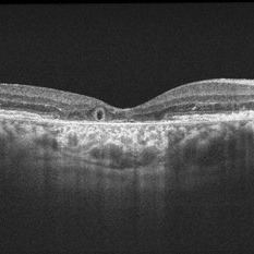 Outer-Retinal-Tubulation
Jun 27 2013 by Jason S. Calhoun
Patient with a history of wet macular degeneration and glaucoma in both eyes. VA is 20/50, right eye, 20/80, left eye. Patient is treated with Eylea in both eyes. Enhanced depth imaging OCT reveals a small like form of a cyst which in fact isn't a cyst at all. This is called outer retinal tubulation in which degenerating photo-receptors may become arranged in a circular or ovoid fashion. This is sometimes misdiagnosed as cystic changes in the retinal pigment epithelium or sub-retinal fluid.
Photographer: Jason S. Calhoun, Mayo Clinic Jacksonville, Florida
Imaging device: ZEISS OCT CIRRUS
Condition/keywords: optical coherence tomography (OCT)
-
 Gonioscopy; Scattered Peripheral Anterior Synechiae
Jul 8 2013 by Jason S. Calhoun
Patient came in for evaluation for glaucoma. Patient also has a history of uveitis. Last flare up was back in 1990. Patient's VA was 20/30, Right eye and 20/40-1, Left eye. Slit Lamp Gonioscopy reveals iris bow with scattered PAS around the angles of the anterior chamber. You can also see pigmentation in the trabecular meshwork. Patient will follow up in 3 months.
Photographer: Jason S. Calhoun, Department of Ophthalmology, Mayo Clinic Jacksonville, Florida
Condition/keywords: gonioscopy, goniosynechiae
-
 Superior Peripapillary Hemorrhage
Jul 13 2013 by Jason S. Calhoun
Patient was seen for acute vision loss in the right eye. Patient has glaucoma. VA was 20/70 in the right eye. Had vitrectomy back in May 2012 for ERM stripping. Also had trabectome with cataract surgery in December of 2012. Fundus photos presents a superior peripapillary Hemorrhage of the optic nerve. Patient will be followed up in one month.
Photographer: Jason S. Calhoun, Department of Ophthalmology, Mayo Clinic Jacksonville, Florida
Imaging device: TOPCON TRC 50-EX
Condition/keywords: peripapillary hemorrhage
-
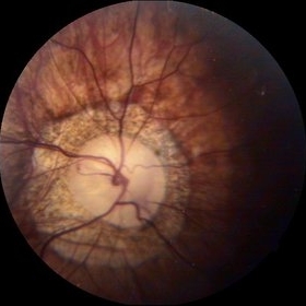
 Glaucomatous Optic Atrophy
Sep 20 2014 by Mehul A Shah
A 65-year-old male presented with loss of vision and found to have glaucomatous optic atrophy.
Photographer: Drashti Netralaya,Dahod
Imaging device: Zeiss ff450
Condition/keywords: glaucomatous atrophy of optic disc
-
 Staphyloma Steroid Induced Glaucoma
Apr 14 2014 by Dipankar Barua, M.Sc
Male patient, 25-years-old. On examination his vision of the right eye is perception of light and left eye is 6/6. His IOP is 30mmHg in right eye and 10 mmHg in left eye. It seems to be a case of staphyloma steroid induced glaucoma.
Photographer: Dipankar Barua
Imaging device: Topcon TRC 50 DX (IA)
Condition/keywords: glaucoma, staphyloma, steroids
-
 Optos Picture With Speculum: Dislocated Natural Lens
Oct 9 2018 by John S. King, MD
55-year-old white female with history of pathologic myopia+, lattice (laser), SB OU (1990s), and dislocated natural lenses OU that had been watched for years. In the fellow eye she developed phacolytic glaucoma and a PPV, PPL was performed. Plan for both eyes are monitoring. I wanted to get a good picture of her lens today with the optos machine, as the other pics had artifact from the lower lid. It worked out well to use a speculum in the left eye. Vision cc is 20/400 J1+ OD and 20/40 J2 OS; aphakic OU; vitreous clear OD; dislocated lens OS (see pic); retinas attached.
Photographer: Maisee Yang
Imaging device: Optos California
Condition/keywords: dislocated crystalline lens, pathologic myopia, scleral buckle, staphyloma
-
 Peripapillary Atrophy
Oct 3 2014 by Mehul A Shah
A 55-year-old patient presented with diminished vision OU on examination patient had glaucoma with peripapillary optic atrophy.
Photographer: Drashti Netralaya,Dahod
Imaging device: Zeiss ff450
Condition/keywords: atrophy, peripapillary
-
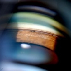
 Krukenberg's Spindle
Jul 13 2013 by Jason S. Calhoun
Slit lamp shows pigment on the lower quadrant of the endothelium due to pigment dispersion glaucoma.
Photographer: Jason S. Calhoun, Department of Ophthalmology, Mayo Clinic Jacksonville, Florida
Imaging device: TOPCON D-90 SL NIKON CAMERA
Condition/keywords: Krukenberg's spindle
-
 Pseudoexfoliation Syndrome Ring
Sep 17 2015 by Jason S. Calhoun
Pseudoexfoliation syndrome ring on the lens.
Photographer: Jason Calhoun, Mayo Clinic Jacksonville, Department of Opthalmolgy
Imaging device: Haag Striet Cannon D7
Condition/keywords: pseudoexfoliation glaucoma
-
 Glaucoma
Feb 9 2015 by Govindarajan Venkatesan
Glaucoma.
Photographer: Govindarajan Venkatesan
Condition/keywords: optic disc
-
 Advanced Stage of Neovascular Glaucoma
Mar 21 2013 by Yusuke Oshima, MD, PhD
An 82-year-old man with a advanced stage of neovascular glaucoma. A slit-lamp photograph illustrates iris ectropion with prominent iris neovascularization.
Photographer: Yusuke Takada, Osaka University Graduate School of Medicine
Condition/keywords: neovascular glaucoma
-
 Myopic Degeneration With RPE Loss
Oct 31 2013 by Jason S. Calhoun
Patient in for follow up for glaucoma. VA is 20/30 in both eyes. Fundus photography shows the RPE loss due to myopic degeneration, nasally, inferiority in the left eye.
Photographer: Jason S. Calhoun, Ophthalmic Photographer, Department of Ophthalmology, Mayo Clinic Jacksonville
Imaging device: TOPCON TRC 50-EX
Condition/keywords: myopic degeneration
-
 Pigmentary Glaucoma, Zonules Visible Inferiorly
Jul 10 2013 by Jason S. Calhoun
Young gentleman with pigmentary glaucoma. Heavy pigment lining trabecular meshwork with goinoscopy. Crisp image of the zonules.
Photographer: Jason S. Calhoun, Department of Ophthalmology, Mayo Clinic Jacksonville, Florida
Condition/keywords: glaucoma, pigment dispersion syndrome of iris
-
 Krukenberg's Spindle
Jul 13 2013 by Jason S. Calhoun
Slit lamp shows pigment on the lower quadrant of the endothelium due to pigment dispersion glaucoma.
Photographer: Jason S. Calhoun, Department of Ophthalmology, Mayo Clinic Jacksonville, Florida
Imaging device: TOPCON D-90 SL NIKON CAMERA
Condition/keywords: Krukenberg's spindle
-
 Acquired Optic Pit Maculopathy
Aug 20 2014 by Andree Henaine-Berra, MD
Optical coherence tomography of the left eye of a 60-year-old man with an acquired optic pit maculopathy and glaucoma. The image shows an enlarged optic disc cup and a macular serous detachment.
Photographer: Andree Henaine-Berra. Asociacion Para Evitar la Ceguera en Mexico. Mexico City.
Imaging device: Heidelberg Spectralis
Condition/keywords: glaucoma, maculopathy, optic pit
-
---thumb.JPG/image-square;max$300,300.ImageHandler) Rubeosis Iridis
Rubeosis Iridis
Jul 8 2013 by Jason S. Calhoun
Patient presents with rubeosis iridis in the right eye due to neovascular glaucoma. VA is 20/40 in the right eye. Will follow up in 3 months.
Photographer: Jason S. Calhoun, Department of Ophthalmology, Mayo Clinic Jacksonville, Florida
-
 Primary Open Angle Glaucoma - Excavated Disc
Oct 7 2013 by Gerardo Garcia-Aguirre, MD
Primary open angle glaucoma - excavated disc,
Condition/keywords: glaucoma, open-angle glaucoma
-
 Uveal Effusion Syndrome
Mar 9 2013 by Gabriela Lopezcarasa Hernandez, MD
A 55-year-old man with glaucoma surgery and severe decrease in visual acuity, with superior nasal occlusion and serous retinal detachment.
Photographer: Araceli Rojas Arriaga, Hospital Angeles Lomas, Mexico
Imaging device: Zeiss FF4
Condition/keywords: idiopathic uveal effusion syndrome
-
 Superior Peripapillary Hemorrhage
Jun 27 2013 by Jason S. Calhoun
Patient was seen for acute vision loss in the right eye. Patient has glaucoma. VA was 20/70 in the right eye. Had vitrectomy back in May 2012 for ERM stripping. Also had trabectome with cataract surgery in December of 2012. Fundus photos presents a superior peripapillary Hemorrhage of the optic nerve. Patient will be followed up in one month.
Photographer: Jason S. Calhoun, Mayo Clinic Jacksonville, Florida
Imaging device: TOPCON TRC 50-EX
Condition/keywords: peripapillary hemorrhage
-
 Staphyloma Steroid Induced Glaucoma
Apr 14 2014 by Dipankar Barua, M.Sc
Male patient, 25-years-old. On examination his vision of the right eye is perception of light and left eye is 6/6. His IOP is 30mmHg in right eye and 10 mmHg in left eye. It seems to be a case of staphyloma steroid induced glaucoma.
Photographer: Dipankar Barua
Imaging device: Topcon TRC 50 DX (IA)
Condition/keywords: glaucoma, staphyloma
 Loading…