 Initializing download.
-
 By Jason S. Calhoun
Mayo Clinic Jacksonville, Florida - Uploaded on Jun 27, 2013.
- Last modified by Jason S. Calhoun on Jun 30, 2013.
- Rating
- Appears in
- Optical Coherence Tomography
- Condition/keywords
- optical coherence tomography (OCT)
- Photographer
- Jason S. Calhoun, Mayo Clinic Jacksonville, Florida
- Imaging device
-
Optical coherence tomography system
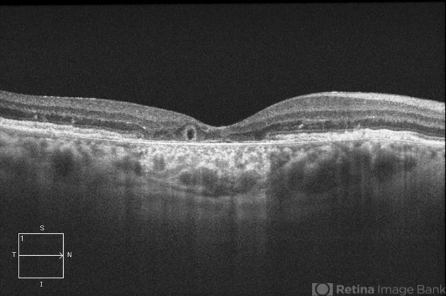
ZEISS OCT CIRRUS - Description
- Patient with a history of wet macular degeneration and glaucoma in both eyes. VA is 20/50, right eye, 20/80, left eye. Patient is treated with Eylea in both eyes. Enhanced depth imaging OCT reveals a small like form of a cyst which in fact isn't a cyst at all. This is called outer retinal tubulation in which degenerating photo-receptors may become arranged in a circular or ovoid fashion. This is sometimes misdiagnosed as cystic changes in the retinal pigment epithelium or sub-retinal fluid.

")


")
")
")


