Search results (310 results)
-
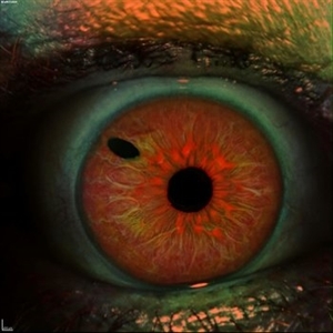
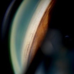
 Candy Stripe Sign
Mar 30 2023 by pedro fernandes souza neto
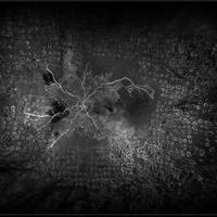
Candy Stripe Sign, patient with proliferative diabetic retinopathy progressing to vitreous hemorrhage and subsequently to ghost cell glaucoma.
Photographer: Marlos Henrique Oliveira Junior, Federal University of Bahia.
Condition/keywords: dehemoglobinized hemorrhage, diabetes, diabetic glaucoma
-
 Bleb Needling, Blood Streaming in Anterior Chamber
Jul 11 2013 by Jason S. Calhoun
Patient who had a bleb needling had a stream of blood start from the bleb superiorly to inferiorly within the anterior chamber.
Photographer: Jason S. Calhoun, Department of Ophthalmology, Mayo Clinic Jacksonville, Florida
Condition/keywords: glaucoma
-
 Iris
Iris
Apr 29 2019 by Stephanie Moolman
Multi-color images after Yag PI of iris.
Photographer: Stephanie Moolman, Dr Marissa Willemse, Pretoria, South Africa
Imaging device: Heidelberg Spectralis
Condition/keywords: glaucoma, iris, multicolor, NdYAG laser, peripheral iridotomy
-
 Dropped Crystalline Lens
Mar 8 2019 by Abdulaziz A. Alshamrani, MD
A 15-year-old female with congenital glaucoma complaining of acute diminution of vision after a blunt trauma.
Condition/keywords: crystalline lens, dropped nucleus, ora serrata
-
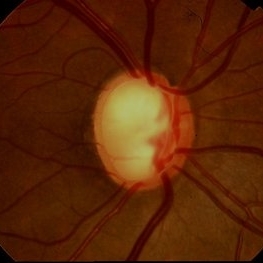 Glaucoma
Feb 9 2015 by Govindarajan Venkatesan
Glaucoma.
Photographer: Govindarajan Venkatesan
Condition/keywords: optic disc
-
 Pseudoexfoliation Syndrome
Sep 17 2015 by Jason S. Calhoun
Pseudoexfoliation on lens.
Photographer: Jason Calhoun, Mayo Clinic Jacksonville, Department of Opthalmolgy
Imaging device: Haag Striet Cannon D7
Condition/keywords: pseudoexfoliation glaucoma, pseudoexfoliation of lens capsule
-
 Advanced Stage of Neovascular Glaucoma
Mar 21 2013 by Yusuke Oshima, MD, PhD
An 82-year-old man with a advanced stage of neovascular glaucoma. A slit-lamp photograph illustrates iris ectropion with prominent iris neovascularization.
Photographer: Yusuke Takada, Osaka University Graduate School of Medicine
Condition/keywords: neovascular glaucoma
-
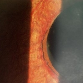
 Bergmeister Papilla
Feb 20 2020 by Nisarg Joshi, MD
Gross pathology photo of a Bergmeister Papilla. It is a remnant of incompletely resorbed hyaloid vasculature from ocular development. This glial tissue is seen emminating from the optic nerve, which also shows glaucomatous cupping. The eye was enucleated due to a choroidal melanoma.
Photographer: Nisarg Joshi, MD, Geisinger Medical Center
Imaging device: Digital camera
Condition/keywords: Bergmeister's Papillae, hyaloid artery, persistent fetal vasculature (PFV)
-
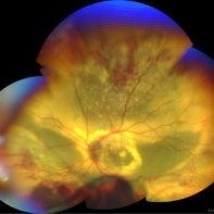
 Central Retinal Vein Occlusion with Retinal Neovascularization
Jan 19 2022 by Olivia Rainey
Ultra-widefield fluorescein angiogram of a 56-year-old male with a Central Retinal Vein Occlusion with Retinal Neovascularization affecting his left eye. The patient presented on 1/19/2022 with scNLP vision in the left eye. The patient has good PRP, however areas of ischemia still remain untreated by laser. He also has severe neovascular glaucoma contributing to his poor vision.
Photographer: Olivia Rainey, OCT-C, COA
Imaging device: Optos California
Condition/keywords: central retinal vein occlusion (CRVO), FA early phase, fluorescein angiogram (FA), hemorrhage, ischemic CRVO, left eye, neovascular glaucoma, Optos, pan-retinal photocoagulation (PRP), retinal ischemia, retinal neovascularization, ultra-wide field imaging
-
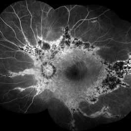
 Coats' disease
Sep 7 2022 by Niloofar Piri, MD
Total exudative RD with extensive subretinal exudates and peripheral telangiectatic vascular anomalies in stage 4 Coats's disease. Patient is a 12 yo who presented with severe eye pain and neovascular glaucoma secondary to the above.
Photographer: Jacob Grodsky, MD
Condition/keywords: Coats' disease
-
 Degeneration Paravenous
Sep 20 2016 by JEFFERSON R SOUSA, Tecg.º (Biomedical Systems Technology)
Female patient, 32-years-old, Asian, appeared at the clinic with a history of glaucoma. 20/20 visual acuity in both eyes. Examination of color photography, pigmentary changes were observed following the vascular arcades only in the left eye. Suggestive of paravenous degeneration.
Photographer: JEFFERSON R SOUSA - Study Center and Ophthalmological Research Dr. Andre M V Gomes, Institute Dr. Suel Abujamra São Paulo-Brazil
Imaging device: Zeiss / VisuCam-500 - Angulation of field photo of 45 Degrees, flash 24.
Condition/keywords: degeneration paravenous
-
 Dislocated Capsular Tension Ring in Vitreous Cavity
Dec 21 2019 by Pablo Baquero Ospina, MD
Fundus photograph of an 52-year-old woman with pseudoexfoliation glaucoma and previous cataract surgery with capsular tension ring. 5 years later she refers floaters.
Photographer: Pablo Baquero, Asociacion Para Evitar la Ceguera en Mexico, Mexico city
Imaging device: Optos/Daytona
Condition/keywords: fundus photograph, pseudoexfoliation glaucoma
-
 Gonioscopy: Pigment Dispersion Glaucoma
Jul 8 2013 by Jason S. Calhoun
Patient with no family history of glaucoma, came in with elevated IOP. During gonioscopy exam. brown pigment overlying the trabecular meshwork. Also, trans-illumination defects on the iris.
Photographer: Jason S. Calhoun, Department of Ophthalmology, Mayo Clinic Jacksonville, Florida
Condition/keywords: gonioscopy, pigment dispersion syndrome of iris
-
 Gonioscopy; Scattered Peripheral Anterior Synechiae
Jul 8 2013 by Jason S. Calhoun
Patient came in for evaluation for glaucoma. Patient also has a history of uveitis. Last flare up was back in 1990. Patient's VA was 20/30, right eye and 20/40-1, left eye. Slit Lamp gonioscopy reveals iris bow with scattered PAS around the angles of the anterior chamber. You can also see pigmentation in the trabecular meshwork. Patient will follow up in 3 months.
Photographer: Jason S. Calhoun, Department of Ophthalmology, Mayo Clinic Jacksonville, Florida
Condition/keywords: gonioscopy, goniosynechiae
-
 Pigmentary Glaucoma, Zonules Visible Inferiorly
Jul 10 2013 by Jason S. Calhoun
Young gentleman with pigmentary glaucoma. Heavy pigment lining trabecular meshwork with goinoscopy. Crisp image of the zonules.
Photographer: Jason S. Calhoun, Department of Ophthalmology, Mayo Clinic Jacksonville, Florida
Condition/keywords: glaucoma, pigment dispersion syndrome of iris
-
 Pseudoexfoliation Syndrome
Sep 17 2015 by Jason S. Calhoun
Pseudoexfoliation syndrome on the lens.
Photographer: Jason Calhoun, Mayo Clinic Jacksonville, Department of Opthalmolgy
Imaging device: Haag Striet Cannon D7
Condition/keywords: pseudoexfoliation glaucoma
-
 Pseudoexfoliation Syndrome
Sep 17 2015 by Jason S. Calhoun
Pseudoexfoliation syndrome on the lens.
Photographer: Jason Calhoun, Mayo Clinic Jacksonville, Department of Opthalmolgy
Imaging device: Haag Striet Cannon D7
Condition/keywords: pseudoexfoliation glaucoma
-
 Pseudoexfoliation Syndrome
Sep 17 2015 by Jason S. Calhoun
Pseudoexfoliation syndrome on the lens.
Photographer: Jason Calhoun, Mayo Clinic Jacksonville, Department of Opthalmolgy
Imaging device: Haag Striet Cannon D7
Condition/keywords: pseudoexfoliation glaucoma
-
 Pseudoexfoliation Syndrome Ring
Sep 17 2015 by Jason S. Calhoun
Pseudoexfoliation syndrome ring on the lens capsule.
Photographer: Jason Calhoun, Mayo Clinic Jacksonville, Department of Opthalmolgy
Imaging device: Haag Striet Cannon D7
Condition/keywords: pseudoexfoliation glaucoma, pseudoexfoliation of lens capsule, pseudoexfoliation syndrome
-
 Pseudoexfoliation Syndrome Ring
Sep 17 2015 by Jason S. Calhoun
Pseudoexfoliation syndrome ring on the lens.
Photographer: Jason Calhoun, Mayo Clinic Jacksonville, Department of Opthalmolgy
Imaging device: Haag Striet Cannon D7
Condition/keywords: pseudoexfoliation glaucoma
-
Spontaneous lens dislocation (Weill Marchesani Syndrome)
Nov 9 2022 by Heitor Nogueira
A 9 year-old Male patient diagnosed with Weill Marchesani presented spontaneous bilateral lens dislocation. Weill-Marchesani syndrome, also known as spherophakia-brachymorphy syndrome and mesodermal dysmorphodystrophy, is an inherited connective tissue disorder characterized by eye lens abnormalities, secondary glaucoma, short stature, brachydactyly, joint stiffness, and cardiovascular defects.
Photographer: Heitor Nogueira, Insituto Penido Burnier, Campinas-SP, Brazil
Condition/keywords: mesodermal dysmorphodystrophy, spherophakia-brachymorphy syndrome, spontaneous lens dislocation, video, Weill Marchesani Syndrome
-
 Toxoplasmic
May 27 2021 by Gabriel Costa Andrade, PhD
Fundus photograph of an 52-year-old woman with glaucoma and choroidal neovascularization due to toxoplasmosis.
Photographer: Gabriel Andrade
Condition/keywords: choroidal neovascularization (CNV), toxoplasmosis
-
 Optos Picture With Speculum: Dislocated Natural Lens
Oct 9 2018 by John S. King, MD
55-year-old white female with history of pathologic myopia+, lattice (laser), SB OU (1990s), and dislocated natural lenses OU that had been watched for years. In the fellow eye she developed phacolytic glaucoma and a PPV, PPL was performed. Plan for both eyes are monitoring. I wanted to get a good picture of her lens today with the optos machine, as the other pics had artifact from the lower lid. It worked out well to use a speculum in the left eye. Vision cc is 20/400 J1+ OD and 20/40 J2 OS; aphakic OU; vitreous clear OD; dislocated lens OS (see pic); retinas attached.
Photographer: Maisee Yang
Imaging device: Optos California
Condition/keywords: dislocated crystalline lens, pathologic myopia, scleral buckle, staphyloma
-
 Neovascular Glaucoma
Dec 4 2015 by Kathy Karsten, COT
Ahmad tube shunt with peaked pupil in the left eye of a 63-year-old woman for neovascular glaucoma.
Photographer: Kathy Karsten, COT
Imaging device: Topcon TRC 50-DX
Condition/keywords: angle neovascularization
-
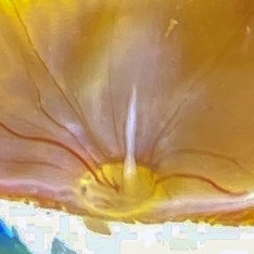
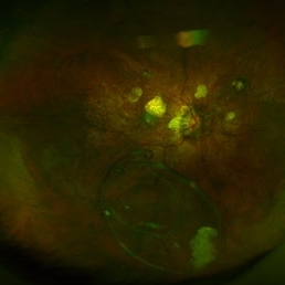
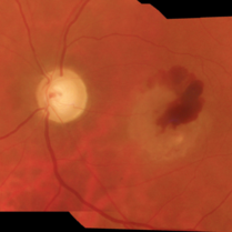
 Acquired Optic Pit Maculopathy
Aug 20 2014 by Andree Henaine-Berra, MD
Autofluorescence image of the left eye of a 60-year-old man with an acquired optic pit maculopathy and glaucoma.
Photographer: Andree Henaine-Berra. Asociacion Para Evitar la Ceguera en Mexico. Mexico City.
Imaging device: Heidelberg Spectralis
Condition/keywords: glaucoma, maculopathy, optic pit
 Loading…