Search results (300 results)
-
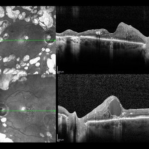
 Acquired Optic Pit Maculopathy
Aug 20 2014 by Andree Henaine-Berra, MD
Optical coherence tomography of the left eye of a 60-year-old man with an acquired optic pit maculopathy and glaucoma. The image shows an enlarged optic disc cup and a macular serous detachment.
Photographer: Andree Henaine-Berra. Asociacion Para Evitar la Ceguera en Mexico. Mexico City.
Imaging device: Heidelberg Spectralis
Condition/keywords: glaucoma, maculopathy, optic pit
-
 Acquired Optic Pit Maculopathy
Aug 20 2014 by Andree Henaine-Berra, MD
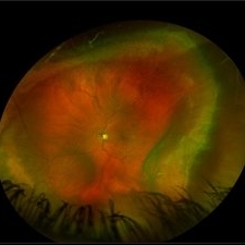
Fundus photograph of the left eye of a 60-year-old man with an acquired optic pit maculopathy and glaucoma. The image shows an enlarged optic disc cup and a macular serous detachment.
Photographer: Andree Henaine-Berra. Asociacion Para Evitar la Ceguera en Mexico. Mexico City.
Imaging device: Heidelberg Spectralis
Condition/keywords: glaucoma, maculopathy, optic pit
-
 Acquired Optic Pit Maculopathy
Aug 20 2014 by Andree Henaine-Berra, MD
Autofluorescence image of the left eye of a 60-year-old man with an acquired optic pit maculopathy and glaucoma.
Photographer: Andree Henaine-Berra. Asociacion Para Evitar la Ceguera en Mexico. Mexico City.
Imaging device: Heidelberg Spectralis
Condition/keywords: glaucoma, maculopathy, optic pit
-
 Acquired Optic Pit Maculopathy
Aug 20 2014 by Andree Henaine-Berra, MD
Optical coherence tomography of the left eye of a 60-year-old man with an acquired optic pit maculopathy and glaucoma. The image shows subretinal fluid extending to the optic nerve and schisis of the outer retinal layers.
Photographer: Andree Henaine-Berra. Asociacion Para Evitar la Ceguera en Mexico. Mexico City.
Imaging device: Heidelberg Spectralis
Condition/keywords: glaucoma, maculopathy, optic pit
-
 Advanced Glaucoma
Aug 21 2023 by Harsh Vardhan Singh, MS
A 64-year-old female with glaucomatous changes in both eye with advanced glaucoma in left eye
Photographer: Harsh Vardhan Singh
Condition/keywords: advanced stage glaucoma, Asymmetric cupping, glaucoma
-
---thumb.jpg/image-square;max$300,300.ImageHandler) Aniridia With Glaucoma
Aniridia With Glaucoma
Jan 2 2014 by David Callanan, MD
32-year-old male, aniridia with glaucoma, 20/200 OU.
Condition/keywords: aniridia, glaucoma
-
---thumb.jpg/image-square;max$300,300.ImageHandler) Aniridia With Glaucoma
Aniridia With Glaucoma
Jan 2 2014 by David Callanan, MD
32-year-old male, aniridia with glaucoma, 20/200 OU.
Condition/keywords: aniridia, glaucoma
-
---thumb.jpg/image-square;max$300,300.ImageHandler) Aniridia With Glaucoma
Aniridia With Glaucoma
Jan 2 2014 by David Callanan, MD
32-year-old male, aniridia with glaucoma, 20/200 OU.
Condition/keywords: aniridia, glaucoma
-
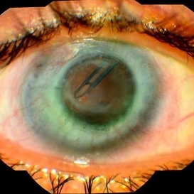
 Bleb Needling, Blood Streaming in Anterior Chamber
Jul 11 2013 by Jason S. Calhoun
Patient who had a bleb needling had a stream of blood start from the bleb superiorly to inferiorly within the anterior chamber.
Photographer: Jason S. Calhoun, Department of Ophthalmology, Mayo Clinic Jacksonville, Florida
Condition/keywords: glaucoma
-
 Central Retinal Vein Occlusion
Aug 17 2023 by Karen Santamaría
Fundus photograph of a female patient with a disc neovascularization secondary to central retinal vein occlusion.
Photographer: Karen Santamaría, Hospital Militar de Especialidades Oftalmológicas - Servicio de glaucoma, Ciudad de México
Imaging device: OCT RS-330 Retina Scan Duo 2 - NIDEK
Condition/keywords: Central vein oclussion, glaucoma
-
 Choroid Detachment
Jul 7 2021 by Patrik Rajs
This eye was a tough one. The patient underwent PPV twice, the second one with silicone oil (SO) for retinal re-detachment. Due to the development of secondary glaucoma, silicone oil evacuation and lavage of the anterior chamber were performed. Because of the high IOP even after the evacuation, the XEN was implanted. The surgery was followed by choroidal detachment presented in the picture on the left side along with the residual silicone bubble superiorly. The retinal tear is captured inferiorly surrounded by laser spots. The second image (on the right) was taken only 7 days later and it shows that choroidal detachment in the eye resolved completely.
Photographer: Patrik Rajs, EYE CLINIC of Jan Evangelista Purkyne University and Masaryk Hospital, Czech Republic, Ústí nad Labem
Condition/keywords: choroid, detachment, glaucoma, retina, silicone oil, tear
-
 Coloboma
Mar 26 2019 by Gary R. Cook, MD, FACS
White female with large coloboma of retina and choroid OD. She also had glaucoma in the eye.
Condition/keywords: coloboma, glaucoma
-
 Diabetic Macular Edema
May 28 2016 by Olivia Rainey
Optical coherence tomography of an 54-year-old female with diabetic macular edema affecting both eyes. Patient has a history of proliferative diabetic retinopathy s/p PRP/PPV/MP/EL, and glaucoma s/p tube shunt in both eyes. There has been a persistence of her macular edema and limited response to antiVEGF therapy, which puts into question whether there is another cause for her edema. Leading the possible causes is her renal insufficiency and fluid retention. Patient was seeing 20/50 in the right eye and 20/80 in the left eye.
Photographer: Olivia Rainey
Imaging device: Heidelberg Spectralis
Condition/keywords: anti-VEGF, diabetic macular edema, edema, glaucoma, optical coherence tomography (OCT), pan-retinal photocoagulation (PRP), proliferative diabetic retinopathy (PDR)
-
 Disc Heme Wedge Defect Blue Reflectance
Oct 5 2015 by Jared Watson
+FHx, HVF defects corresponding to RNFL thinning and ONH appearance.
Photographer: Jared Watson COT
Imaging device: Heidelberg Spectralis
Condition/keywords: glaucoma
-
 Fundus Showing Cupping of Optic Disc - Unedited Image from Smartphone
Nov 9 2018 by John Akkara
Optic disc cupping in right eye of a young glaucoma patient.
Photographer: Dr John Davis
Imaging device: HopeScope - Smartphone Fundus Camera
Condition/keywords: cupped out disc, glaucoma, inferior notch, notching
-
 Fundus Showing Cupping of Optic Disc - Unedited Image from Smartphone
Nov 9 2018 by John Akkara
Optic disc cupping in right eye of a young glaucoma patient.
Photographer: Dr. John Davis
Imaging device: HopeScope - Smartphone Fundus Camera
Condition/keywords: cupped out disc, glaucoma, inferior notch, notching
-
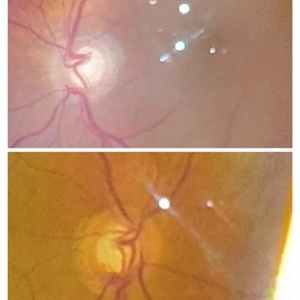
 Glaucoma
Feb 8 2018 by JEFFERSON R SOUSA, Tecg.º (Biomedical Systems Technology)
Male patient, 61-years-old in follow-up of glaucoma has several years. She performed trabeculectomy surgery with a tube implant.
Photographer: JEFFERSON R SOUSA - Study Center and Ophthalmological Research Dr. Andre M V Gomes, Dr. Suel Abujamra Institute São Paulo-Brazil
Imaging device: Fundus camera Acquisition of the image in the Camera background Topcon TRC-50 Dx - IA, field photo of 50 Degrees. Composition manual adjustment.
Condition/keywords: glaucoma
-
 Glaucoma
Feb 9 2015 by Govindarajan Venkatesan
Glaucoma.
Photographer: Govindarajan Venkatesan
Condition/keywords: optic disc
-
---thumb.jpg/image-square;max$300,300.ImageHandler) Glaucoma
Glaucoma
-
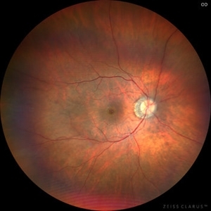 Glaucoma with full thickness macular hole
Jul 31 2023 by Harsh Vardhan Singh, MS
58-year-old male with open angle glaucoma & full thickness macular hole
Photographer: Dr Harsh Vardhan Singh, AIIMS, Guwahati
Imaging device: Zeiss Clarus 700
Condition/keywords: full thickness macular hole, glaucoma
-
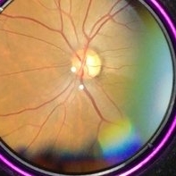
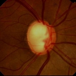
 Glaucomatous optic disc
Mar 5 2023 by Kalyan Singh
Chronic history of progressive diminution of vision. No history of previous glaucoma treatment.
Photographer: Kalyan Singh, GSVM medical college, Kanpur
Imaging device: Smartphone (1 plus 10 R)
Condition/keywords: glaucoma
-
 Iris
Iris
Apr 29 2019 by Stephanie Moolman
Multi-color images after Yag PI of iris.
Photographer: Stephanie Moolman, Dr Marissa Willemse, Pretoria, South Africa
Imaging device: Heidelberg Spectralis
Condition/keywords: glaucoma, iris, multicolor, NdYAG laser, peripheral iridotomy
-
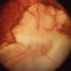
 Limited Choroidal Hemorrhage S/P Glaucoma Valve Implant OS; Retinoschisis
Aug 21 2023 by Angela Rico
A 52 year old Female presents to office S/P Glaucoma Valve Implant with IOP: 5mmHg OS
Photographer: Angela Rico M.D.
Condition/keywords: choroidal hemorrhage, glaucoma, hypotony, retinoschisis
 Loading…